3848
A continuous-time random-walk diffusion model for predicting tumor consistency and extent of resection in patients with pituitary adenomas1Department of Radiology, The First Affiliated Hospital of Nanjing Medical University, Nanjing, China, 2United Imaging Healthcare, Shanghai, China
Synopsis
Keywords: Tumors (Pre-Treatment), Tumor, Diffusion; Pituitary adenoma
Motivation: Preoperative evaluation of the consistency of pituitary adenomas (PAs) plays a significant role in the surgical strategy. However, previous studies concerning the assessment of tumor consistency of PAs were controversial1-4.
Goal(s): To evaluate the ability of the continuous-time random-walk (CTRW) diffusion model to predict the consistency and extent of resection (EOR) of PAs.
Approach: The CTRW diffusion model relaxes a priori distributions of waiting times and distance increments of water molecular diffusion, providing a realistic description of the complex structures of biological tissues5.
Results: CTRW diffusion model could provide information about the tumor consistency and EOR of PAs.
Impact: CTRW diffusion model provides an imaging dimension for characterizing tissue microstructure of PAs and may serve as a promising tool to predict the tumor consistency and EOR in patients with PAs.
Introduction
Pituitary adenomas (PAs) account for approximately 10-25% of intracranial neoplasms6. Endoscopic transsphenoidal surgery has been accepted as a preferred minimally invasive surgical approach to remove them. Extent of resection (EOR) has long been considered an important metric for clinical outcomes including prognosis of endocrinopathy and visual compromise and rates of recurrence. Besides tumor size and invasiveness, tumor consistency is a key determinant of EOR7-8. Preoperative knowledge of tumor consistency may aid surgeons in operative strategy selection and early risk stratification. The tumor consistency of PAs has been investigated in many previous studies based on conventional T2-weighted imaging (T2WI) and diffusion-weighted imaging (DWI), with controversial results1-3. Therefore, the utility of MRI in assessing tumor consistency of pituitary macroadenoma has not yet been established. Unlike other non-Gaussian diffusion models, the CTRW diffusion model recognizes intra-voxel diffusion heterogeneity in both time and space. This study aims to assess the performance of CTRW model in predicting the consistency and EOR of PAs.Methods
53 patients with pituitary macroadenoma were included. All MRI examinations including T1WI, coronal T2WI, contrast-enhanced T1WI, and multi-b-values DWI were performed on a 3.0 T MR scanner (uMR 770, United Imaging Healthcare, Shanghai, China). For DWI, scanning parameters were as follows: TR=2300ms, TE=131.5ms, FOV=200×200mm, matrix = 384×384, thickness=4mm, 15 b-values (0, 20, 30, 50, 80, 100, 120, 150, 200, 250, 300, 500, 700, 1000, 1500, 2000 s/mm2). Consistency of macroadenomas was evaluated by two neurosurgeons after surgical treatment and was classified into soft or hard groups. Regions of interest (ROIs) were manually delineated in the macroadenomas on EPI images with b = 0 s/mm2. Model fitting of the DWI data was performed based on the formula as follows: S/S0=Eα[(-(bDm))β], where Dm is an anomalous diffusion coefficient, α and β are parameters related to temporal and spatial diffusion heterogeneity, respectively, and Eα is a Mittag-Leffler function. Differences between CTRW model parameters were compared between the soft and hard groups using independent t-tests. Predictors of total or near-total resection were analyzed using appropriate univariate analyses, followed by multivariate logistic regression analysis. Receiver-operating-characteristic (ROC) analysis was adopted to evaluate the performance of clinical and imaging variables for discrimination.Results
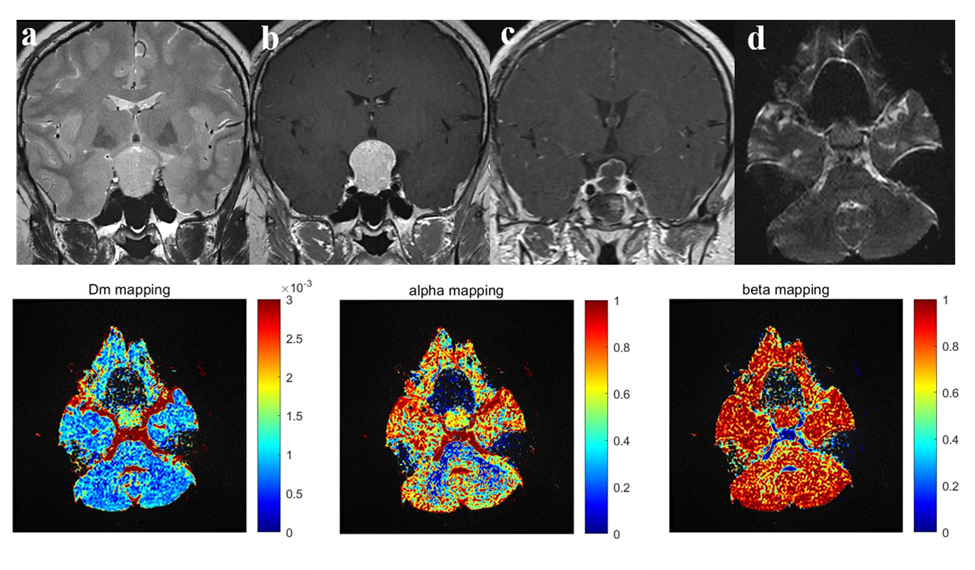
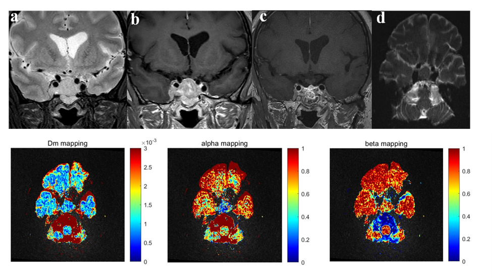
Among the 53 patients, tumor consistencies confirmed by surgery were hard macroadenoma in 17 patients and soft macroadenoma in 36 patients. The CTRW model parameters were found to be significantly lower in the hard PAs than the soft PAs (all P<0.05). In univariable analyses, patients who achieved total or near-total resection had lower Knosp grade (P=0.006), higher α (P<0.001), higher β (P<0.001) and higher ADC (P=0.032). Multivariable logistic regression analysis showed that Knosp grade (OR, 0.285; 95% CI, 0.109–0.743; P=0.007), α (OR, 1.102; 95% CI, 1.102-1.192; P=0.017) and β (OR, 1.202; 95% CI, 1.042-1.386; P=0.012) were independent predictors for achieving total or near-total resection after adjusting for the tumor diameter and volume of the PAs. Two typical cases—one soft and one hard PA—are shown in Figures 1 and 2, respectively. The variables of Knosp grade, α and β achieved an area under the ROC curve (AUC) of 0.766, 0.875 and 0.829 for predicting total or near-total resection, respectively. By the combination of Knosp grade, α and β, the AUC increased to 0.941 (95% CI, 0.840-0.987, sensitivity, 86.5%; specificity, 93.8%), significantly higher than that of Knosp grade alone (P=0.027).Discussion
The challenge of non-invasive evaluation for consistency of pituitary adenoma could be mainly attributed to the complexity of tumor composition. The CTRW model incorporates intra-voxel diffusion heterogeneity into the classical Fick’s equation by using two fractional order parameters, α and β. The β parameter was intended to account for the non-Gaussian distribution of diffusion displacement and has been related to the heterogeneous diffusion jump length associated with intravoxel spatial heterogeneity. The temporal parameter α reflects the temporal heterogeneity of the diffusion process, which means the likelihood of the water molecule to be “trapped” or “released” while it diffuses through the complex tissue structure. Although the spatial and temporal heterogeneity both originate from the underlying tissue structural heterogeneity, they reflect two independent and complementary aspects of diffusion heterogeneities5,9. According to the preliminary results, the consistency of PAs showed a strong correlation with the components of short α and β, which may be attributed to increased collagen content. Furthermore, we also found that the use of the CTRW model may enable neurosurgeons to better plan effective approaches and avoid multistage surgical procedures.Conclusion
This study demonstrates the potential utility of CTRW diffusion model in predicting tumor consistency and EOR in patients with PAs.Acknowledgements
No acknowledgement found.References
- Pierallini A, Caramia F, Falcone C, et al. Pituitary macroadenomas: Preoperative evaluation of consistency with diffusion-weighted MR imaging-initial experience. Radiology 2006;239:223–231.
- Smith KA, Leever JD, Chamoun RB. Prediction of consistency of pituitary adenomas by magnetic resonance imaging. J Neurol Surg B Skull Base 2015;76:340–343.
- Thotakura AK, Patibandla MR, Panigrahi MK, et al. Is it really possible to predict the consistency of a pituitary adenoma preoperatively? Neurochirurgie 2017;63:453–457.
- Su CQ, Zhang X, Pan T, et al. Texture Analysis of High b-Value Diffusion-Weighted Imaging for Evaluating Consistency of Pituitary Macroadenomas. J Magn Reson Imaging. 2020;51(5):1507-13.
- Karaman MM, Sui Y, Wang H, et al. Differentiating low- and high-grade pediatric brain tumors using a continuous-time random-walk diffusion model at high b-values. Magn Reson Med. 2016;76(4):1149-57.
- Alimohamadi M, Sanjari R, Mortazavi A, et al. Predictive value of diffusion-weighted MRI for tumor consistency and resection rate of nonfunctional pituitary macroadenomas. Acta Neurichir 2014;156: 2245–2252.
- Van Gerven L, Qian Z, Starovoyt A, et al. Endoscopic, Endonasal Transsphenoidal Surgery for Tumors of the Sellar and Suprasellar Region: A Monocentric Historical Cohort Study of 369 Patients. Front Oncol. 2021;11:643550.
- Youssef AS, Agazzi S, van Loveren HR. Transcranial surgery for pituitary adenomas. Neurosurgery 2005;57(1 Suppl):168–175. Karaman MM, Zhang J, Xie KL, et al. Quartile histogram assessment of glioma malignancy using high b-value diffusion MRI with a continuous-time random-walk model. NMR Biomed. 2021;34(4): e4485.
Figures