3841
The association between the intravoxel incoherent motion diffusion-weighted imaging(IVIM) with the DSC and 3DASL imaging in Glioma Recurrence1Department of Radiology, The First Medical Center, Chinese PLA General Hospital, Beijing, China
Synopsis
Keywords: Tumors (Post-Treatment), Diffusion/other diffusion imaging techniques, Glioma;recurrence
Motivation: Diffusion-weighted imaging and perfusion-weighted imaging have shown promising accuracy in differentiating glioma recurrence and treatment-related changes. Intravoxel incoherent motion (IVIM) MRI enables the simultaneous acquisition of diffusion and perfusion parameters, providing true diffusion and capillary perfusion in the tumor.
Goal(s): To evaluate the diagnostic performance of IVIM-derived parameters in glioma recurrence patients with DSC and 3DASL perfusion-weighted imaging.
Approach: The correlation between IVIM parameter and perfusion-MRI parameters was evaluated in regions of contrast enhancement with Pearson’s correlation analysis.
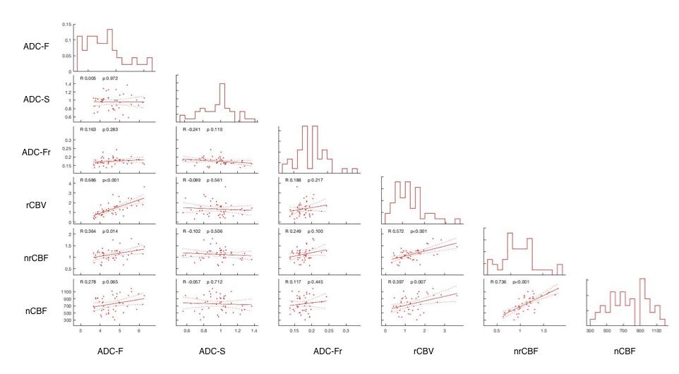
Results: ADC-F within T1-enhanced lesions positively correlated with relative cerebral blood volume (rCBV),nrCBF, and nCBF. Additionally,ADC-F was statistically significantly associated with rCBV and nrCBF.
Impact: IVIM has the same
diagnostic efficacy for recurrent glioma with the perfusion-MRI parameters and
can used as a reliable alternative diagnostic sequence for glioma
recurrence.
Introduction
Gliomas are the most common primary neoplasms of the brain. [1]The Recurrence of gliomas is histologically determined based on the presence of cellular anaplasia and nuclear atypia, cell density, mitoses, microvascular proliferation, and necrosis. The diffusion property has been reported to reflect cell density, and the perfusion property is largely determined by the microvascular proliferation or angiogenesis of tumors.[2] Le Bihan et al[3] developed a method to separate diffusion and perfusion parameters based on an intravoxel incoherent motion (IVIM) model. The IVIM MRI allows the simultaneous acquisition of diffusion and perfusion parameters which reflect true diffusion and capillary perfusion.[4]Thus, we evaluate the diagnostic performance of IVIM-derived parameters in glioma recurrence patients with DSC and 3DASL perfusion-weighted imaging.Materials ang Methods
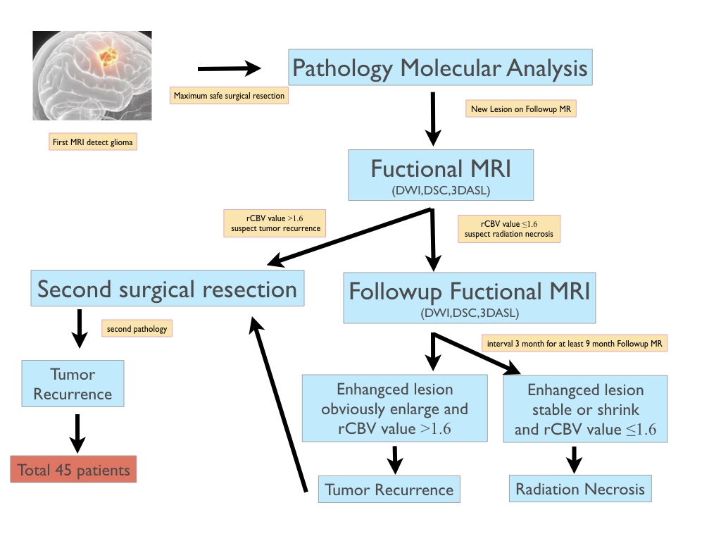
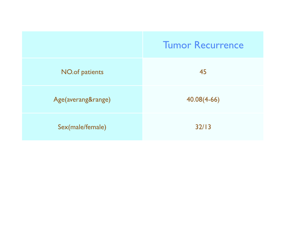
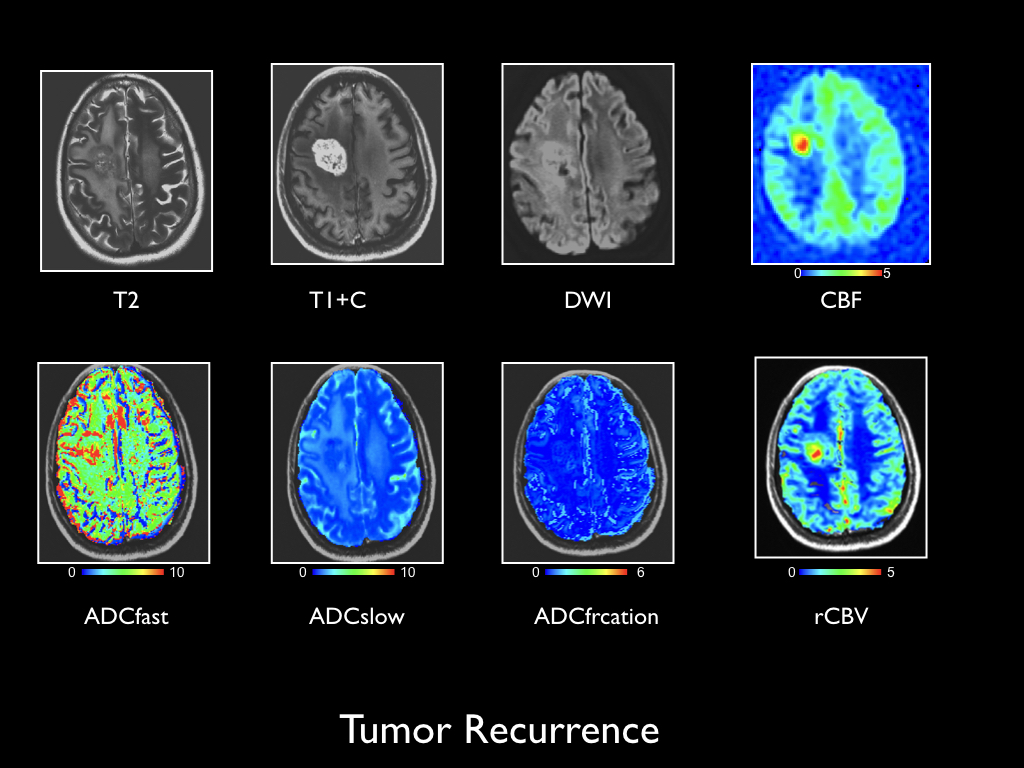
A total of 45 histologically confirmed glioma recurrence patients were enrolled in this retrospective study. (Fig.1) Gliomas with enhancement after standard treatment performed MR scanning which included IVIM, DSC, and ASL. (Fig.2)The IVIM DWI was performed before gadolinium injection using 11 different b-values: 0, 50, 100, 150, 200, 300, 500, 800, 1000, 2000 and 3000s/mm2. D is the diffusion parameter, D* represents perfusion-related diffusion and f is the perfusion fraction. Cerebral blood volume (rCBV) was calculated by DSC-MRI images Then it was performed by a bi-directional contrast agent leakage correction algorithm (Leu 2016), normalized by the average CBV value in contralateral, normal-appearing white matter (NAWM). Cerebral blood flow(nCBF) and relative cerebral blood flow(nrCBF) were calculated by 3D-ASL perfusion images. The correlation between IVIM parameters and perfusion-MRI parameters was evaluated in regions of contrast enhancement with Pearson’s correlation analysis. (Fig.3)Results
A statistically significant correlation was observed between ADC-F and rCBV within areas of enhancing tumor (R2=0.686, P=0.0043), the statistically significant correlation between ADC-F and nrCBF was also observed (R2=0.364,P=0.014). No statistical association was observed between ADC-F and nCBF although the trend is positive correlation within areas of enhancing tumor(R2=0.278,P=0.065).(Fig.4)Discussion and Conclusion
Median measurements within tumor regions across a large number of patients demonstrated a positive linear association between IVIM and perfusion-MRI in areas of contrast enhancement. These findings suggest that IVIM has the same diagnostic efficacy for recurrent glioma with the perfusion-MRI parameters and can be used as a reliable alternative diagnostic sequence for glioma recurrence.Acknowledgements
No acknowledgement found.References
[1] OSTROM Q T, CIOFFI G, WAITE K, et al. CBTRUS Statistical Report: Primary Brain and Other Central Nervous System Tumors Diagnosed in the United States in 2014–2018[J]. Neuro-Oncology, 2021, 23(Supplement_3): iii1–iii105. DOI:10.1093/neuonc/noab200.
[2] SUGAHARA T, KOROGI Y, KOCHI M, et al. Usefulness of diffusion-weighted MRI with echo-planar technique in the evaluation of cellularity in gliomas[J]. Journal of Magnetic Resonance Imaging, J Magn Reson Imaging, 1999, 9(1): 53–60. DOI:10.1002/(sici)1522-2586(199901)9:1<53::aid-jmri7>3.0.co;2-2.
[3] LE BIHAN D, BRETON E, LALLEMAND D, et al. Separation of diffusion and perfusion in intravoxel incoherent motion MR imaging.[J]. Radiology, Radiology, 1988, 168(2): 497–505. DOI:10.1148/radiology.168.2.3393671.
[4] SHIM W H, KIM H S, CHOI C-G, et al. Comparison of Apparent Diffusion Coefficient and Intravoxel Incoherent Motion for Differentiating among Glioblastoma, Metastasis, and Lymphoma Focusing on Diffusion-Related Parameter[J]. PLOS ONE, PLoS One, 2015, 10(7): e0134761. DOI:10.1371/journal.pone.0134761.
Figures