3839
Application of Contrast-Enhanced MULTIPLEX T1 and aT1-weighted Imaging in the Diagnosis of Brain Metastases1Department of Radiology, The Affiliated Drum Tower Hospital of Nanjing University Medical School, Nanjing University, Nanjing, China, 2Department of Geriatrics,The Affiliated Drum Tower Hospital of Nanjing University Medical School, Department of Geriatrics,The Affiliated Drum Tower Hospital of Nanjing University Medical School, Nanjing, China, 3Central Research Institute, United Imaging Healthcare Group Co., Ltd, Shanghai, China
Synopsis
Keywords: Tumors (Pre-Treatment), Tumor, MULTI-parametric MR imaging with fLEXible design(MULTIPLEX); contrast-enhanced(CE); gradient recalled-echo(GRE);brain tumor; intracranial metastasis
Motivation: Early monitoring for intracranial metastatic lesions through non-invasive methods will directly impact patient prognosis and quality of life.
Goal(s): The aim of this study was to evaluate the performance of MULTIPLEX in detecting brain metastases.
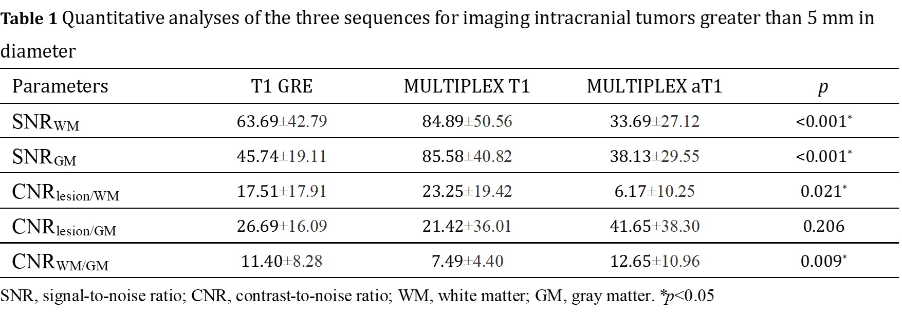
Approach: Signal-to-noise ratios (SNRs) for gray matter (GM) and white matter (WM), contrast-to-noise ratios (CNRs) for lesion/GM, lesion/WM, and GM/WM were quantitatively compared.
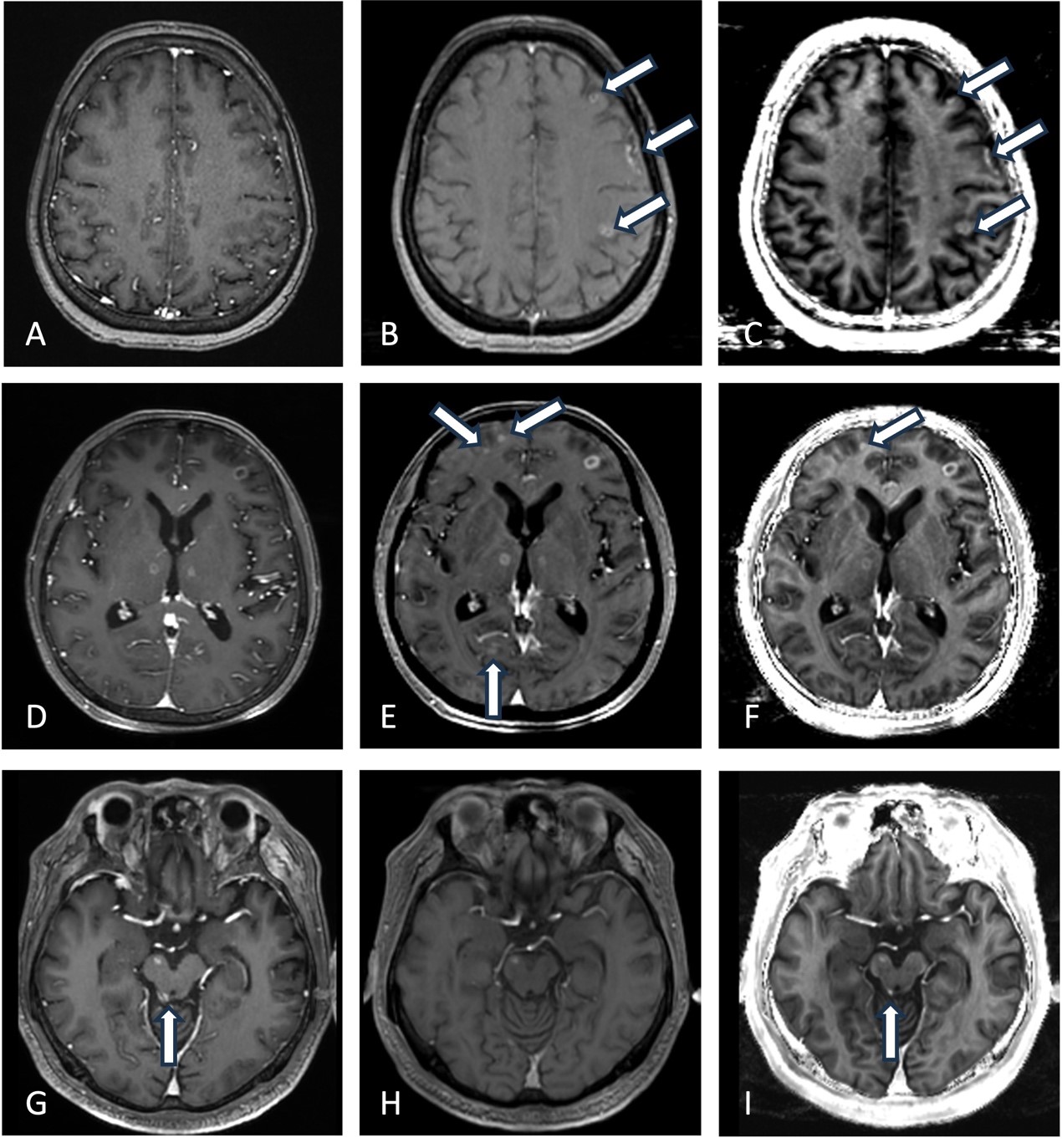
Results: MULTIPLEX aT1 imaging is preferred for depicting lesions containing hemorrhage or calcifications.
Impact: The combination of MULTIPLEX T1 and aT1 sequences enhances the detection of brain metastatic lesions compared to T1-GRE imaging, which could be helpful for detecting of small metastatic tumors.
Background
With the advancement of systemic therapies for lung cancer, the incidence of brain and meningeal metastases increases with prolonged patient survival1-2. Early monitoring for intracranial metastatic lesions through non-invasive methods will directly impact patient prognosis and quality of life3-5. The use of non-invasive high-resolution magnetic resonance imaging combined with new scanning sequences is beneficial for the visualization and diagnosis of brain and meningeal metastatic lesions6.MULTIPLEX can not only provide multi-contrast images but also offer quantitative parameters, which aids in the visualization of tumors with hemorrhage and calcification.Purpose
The aim of this study was to compare the performance of contrast-enhanced (CE) MULTIPLEX T1-weighted image, aT1-weighted image, and conventional T1-weighted gradient recalled echo (GRE) imaging in detecting brain metastases.Methods
Forty-nine patients suspected of intracranial metastases were enrolled in this study. Contrast-enhanced MULTIPLEX T1, aT1, and conventional T1 GRE imaging were separately reviewed. Signal-to-noise ratios (SNRs) for gray matter (GM) and white matter (WM), contrast-to-noise ratios (CNRs) for lesion/GM, lesion/WM, and GM/WM were quantitatively compared. Two radiologists determined the total number of enhancing lesions by consensus. Intraclass correlation coefficients (ICCs) between the two radiologists for the presence of metastases, qualitative ratings for image quality, and diagnostic confidence of enhancing lesions for each sequence were assessed.Results
Among the three sequences, SNRs between lesions and surrounding parenchyma were highest using MULTIPLEX T1 imaging, but CNRWM/GM was the lowest with MULTIPLEX T1 imaging(P<0.05). SNRWM for T1 GRE images were higher than those for MULTIPLEX aT1 imaging (P<0.001). CNRlesion/WM for MULTIPLEX T1 imaging were higher than those for MULTIPLEX aT1 imaging (P=0.007), while other CNRs between lesions and surrounding parenchyma were similar for 3 sequences (P>0.05). Significantly more brain metastases were detected with MULTIPLEX T1 imaging (n=186) compared with MULTIPLEX aT1 imaging (n=172) and T1 GRE imaging(n=159).Furthermore, all detected lesions with discrepant findings among the three sequences had a diameter of less than 5mm. The ICCs were 0.965 for MULTIPLEX T1 imaging, 0.954 for MULTIPLEX aT1 imaging and 0.946 for T1 GRE imaging. Qualitative scores for lesion imaging were similar for 3 sequences(P>0.05). The diagnostic confidence of enhancing lesions using MULTIPLEX T1 imaging were significantly higher than those obtained with MULTIPLEX aT1and T1 GRE imaging (P=0.027 and P=0.019, respectively),while the aT1 sequence provided clearer visualization of lesions containing hemorrhage or calcifications.
Conclusion
MULTIPLEX aT1 imaging is preferred for depicting lesions containing hemorrhage or calcifications. The combination of MULTIPLEX T1 and aT1 sequences enhances the detection of brain metastatic lesions compared to T1-GRE imaging, which could be helpful for detecting of small metastatic tumors.Acknowledgements
No acknowledgement found.References
1. Ye Y, Lyu J, Hu Y, et al. MULTI-parametric MR imaging with fLEXible design (MULTIPLEX). Magn Reson Med. 2022 Feb;87(2):658-673.
2.Leupold J, Kiselev VG. On the noise in "augmented T1-weighted steady state magnetic resonance imaging". NMR Biomed. 2023;36(5):e4886.
3.Fu Q, Cheng QG, Kong XC, et al. Comparison of contrast-enhanced T1-weighted imaging using DANTE-SPACE, PETRA, and MPRAGE: a clinical evaluation of brain tumors at 3 Tesla. Quant Imaging Med Surg. 2022;12(1):592-607.
4. Kaufmann TJ, Smits M, Boxerman J, et al. Consensus recommendations for a standardized brain tumor imaging protocol for clinical trials in brain metastases. Neuro Oncol. 2020;22(6):757-772.
5.Oh J, Choi SH, Lee E, et al. Application of 3D Fast Spin-Echo T1 Black-Blood Imaging in the Diagnosis and Prognostic Prediction of Patients with Leptomeningeal Carcinomatosis. AJNR Am J Neuroradiol. 2018;39(8):1453-1459.
6. Seong M, Park S, Kim ST, et al. Diagnostic accuracy of MR imaging of patients with leptomeningeal seeding from lung adenocarcinoma based on 2017 RANO proposal: added value of contrast-enhanced 2D axial T2 FLAIR. J Neurooncol. 2020;149(2):367-372.
Figures