3829
Quick, Quiet and Quantitative Magnetization Transfer Imaging1King's College London, London, United Kingdom
Synopsis
Keywords: Quantitative Imaging, Magnetization transfer, MS, ZTE
Motivation: Quantitative magnetization transfer (qMT) imaging has been long known to provide valuable information for MS imaging. To promote the widespread adoption of this technique faster and efficient scan protocols are needed.
Goal(s): To develop a patient-friendly, rapid, and reliable quantitative magnetization transfer scan.
Approach: Our sequence consists of an inversion pulse followed by a zero-echo time (ZTE) readout. We observe magnetization transfer (MT) exchange effects between a fully inverted free water pool and a partially inverted bound water pool.
Results: We show the feasibility of the method by producing quantitative maps of MT parameters.
Impact: Fast patient-friendly quantitative scans will enable informative and frequent monitoring of MS. This is useful for personalised
management of therapeutic courses and for drug development. We demonstrate a quick, quiet quantitative MT scan
targeted to multiple sclerosis.
Introduction
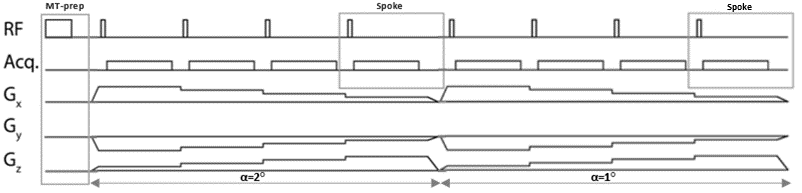
Quantitative MRI (qMRI) provides unique and specific information about brain tissues1. A common drawback of qMRI is the long acquisition time and poor repeatability of the results1. In common with other diseases, it is important to track the evolution of multiple sclerosis over time. Specific biomarkers are sought especially for the progressive disease2. In this abstract, we present preliminary results towards rapid quantitative MT (qMT) imaging using an MT-prepared ZTE sequence (see Figure 1).The advantages of ZTE are quiet scans, due to slow gradient changes and fast 3D radial acquisitions. The contrast of the sequence is altered from the native T1-weighting using preparation pulses3.
Methods
SequenceWe used a 3T GE Premier scanner (GE Healthcare, Chicago, USA) equipped with a 48-channel head coil. The trajectory used curved spokes with incoherent k-space sampling using a 2D golden means pattern4. The sequence consists of an inversion pulse followed by two ZTE readouts; see Figure 1. Dummy sequence repetitions are played before the acquisition to establish an overall steady state. The inversion pulse fully inverts the free water pool and saturates the bound pool to 24% of its equilibrium magnetization5. Other parameters were flip angles 2° and 1°, for the first and second readout segment respectively, spokes per segment, SPS = 384, FOV = 224mm isotropic with 1.4mm isotropic voxel size, readout bandwidth +/-20.83kHz, TR = 2.172 ms, pulse-width, tp = 24 µs. The total scan time was 2 minutes and 59 seconds.
Reconstruction
We used Riesling6 to reconstruct 16 frames from the ZTE readout by binning the data every 48 spokes. The reconstruction used the Alternating Directions Method-of-Multipliers (admm) with Total Variation regularization along the time dimension (tvt) with regularization parameter, λ = 0.01.
Model, simulation and fitting
We simulated our sequence following the theory of homogenised Bloch equations presented by Malik et al7. This simulation method is particularly suited to steady-state (SS) ZTE imaging. In fact, SS solutions are cumbersome to find by algebraic manipulation and direct integration of the equations is slow. We assumed the sequence to be perfectly spoiled and only simulated the two longitudinal components of the magnetization. We also assumed an on-resonance absorption lineshape value of the bound pool, G0 = 14 µs-1 and T1f = T1b for the free and bound pool relaxation times8. Our MT model was characterised by the semisolid fraction fs (defined such that fs + ff = 1) and the exchange rate k such that kfs = kff 9. The parameter estimates were then extracted voxel-wise with non-linear least-squares fits from Scipy (scipy.curve_fit)10. The fit parameters were fs, T1f, k and B1.
Results and discussion
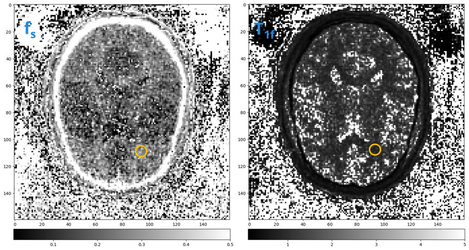
Figure 4 shows the expected recovery of the magnetization throughout the reconstructed frames starting from full inversion of the signal up to the equilibrium value. We successfully generated fs and T1f maps from our MT-prepared ZTE sequence; see Figures 2 and 3. The parameter maps, albeit noisy, show values in the expected range in brain tissue1,7. WM is expected to have the highest values. An analysis of the ROIs in Figures 3 and 4 returned fs = 0.284±0.020 and T1f = 0.897±0.048 s-1. It was impossible to extract estimates for GM and CSF. Our B1+ and k maps were dominated by noise but the values are within plausible ranges, k = (0.1, 20.0) and B1+ = (0.5, 2.0).We are now working on Cramér–Rao lower-bound optimization to evaluate theoretically optimal sequence parameters11. Additionally, we are also developing a model-based subspace reconstruction to further improve image quality12. This method should allow convenient extraction of parameter maps and the optimal use of all the information from the ZTE transient. All improvements rely on accurate simulations, we are therefore investigating if the assumptions in our model are accurate enough. For example, the saturation of the bound pool after the inversion pulse and the equivalence of the relaxation times of the free and bound pool7,8.
Conclusion
We have demonstrated the feasibility of qMT using a magnetization-prepared ZTE sequence. We extracted quantitative semisolid fraction and free-water relaxation time maps which albeit noisy provide the expected contrast of white matter. This work contributes to the development of imaging in MS by proposing a scan which is silent, thus patient-friendly, fast, and rich in information about the disease.Acknowledgements
No acknowledgement found.References
1. E. N. York, M. J. Thrippleton, R. Meijboom, D. P. J. Hunt, and A. D. Waldman, ‘Quantitative magnetization transfer imaging in relapsing-remitting multiple sclerosis: a systematic review and meta-analysis’, Brain Communications, vol. 4, no. 2, p. fcac088, Apr. 2022, doi: 10.1093/braincomms/fcac088.
2. T. Kuhlmann et al., ‘Multiple sclerosis progression: time for a new mechanism-driven framework’, The Lancet Neurology, vol. 22, no. 1, pp. 78–88, Jan. 2023, doi: 10.1016/S1474-4422(22)00289-7.
3. E. Ljungberg et al., ‘Silent zero TE MR neuroimaging: Current state-of-the-art and future directions’, Progress in Nuclear Magnetic Resonance Spectroscopy, vol. 123, pp. 73–93, Apr. 2021, doi: 10.1016/j.pnmrs.2021.03.002.
4. Chan, R.W., Ramsay, E.A., Cunningham, C.H., Plewes, D.B., 2009. Temporal stability of adaptive 3D radial MRI using multidimensional golden means. Magnetic Resonance in Medicine 61, 354–363. https://doi.org/10.1002/mrm.21837 .
5. L. A. Reynolds et al., ‘Nonaqueous magnetization following adiabatic and selective pulses in brain: T1 and cross-relaxation dynamics’, NMR in Biomedicine, vol. 36, no. 8, p. e4936, 2023, doi: 10.1002/nbm.4936.
6. T. C. Wood, E. Ljungberg, and F. Wiesinger, ‘Radial Interstices Enable Speedy Low-volume Imaging’, Journal of Open Source Software, vol. 6, no. 66, p. 3500, Oct. 2021, doi: 10.21105/joss.03500.
7. S. J. Malik, R. P. A. G. Teixeira, D. J. West, T. C. Wood, and J. V. Hajnal, ‘Steady-state imaging with inhomogeneous magnetization transfer contrast using multiband radiofrequency pulses’, Magnetic Resonance in Medicine, vol. 83, no. 3, pp. 935–949, 2020, doi: 10.1002/mrm.27984.
8. M. Gloor, K. Scheffler, and O. Bieri, ‘Quantitative magnetization transfer imaging using balanced SSFP’, Magnetic Resonance in Medicine, vol. 60, no. 3, pp. 691–700, 2008, doi: 10.1002/mrm.21705.
9. Y. Wang, P. van Gelderen, J. A. de Zwart, and J. H. Duyn, ‘B0-field dependence of MRI T1 relaxation in human brain’, NeuroImage, vol. 213, p. 116700, Jun. 2020, doi: 10.1016/j.neuroimage.2020.116700.
10. P. Virtanen et al., ‘SciPy 1.0: fundamental algorithms for scientific computing in Python’, Nat Methods, vol. 17, no. 3, Art. no. 3, Mar. 2020, doi: 10.1038/s41592-019-0686-2.
11. R. P. A. G. Teixeira, S. J. Malik, and J. V. Hajnal, ‘Joint system relaxometry (JSR) and Crámer-Rao lower bound optimization of sequence parameters: A framework for enhanced precision of DESPOT T1 and T2 estimation’, Magnetic Resonance in Medicine, vol. 79, no. 1, pp. 234–245, 2018, doi: 10.1002/mrm.26670.
12. X. Wang, Z. Tan, N. Scholand, V. Roeloffs, and M. Uecker, ‘Physics-based reconstruction methods for magnetic resonance imaging’, Phil. Trans. R. Soc. A., vol. 379, no. 2200, p. 20200196, Jun. 2021, doi: 10.1098/rsta.2020.0196.
Figures