3828
Single-Shot $$$T_1$$$ Mapping using an Interleaved SMS IR-FLASH Sequence and a NLINV Subspace Reconstruction1Institute of Biomedical Imaging, Graz University of Technology, Graz, Austria, 2Institute for Diagnostic and Interventional Radiology, University Medical Center Göttingen, Göttingen, Germany, 3DZHK (German Centre for Cardiovascular Research), Partner Site Göttingen, Göttingen, Germany, 4Department of Radiology, Harvard Medical School, Boston, MA, United States
Synopsis
Keywords: Quantitative Imaging, Image Reconstruction, Cardiac, Myocardial, T1 mapping, Single-Shot Sequence, qMR, NLINV, Subspace
Motivation: Volumetric quantification of$$$~T_1~$$$is highly desirable but limited by long acquisition times especially for motion-affected organs, such as heart. Therefore, conventional cardiac$$$~T_1~$$$mapping techniques are limited to single slices and suffer from low spatial resolution.
Goal(s): Develop a technique for rapid$$$~T_1~$$$mapping of multiple slices at high spatial resolution.
Approach: A radial slice-interleaved SMS readout was developed for single-shot IR-FLASH acquisition. NLINV subspace reconstruction is further proposed to jointly estimate subspace coefficient maps and coil sensitivities for all slices.
Results: Accurate$$$~T_1~$$$maps were achieved in a phantom and brain experiment for 9 slices within 4$$$~$$$seconds. Six cardiac short-axis myocardial$$$~T_1~$$$maps were reconstructed with an in-plane resolution of $$$1.25~mm^2$$$.
Impact: Multi-slice $$$T_1$$$ mapping has been achieved with a slice-interleaved radial SMS IR-FLASH acquisition and subspace NLINV reconstruction. This development may facilitate high resolution whole-heart myocardial $$$T_1$$$ mapping within a short breathhold (4 seconds).
Introduction
$$$T_1~$$$as a biomarker is of high interest in clinical applications$$$^1$$$, but conventionally its accessibility is limited by time-consuming acquisitions$$$^2$$$. Those become especially challenging in motion-affected organs such as the heart. Conventional myocardial $$$T_1$$$ mapping therefore utilizes gated acquisitions over multiple heartbeats during a breath-hold and has been restricted on single-slice applications.In this abstract, we develop a time-efficient multi-slice$$$~T_1~$$$mapping technique. We combine a single-shot IR-FLASH sequence incorporating radial slice-interleaved SMS readouts with NLINV subspace reconstruction to achieve acquisition times of 4 seconds for 6 or 9 slices. The technique is demonstrated in a phantom, brain and cardiac experiment.
Methods
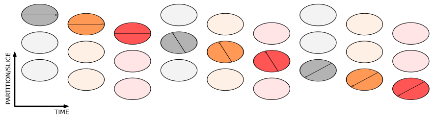
SequenceThe proposed multi-slice IR-FLASH sequence starts with a non-selective inversion followed by interleaved SMS excitations and radial readouts. The $$$n$$$ slice-interleaved SMS acquisitions with a multiband factor $$$m$$$ are specified as SMS$$$n$$$x$$$m$$$. Each of the $$$n$$$ SMS groups is sampled with the same pattern, with distinct samples in partition dimension utilizing an angle based on the golden-ratio between two consecutive spokes per group. (Figure$$$~1$$$)
Reconstruction
The underlying Look-Locker signal model in voxel$$$~r$$$
$$M(t,r)~=~M_{ss}(r)~-~(M_{ss}(r)~+~M_{0}(r))~e^{-t/T_1^*(r)}$$
is linearized in a subspace spanned by $$$N_c=4$$$ basis functions
$$M(\boldsymbol{x_p}(r),t)~\approx~\sum_c^{N_c}~\boldsymbol{x_c}(r)~B_c(t),$$
with the model parameters $$$\boldsymbol{x_p}(r)=(M_0, M_{ss}, T_1^*)^T$$$ and the respective subspace coefficients $$$\boldsymbol{x_c}(r)$$$. The basis functions are determined with a PCA from a simulated dictionary with different physical parameter combinations$$$^3$$$.
Coefficient maps $$$\boldsymbol{x_c}$$$ are reconstructed independently per SMS groups with the developed algorithm, a subspace version of SMS-NLINV$$$^{4,5}$$$.
The nonlinear forward operator
$$\textit{F}:~\boldsymbol{x}~=~(\boldsymbol{x_c},~\boldsymbol{c})~\to~\mathcal{P}~{\Xi} \mathcal{F}~\mathcal{D}~\mathcal{B}~(\boldsymbol{x_c}~\odot~\boldsymbol{c})$$
maps the unknown coil sensitivities $$$\boldsymbol{c}$$$ and coefficient maps $$$\boldsymbol{x_c}$$$ to the acquired k-space data $$$\boldsymbol{y}$$$. Here, $$$\odot$$$ is the element-wise product, $$$\mathcal{B}$$$ the basis operator, $$$\mathcal{D}$$$ a temporal mask selecting diastolic data, $$$\mathcal{F}$$$ the Fourier transform, $$$\Xi$$$ the SMS-encoding matrix and $$$\mathcal{P}$$$ the radial sampling pattern.
The resulting nonlinear inverse problem
$$\hat{\boldsymbol{x}}~=~\arg~\min_{\boldsymbol{x}}~||F(\boldsymbol{x})-\boldsymbol{y}||^2_2~+~\alpha~||\boldsymbol{x_c}||^2_2~+~\alpha~||W\boldsymbol{c}||^2_2$$
incorporates a Sobolev weighting matrix$$$~W$$$ and is solved by an iteratively regularized Gauss Newton method$$$^4$$$.
After reconstruction of $$$\boldsymbol{x_c}$$$, the physical parameters are pixel-wise fitted in the linear subspace
$$\hat{\boldsymbol{x_p}}~=~\arg~\min_{\boldsymbol{x_p}}~||\mathcal{B}^H~\mathcal{DB}\boldsymbol{x_c}~-~\mathcal{B}^H~\mathcal{D}\mathcal{M}(\boldsymbol{x_p})||_2^2~.$$
Finally, the Look-Locker correction $$$T_1~=~M_0/M_{ss}~T_1^*~+~2~T_d$$$ with delay time $$$T_d$$$ is applied to obtain the $$$T_1~$$$map$$$^6$$$.
All reconstruction steps were implemented in the Berkeley advanced reconstruction toolbox (BART)$$$^7$$$.
Acquisition
The evaluation of the sequence was performed in a NIST-phantom and brain with SMS3x3, and demonstrated for myocardial mapping with SMS2x3.
All MRI experiments were performed on a 3T Magnetom Vida (Siemens Healthineers, Erlangen, Germany). Each IR-FLASH consists of 1080 spokes acquired at $$$T_R=3.6~ms$$$.
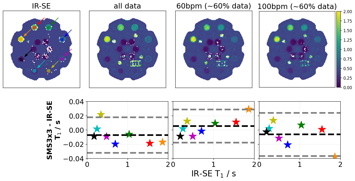
SMS3x3 was quantitatively evaluated in the NIST-phantom where reference values were determined with a gold-standard IR-SE sequence. To validate the gated IR-FLASH sequence, temporal patterns $$$\mathcal{D}$$$ corresponding to heart-rates of 60 and 100 bpm are defined, with data reduction matching the cardiac example.
Cardiac measurements were conducted during exhaled breath-hold and the ECG-trigger was set to start the inversion in early diastolic phase. Systolic data ($$$\approx 40\%$$$) was discarded in order to exclude motion-affected samples$$$^8$$$.
All measurements were performed with a slice thickness of $$$6~mm$$$ and slice gap for SMS$$$n$$$x$$$m$$$ of$$$~50\%$$$. The in-plane resolution was chosen to be $$$0.86~mm^2$$$ and $$$1.25~mm^2~$$$(cardiac).
Results & Discussion
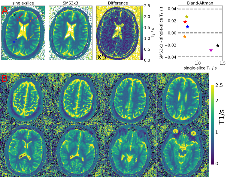
Figure$$$~2$$$ presents the NIST-phantom experiment. The reconstruction of the SMS3x3 IR-FLASH data is compared to a $$$T_1~$$$gold-standard mapping (IR-SE). In the corresponding Bland-Altman plot the bias across a wide range of $$$T_1$$$ is lower than 10 ms. The proposed technique can acquire accurate $$$T_1~$$$maps of $$$9~$$$slices in the phantom.The results of the brain measurements are presented in Figure$$$~3$$$. One representative slice of the SMS3x3 stack is quantitatively compared to the corresponding single-slice IR-FLASH reference. A difference of $$$\approx~2\%~$$$in the ROI analysis can be observed in white and gray matter. Figure$$$~3B$$$ shows a similar quality across the whole volume demonstrating the applicability of SMS3x3 to estimate 9$$$~T_1~$$$maps of different slices in the brain using the proposed method.
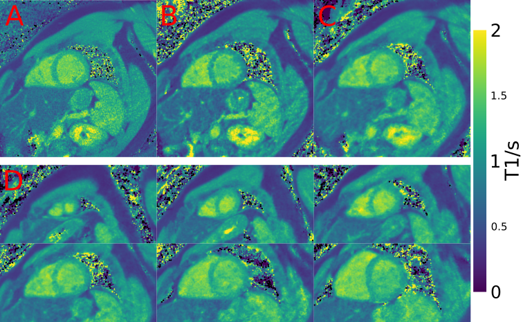
In Figure$$$~4$$$, a reference cardiac single-slice$$$~T_1~$$$map is compared to the extracted corresponding slice from a SMS1x3 and the SMS2x3 acquisition. The resolution is decreased compared to the brain due to the constraint of balancing the available data per partition/slice and the spatial coverage of the larger heart volume. In Figure 4D$$$~T_1~$$$maps of a whole-heart are presented with an in-plane resolution of $$$1.25~mm^2$$$. While the sharpness and quality decreases, the rapid acquisition of up to 6 $$$T_1~$$$maps in a single-shot is promising.
Conclusion
In this work, we combined a single-shot IR-FLASH acquisition incorporating an interleaved SMS excitation and radial readout with a subspace-based NLINV reconstruction. With the proposed method, multi-slice $$$T_1~$$$maps were reconstructed from datasets acquired within $$$4~s$$$. The accuracy is validated in a phantom and brain experiment. The cardiac results demonstrate promising results for single-shot whole-heart myocardial $$$T_1~$$$mapping.Acknowledgements
Funded by the DZHK (German Centre for Cardiovascular Research). Funded by the Deutsche Forschungsgemeinschaft (DFG, German Research Foundation) under Germany’s Excellence Strategy - EXC 2067/1- 390729940References
1. Kellman P and Hansen MS. T1-mapping in the heart: accuracy and precision. J Cardiovasc Magn Reson. 2014;16(1):2.
2. Cheng H, Stikov N, Ghugre N, Wright G. Practical medical applications of quantitative MR relaxometry. J Magn Reson Imaging 2012;36(4):805-24.
3. Wang X, Tan Z, Scholand N, Roeloffs V, Uecker M. Physics-based Reconstruction Methods for Magnetic Resonance Imaging.Philos Trans A Math Phys Eng Sci. 2021;379(2200):20200196.
4. Uecker M, Hohage T, Block KT, Frahm J. Image reconstruction by regularized nonlinear inversion-joint estimation of coil sensitivities and image content.Magn Reson Med. 2008;60(3):674-82.
5. Rosenzweig S, Holme HCM, Wilke R, Voit D, Frahm J, Uecker M. Simultaneous multi-slice MRI using cartesian and radial FLASH and regularized nonlinear inversion: SMS-NLINV.Magn Reson Med. 2018;79(4):2057-66.
6. Deichmann R. Fast high-resolution T1 mapping of the human brain. Magn Reson Med. 2005;54(1):20-7.
7. Uecker M, Virtue P, Ong F, Murphy M, Alley M, Vasanawala S, Lustig M. Software Toolbox and Programming Library for Compressed Sensing and Parallel Imaging. ISMRM Workshop on Data Sampling and Image Reconstruction, Sedona 2013.
8. Wang X, Joseph AA, Kalentev O, Merboldt KD, Voit D, Roeloffs V, van Zalk M, Frahm J. High-resolution myocardial T1 mapping using single-shot inversion recovery fast low-angle shot MRI with radial undersampling and iterative reconstruction. Br J Radiol. 2016;89(1068):20160255
Figures