3827
Spectral Localization by Imaging based T2 Relaxation Under Spin Tagging to measure the distribution of oxygen extraction fraction in human brain1Radiology, University of Kansas Medical Center, Kansas City, KS, United States, 2Hoglund Biomedical Imaging Center, University of Kansas Medical Center, Kansas City, KS, United States, 3Neurology, University of Kansas Medical Center, Kansas City, KS, United States, 4Guy’s and St Thomas’ NHS Foundation Trust, London, United Kingdom, 5FMRIB Division, Wellcome Centre for Integrative Neuroimaging, University of Oxford, Oxford, United Kingdom
Synopsis
Keywords: Quantitative Imaging, Quantitative Imaging, Oxygenation, oxygen extraction fraction (OEF), T2-relaxation-under-spintagging (TRUST), Spectral Localization by Imaging (SLIM)
Motivation: Reliable quantification and region/tissue specific mapping of cerebral oxygen extraction fraction (OEF) are critical for studying cerebral oxygen metabolism in health and disease.
Goal(s): To develop Spectral Localization by Imaging (SLIM) based TRUST techniques to overcome limitations of current TRUST techniques with limited spatial resolution or prolonged scan time for clinical applications at 3T.
Approach: SLIM-TRUST was developed with spatial-encoding and multi-echo for efficient sampling of venous blood transit time. SLIM approach was applied to quantify tissue-type/region-specific OEF.
Results: SLIM-TRUST provided reliable quantification of blood T2 values originated from GM and WM, and lateral and medial regions.
Impact: SLIM‐TRUST allows noninvasive measurement of spatial distribution of OEF, including GM/WM and various brain regions, suitable for clinical applications. The combination of SLIM and multi-echo TRUST approaches also promises reliable assessment of cerebral oxygen metabolism in clinically feasible scan time.
INTRODUCTION
T2‐relaxation‐under‐spin‐tagging (TRUST)1 allows the direct, non‐invasive assessment of cerebral oxygen extraction fraction (OEF), the relative difference in oxygen concentration between arterial and venous blood, from a slab of the brain. Currently available TRUST techniques, including spatially-localized TRUST, present limitations in spatial specificity and/or substantially longer scan time compared to other MRI techniques.2,3 To overcome these limitations, we sought to develop Spectral Localization by Imaging based TRUST (SLIM-TRUST), which employs the combination of SLIM and multi-echo TRUST to improve both data acquisition and processing of TRUST. SLIM-TRUST applies the concept of SLIM, which incorporates high-resolution anatomical MRI during reconstruction to enhance spatial resolution beyond conventional Fourier-based reconstruction.4,5 In addition, SLIM-TRUST uses multiple inversion delays to characterize the source of venous signals in TRUST data acquisition. In this study, we have incorporated multiple sampling of the transit time through a multi-echo approach without increasing the total scan time.METHODS
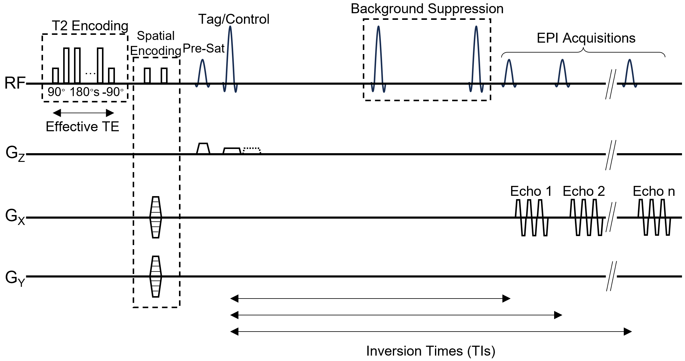
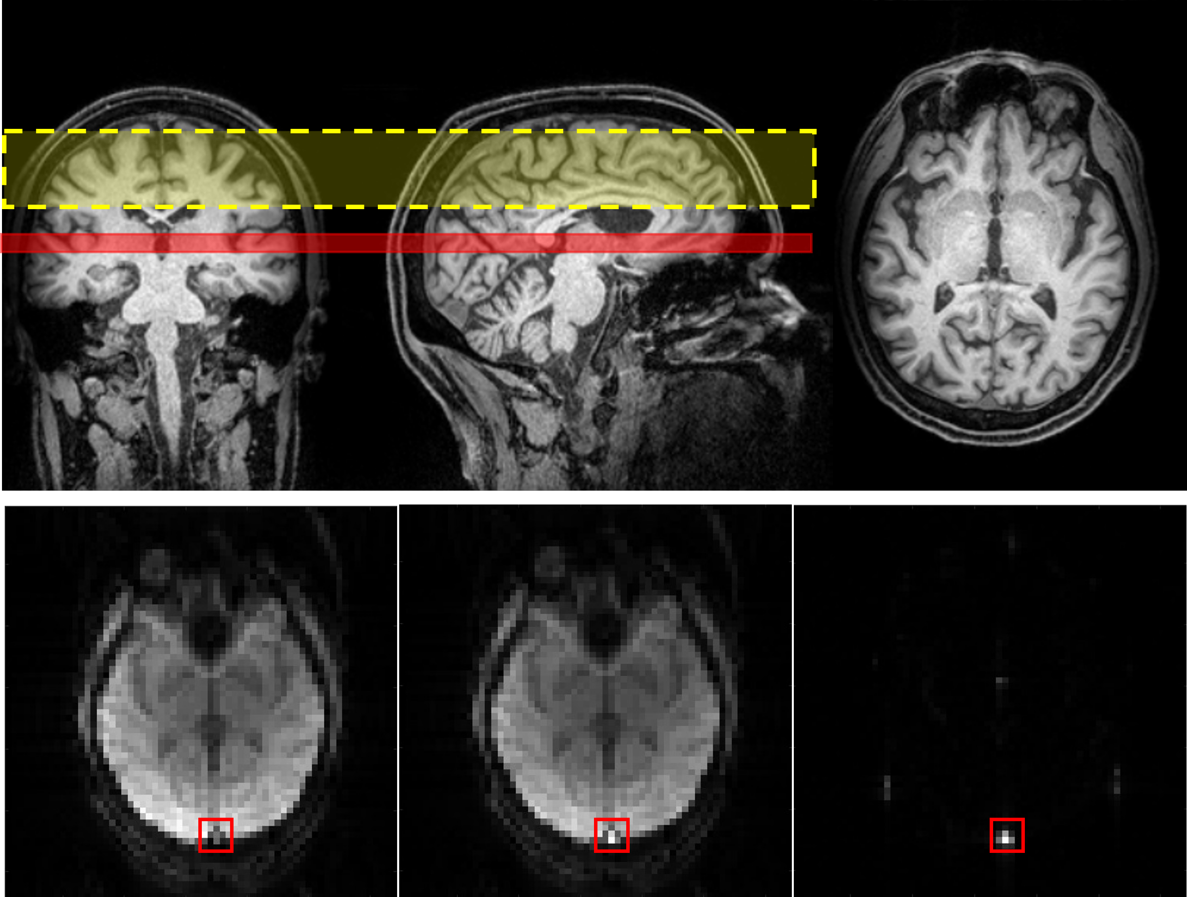
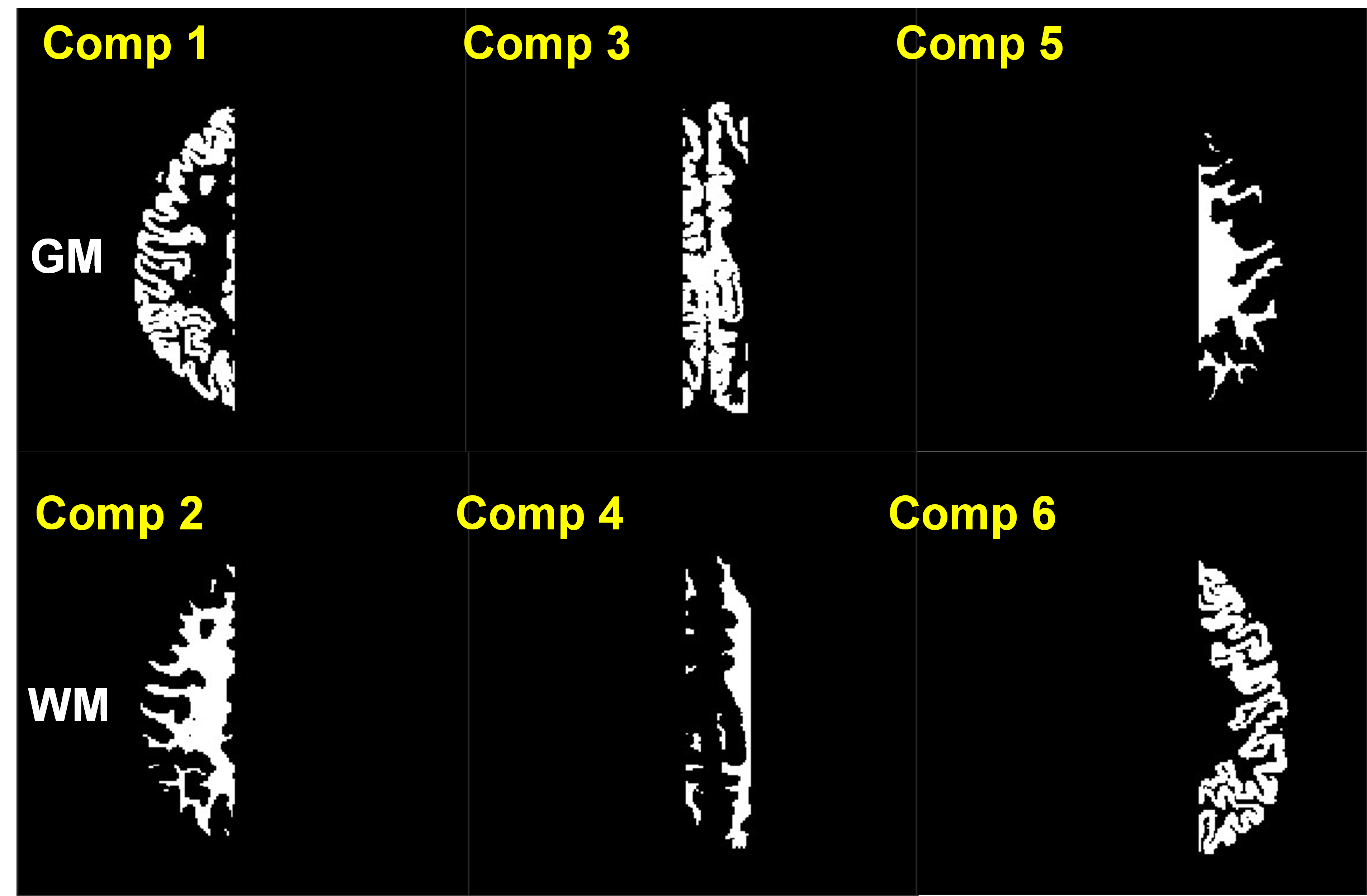
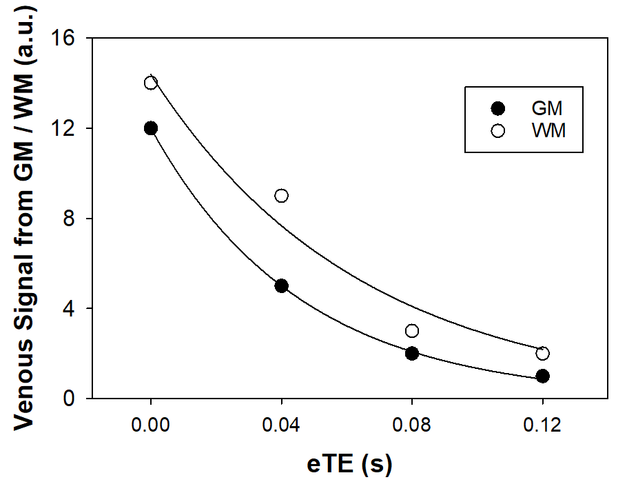
All SLIM-TRUST data were acquired from six healthy volunteers (4 male/2 female, age: 46±13 years) using a TRUST sequence with spatial encoding3 and multi-echo TI sampling (Fig. 1) (EPI TE/TR = 20/3500 ms, matrix = 64×64, FOV = 240×240 mm2, slice thickness = 10 mm, inversion slab thickness = 40-50 mm, TI = 1400, 1805, 2210, 2615 ms). Background suppression was performed prior to the slice excitation, and phase encoding of blood signals was achieved during the spin preparation3 . A total of 560 sets of EPI data were acquired from each subject to measure real/imaginary, spin tagging/control, phase encoding of blood signals (7x5), and effective TE (eTE) sampling (0, 40, 80, 120 ms) using a 32-channel head receive coil at 3T (Prisma, Siemens). Standard TRUST data were also acquired with five eTE’s as a global reference of blood T2.All SLIM-TRUST data processing, including MRI reconstruction with adaptive coil combination, was performed using in-house written MATLAB routines. For TRUST data processing, the sagittal sinus voxels were located from the pair-wise subtraction of spin-tagging control conditions. (Fig. 2) Two acquisitions of cosine and sine modulated venous signals per phase encoding were combined as a single complex number before SLIM reconstruction. SLIM reconstruction on spatially encoded TRUST data was performed using brain tissue compartments created using tissue segmentation masks generated from T1-weighted MRI (MPRAGE) (Fig. 3). Compartmental blood T2 values were calculated using an exponential fit (Fig. 4). Standard partial Fourier reconstruction was also performed to generate R2 maps of venous blood.
RESULTS AND DISCUSSION
The average blood T2 values originated from left lateral, medial, and right lateral brain regions were 58±14, 54±16, 55±6 ms for gray matter (GM) and 61±14, 62±23, 69±15 ms for white matter (WM), respectively. Global values of venous blood T2 were 60±6 ms for SLIM-TRUST and 69±6 ms for conventional TRUST. Overall, these values were consistent with previous findings from spatially-localized TRUST and references therein.2 The estimated OEF values of venous blood originated from GM and WM were approximately 40%, consistent with the average OEF (approximately 40%) using conventional TRUST or other spatially-localized TRUST methods.2 Overall T2 values for the GM originated blood were shorter than those from WM, consistent with higher oxygen consumption in GM than in WM. This study provides the first demonstration of tissue-type specific venous blood T2 (i.e., oxygenation) measurements using in vivo MRI.In conclusion, SLIM-TRUST could provide a new method that enables the measurement of tissue-type specific venous oxygenation levels, which has not been reported to date. (Fig. 4) While we used 7x5 k-space data points for SLIM reconstruction in this study, further reduction of the number of k-space data points to 5x5 or 4x4 should be readily achievable, depending on the number of tissue compartments, which will result in significant reduction in the total scan time. The combination of SLIM and multi‐echo TRUST techniques promises effective spatial localization of specific tissue-types or brain regions with any user-defined shapes, as well as significantly shorter scan time. Further optimization and characterization of SLIM-TRUST are needed for reliable quantification of tissue-type or regional differences in OEF measures for clinical studies in order to assess alterations in oxygen metabolism of brain tissue or regions in aging, neurodegeneration, and various neurological or cerebrovascular conditions.
Acknowledgements
This study is partially supported by NIH R01 AG060050 (to Lee P.).References
1. Lu H, Ge Y. Quantitative evaluation of oxygenation in venous vessels using T2‐relaxation‐under‐spin‐tagging MRI. Magn Reson Med (2008) 60: 357–363.
2. O'Brien C, Okell TW, Chiew M, Jezzard P. Volume-localized measurement of oxygen extraction fraction in the brain using MRI. Magn Reson Med (2019) 84: 1412-23
3. O'Brien C, Okell TW, Chiew M, Jezzard P. Oxygen Extraction Fraction Mapping using Remote Sensing: Spatially Encoded T2-Relaxation-Under-Spin-Tagging (SE-TRUST). Proc Int Soc Magn Reson Med (2020) 28: 35
4. Adany P, Choi IY, Lee P. B0-adjusted and sensitivity-encoded spectral localization by imaging (BASE-SLIM) in the human brain in vivo. Neuroimage (2016) 134: 355-364
5. Lee P, Adany P, Choi IY. Imaging based magnetic resonance spectroscopy (MRS) localization for quantitative neurochemical analysis and cerebral metabolism studies. Anal Biochem (2017) 529: 40-47Figures