3826
Feasibility of High-Speed R2 and R2’ Mapping Via Alternating SSFP With View-Sharing1School of Electronic and Electrical Engineering, Kyungpook National University, Daegu, Korea, Republic of, 2Kmedi-hub, Daegu, Korea, Republic of
Synopsis
Keywords: Quantitative Imaging, Relaxometry
Motivation: Quantifying R2 and R2' provide important implications about physiologic and functional states of tissues.
Goal(s): To explore feasibility of an accelerated R2 and R2’ mapping strategy.
Approach: A view-sharing method is integrated into the alternating nonbalanced SSFP-based technique enabling simultaneous R2 and R2’ mapping.
Results: : In both phantom and brain scans, the parametric maps obtained via the presented scan acceleration approach are overall in good agreement with those derived from a reference method.
Impact: Upon further evaluation, the method may be a useful means in a wide range of neuroimaging applications.
Introduction
The two transverse relaxation rate constants, typically represented by R2 and R2', provide important implications about physiologic and functional states of tissues, yet in very different scales to each other. Hence, quantifying both parameters is of great interest in a number of neuroimaging studies, for example, understanding the BOLD mechanism1 and quantifying iron deposits in deep gray matter structures2. An alternating nonbalanced SSFP-based technique (termed ‘AUSIFDE’) enabling simultaneous 3D mapping of R2 and R2’ across the entire brain3 has recently been introduced as a scan-time practical alternative to conventional spin-echo based methods4. The purpose of this work was to enhance imaging efficiency of the AUSIFDE pulse sequence via a view-sharing strategy.Methods
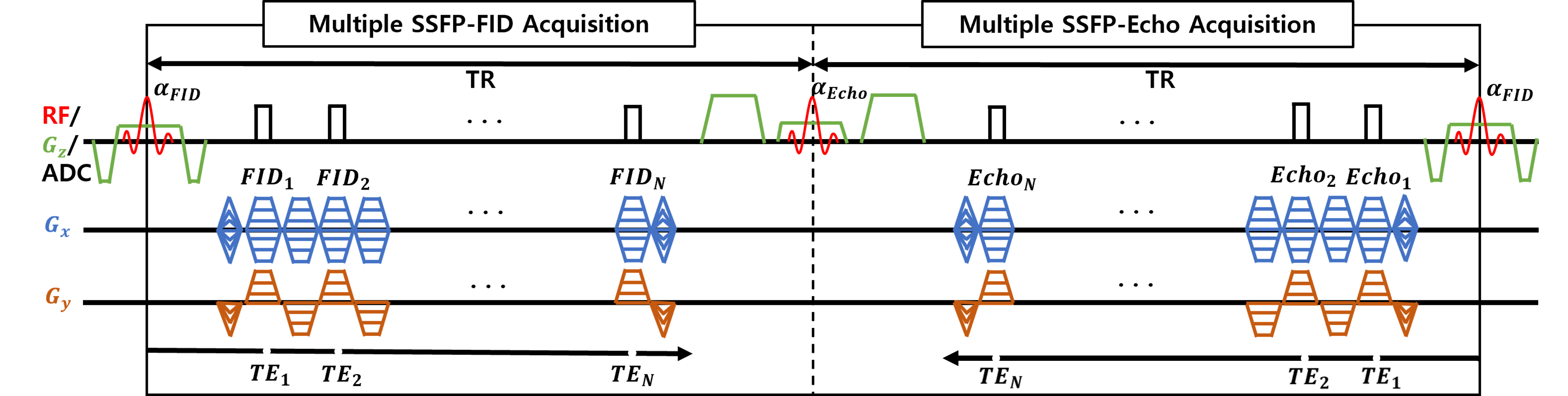
Pulse sequence configuration:Figure 1 shows a timing diagram of the 2D AUSIFDE pulse sequence. Here, SSFP-FID and SSFP-ECHO modules are alternately applied to selectively capture SSFP signals following zeroth and first pathways, respectively, while multiple gradient-recalled echo signals are acquired in each time-of-repetition (TR). Given that the temporal evolution of multi-echo signals within TR is expressed as R2+R2’ and R2-R2’ for SSFP-FID and SSFP-ECHO, respectively, the method allows estimation of both transverse relaxation parameters in a single data acquisition. Furthermore, a radial k-space sampling scheme was employed so as to achieve scan acceleration based on radial view sharing detailed in the following subsection.
View-sharing and image reconstruction:
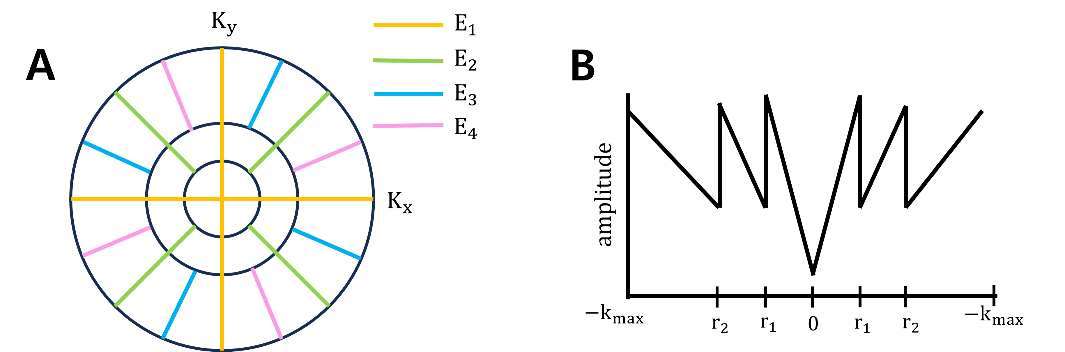
The underlying principle of the view-sharing approach is to exploit the property of radial encoding that central k-space data are inherently oversampled5. Specifically, for a given echo time (TE), corresponding image contrast may be obtained by filling the central portion of k-space with radial views collected at that TE, while complementing outer regions of k-space progressively with spokes acquired at neighboring TEs, as illustrated in Fig. 2a. With this strategy, scan times can be shortened by as many times as the number of echoes collected at various TEs. View-shared k-space for each TE along the echo train was constructed, and corresponding images were then produced by gridding reconstruction. Here, different sampling rates along the radial direction of k-space was accounted for in computing the density compensation function (DCF) (Fig. 2b).
Experiments and analysis:
All experiments in this study were performed at 3T (Siemens Skyra). Data in a phantom and in vivo brain were acquired using the 2D AUSIFDE pulse sequence with the following scan parameters: field-of-view = 240 x 240 mm2, slice thickness = 5 mm, TR = 33 ms, inter-echo spacing = 3.18 ms, number of readout samples = 160, number of views = 240, flip angles = 50°. In this preliminary study, view-sharing-based image reconstruction was simulated by retrospectively decimating sampled data in k-space. Additionally, trajectory errors in k-space resulting from gradient delays were estimated based on calibration data separately acquired, and the AUSFIDE data were corrected accordingly using the method by Block and Uecker6. Data were also collected using the GESFIDE pulse sequence for comparison with AUSIFDE-derived R2 and R2’ maps.
Results
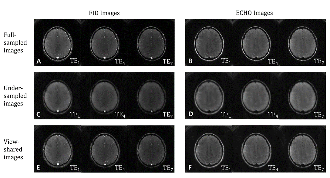
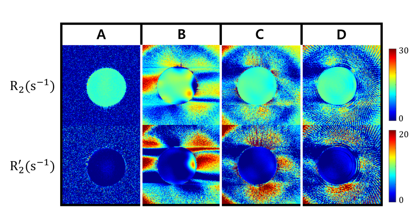
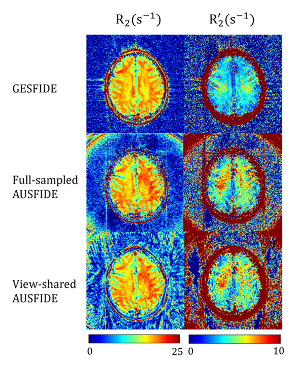
Figure 3 shows three sets of SSFP-FID (Fig. 3 left column) and SSFP-ECHO (Fig. 3 right column) images at three different TEs, reconstructed with fully sampled (Figs. 3A, 3B), undersampled (Figs. 3 C, 3D), and view-shared (Figs. 3E, 3F) k-space datasets. The undersampling factor of four in the middle case was chosen equal to the number of echoes composing the view-shared k-space, resulting in significant image blurring, while the images reconstructed from full and view-shared data are visually comparable. Figure 4 displays four sets of R2 and R2’ maps acquired in a phantom. See figure captions for detailed descriptions. Figure 5 shows R2 and R2’ maps of the human brain, derived using GESFIDE and AUSFIDE with fully sampled and view-shared k-space datasets. In both phantom and brain data, the parametric maps obtained using AUSFIDE with full sampling and view-sharing both are overall in good agreement with those in GESFIDE.Discussion and Conclusion
The results suggest feasibility of accelerated R2 and R2’ mapping via view-shared AUSFIDE imaging. However, the presented method needs further improvement to reduce ringing (Fig. 4D) and shading (Fig. 4C) artifacts, likely resulting from regional discontinuities in k-space, and incomplete correction of k-space trajectory mismatch, in combination with phase errors accumulated along the echo train. We are currently focused on scrutinizing these issues, and upon further evaluation the method may be a useful means in a wide range of neuroimaging applications.Acknowledgements
No acknowledgement found.References
1. Yablonskiy DA, Sukstanskii AL, He X. Blood oxygenation level-dependent (BOLD)-based techniques for the quantification of brain hemodynamic and metabolic properties – theoretical models and experimental approaches. NMR in Biomedicine 2013;26:963-986.
2. Sedlacik J, Boelmans K, Lobel U, Holst B, Siemonsen S, Fiehler J. Reversible, irrerversible and effective transverse relaxation rates in normal aging brain at 3T. NeuroImage 2014;84:1032-1041.
3. Hyunyeol Lee, Felix W. Wehrli. Alternating unbalanced SSFP for 3D R'2 mapping of the human brain. Magn Reson Med. 2021;85:2391-2402.
4. Wendy Ni,Thomas Christen, Zungho Zun, Greg Zaharchuk. Comparison of R2’ Measurement Methods in the Normal Brain at 3T. Magn Reson Med. 2015;73(3):1228–1236.
5. Hee Kwon Song, Lawrence Dougherty. K-space Weighted Image Constrast (KWIC) for Contrast Manipulation in Projection Reconstruction MRI. Magn Reson Med. 2000;44:825-832.
6. Block KT, Uecker M. Proceedings of the 19th scientific meeting, International Society for Magnetic Resonance in Medicine 2011; Montréal. pp. 2816.
Figures