3823
Simultaneous multi-slice MR-STAT for robust high-resolution full-brain relaxometry1Computational Imaging Group, Department of Radiotherapy, UMC Utrecht, Utrecht, Netherlands
Synopsis
Keywords: Quantitative Imaging, Quantitative Imaging
Motivation: MR-STAT is a framework that enables simultaneous T1, T2 and proton-density mapping. Currently, MR-STAT acquires slices sequentially which is relatively inefficient in terms of SNR and time.
Goal(s): Increase the scan-efficiency of MR-STAT while limiting motion-sensitivity
Approach: We use a simultaneous multi-slice (SMS) acquisition to increase the scan efficiency of MR-STAT and use a two-step approach for the reconstruction: an SMS-SENSE reconstruction followed by a conventional slice-by-slice MR-STAT reconstruction.
Results: Phantom and in-vivo results showed that a four-fold increase in encoded slices from 28 to 112 was possible achieving full-brain high-resolution T1, T2 and proton density maps in 5 minutes scan time.
Impact: MR-STAT combined with a simultaneous multislice acquisition enables a 4-fold increase in scan efficiency. This can be used to increase the resolution in the slice-direction and allow the detection of smaller brain structures while not increasing scan time.
Introduction
MR-STAT is a framework that estimates quantitative multi-parametric maps directly from the time-domain data acquired in a single-scan that features cartesian gradient encoding and a varying flip-angle train(1,2). The quantitative parameter maps are obtained by solving a large-scale non-linear inversion problem directly for simultaneous spatial localization and parameter estimation. In a conventional MR-STAT experiment, slices are acquired sequentially (10 s/slice) which makes the acquisition relatively inefficient in terms of SNR and time. To improve scan-efficiency, an extension of MR-STAT to 3D has been presented(3). However, the translation to 3D imaging currently increases the sensitivity to both bulk motion and flow due the longer time-scale of spatial and parameter encoding. In this work, we present an extension of the MR-STAT framework to use simultaneous multi-slice (SMS or multiband) (4,5) with the aim to increase the scan-efficiency of MR-STAT while limiting the sensitivity to motion.Methods
The SMS MR-STAT acquisition consisted of a Cartesian gradient-spoiled sequence with a time-varying flip-angle train and a linear k-space filling. This linear k-space filling was repeated five-times to yield sufficient T1 and T2-encoding. A two-step approach was used to incorporate SMS into the MR-STAT framework by performing an SMS-SENSE reconstruction followed by a standard slice-by-slice MR-STAT reconstruction. Here, the SENSE-reconstruction was performed for each of the k-spaces to resolve the spatial aliasing resulting from the SMS-acquisition (6). The resulting data was Fourier-transformed back to the k-space and was used as input to the time-domain MR-STAT reconstruction. Importantly, the SENSE-reconstruction spatially decouples the simultaneously excited slices which reduces both the memory-requirements and reconstruction time of the MR-STAT reconstruction. Quantitative parameters can thus subsequently be estimated in separate slice-by-slice MR-STAT reconstructions for each slice (3). Figure 1 shows a schematic representation of the SMS MR-STAT reconstruction.Experiments were performed at 3T and 7T with flip-angle trains optimized for low-SAR (root-mean-squared flip-angle = 35 degrees) using the BLAKJac framework (7). Receive coil sensitivities were obtained from a low-resolution RF-spoiled gradient-echo sequence and a 32-channel receive array was used at both field strengths. B1+-mapping was performed using a DREAM B1+-mapping sequence (8) which enabled the correction of B1+-inhomogeneities during the MR-STAT reconstruction.
At 3T, the effect of the SMS acquisition on the quantitative parameters was assessed on gel-vials with known T1/T2 (Eurospin T05). Here, two cases were explored: a standard 2D MR-STAT acquisition with 28 slices and a 4.5 mm slice-spacing and an SMS MR-STAT acquisition with SMS-factor = 4 and 112 slices and a 1.2 mm slice-spacing. In-vivo scans were performed using a standard 2D MR-STAT acquisition and an SMS MR-STAT acquisition with SMS-factor = 4. All acquisitions at 3T featured a slice-thickness of 3 mm and the same scan time of 5:26 [min:s]. In-vivo experiments at 7T were performed to explore the partial volume effects of slice-thickness in SMS MR-STAT. Here, we used the higher SNR at 7T allows to acquire 1.2 mm thick slices which were compared with 3 mm thick slices. Sequence parameters for all acquisitions can be found Table 1.
Results
Figure 2A shows the MR-STAT reconstructions for the phantom experiments. Here, the SMS MR-STAT scans (112 slices) show similar quantitative values and only a limited SNR penalty compared to the reference MR-STAT scan (28 slices) which can also be seen in Figure 2B. The in-vivo results in Figure 3 highlight the increase in through-plane resolution that can be obtained using SMS MR-STAT while keeping the same total scan time. The thin-slice 7T results in Figure 4 show smaller structures in the cerebellum and at tissue boundaries as the reduction of partial volume effects reduces blurring of small details.Conlusion and Discussions
We demonstrated that a combination of simultaneous multi-slice and MR-STAT enables a four-fold increase in the number of slices for a 2D-MR-STAT scan with a 5-minute scan time. SMS MR-STAT was enabled by combining the MR-STAT framework with a SENSE-reconstruction to resolve for spatial aliasing and allow for decoupling of the MR-STAT problems. Our 7T results showed that thinner slices enable visualization of smaller brain structures. This would also be beneficial at 3T but might be challenging due to SNR constraints. To mitigate this, the SENSE-reconstruction step can be combined with denoising regularization by including, e.g., total-variation penalty (9).We expect that SMS MR-STAT could be applied for fast parameter mapping in patients that might move during scanning, e.g. infants, as this SMS approach is expected to be less motion-sensitive than a 3D approach due to short acquisition time per set of slices (10 s/set of slices).
Acknowledgements
This work has been financed by NWO grant number 18951References
1. Sbrizzi A, Heide O van der, Cloos M, et al. Fast quantitative MRI as a nonlinear tomography problem. Magn. Reson. Imaging 2018;46:56–63.
2. van der Heide O, Sbrizzi A, Luijten PR, van den Berg CAT. High-resolution in vivo MR-STAT using a matrix-free and parallelized reconstruction algorithm. NMR Biomed. 2020;33:1–16.
3. Liu H, van der Heide O, Versteeg E, et al. A three-dimensional Magnetic Resonance Spin Tomography in Time-domain protocol for high-resolution multiparametric quantitative magnetic resonance imaging. NMR Biomed. 2023:1–15.
4. Larkman DJ, Hajnal J V, Herlihy AH, Coutts GA, Young IR, Sta Ehnholm G. Use of multicoil arrays for separation of signal from multiple slices simultaneously excited. Wiley Online Libr. 2001.
5. Ye H, Ma D, Jiang Y, et al. Accelerating magnetic resonance fingerprinting (MRF) using t-blipped simultaneous multislice (SMS) acquisition. Magn. Reson. Med. 2016;75:2078–2085.
6. Zahneisen B, Ernst T, Poser BA. SENSE and simultaneous multislice imaging. Magn. Reson. Med. 2015;74:1356–1362.
7. Fuderer M, van der Heide O, Liu H, van den Berg CAT, Sbrizzi A. Efficient performance analysis and optimization of transient-state sequences for multi-parametric MRI. NMR Biomed. 2022.
8. Nehrke K, Börnert P. DREAM-a novel approach for robust, ultrafast, multislice B1 mapping. Magn. Reson. Med. 2012;68:1517–1526.
9. Knoll F, Bredies K, Pock T, Stollberger R. Second order total generalized variation (TGV) for MRI. Magn. Reson. Med. 2011;65:480–491.
Figures
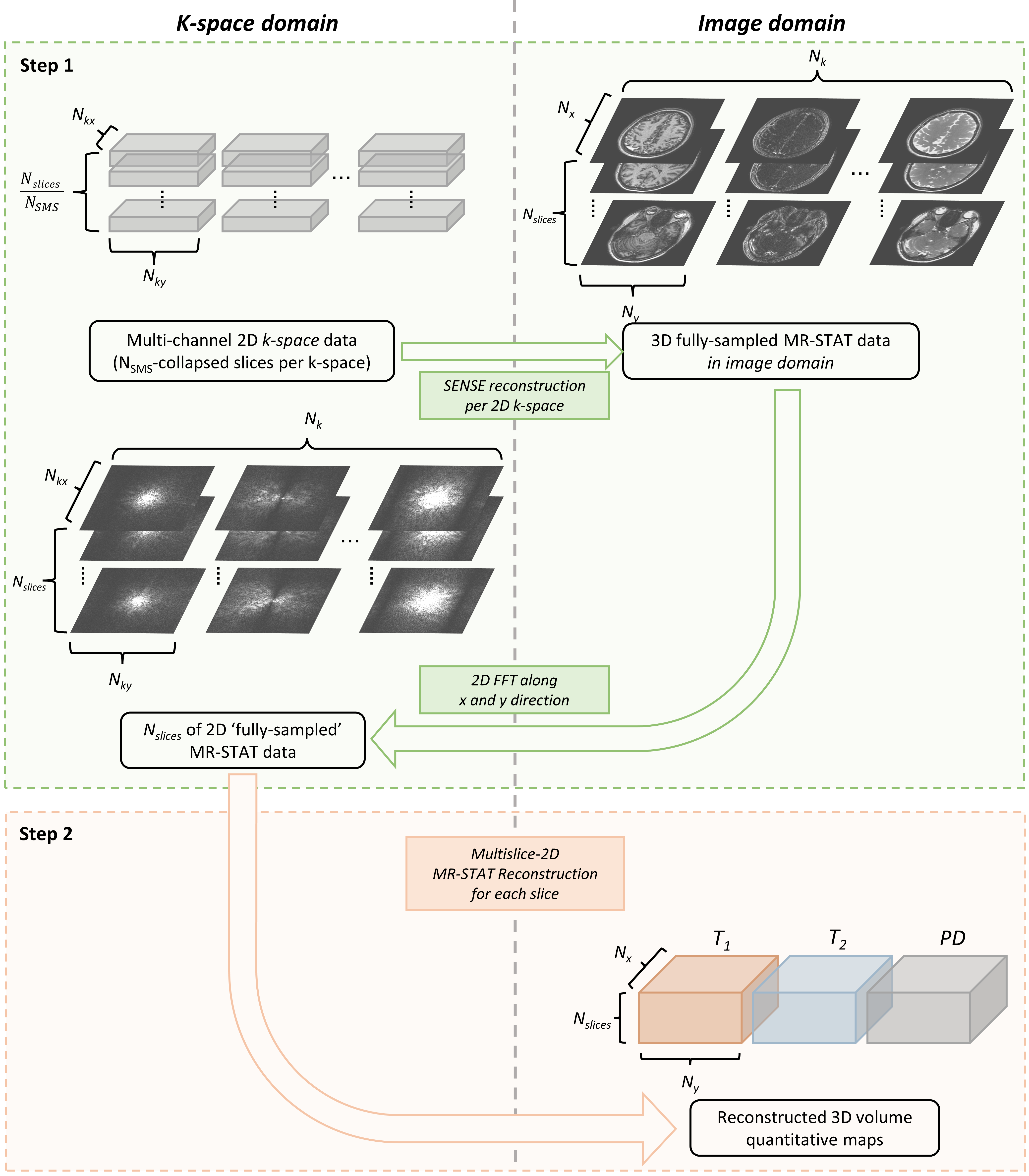
Figure 1 Schematic representation of two-step reconstruction used for SMS MR-STAT. Here, the first step consists of a SENSE reconstruction to resolve spatial aliasing and yield a fully-sampled 2D k-space for each slice and k-space. These k-spaces are used as input to the MR-STAT reconstruction to produce the quantitative maps.
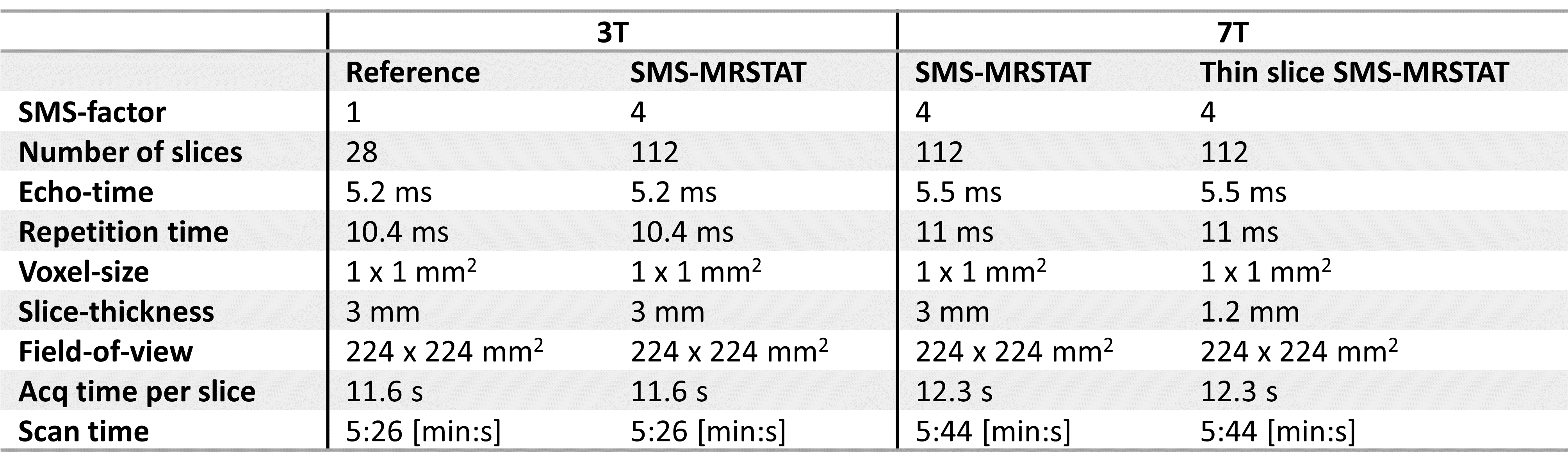
Table 1 Sequence parameters for the SMS MR-STAT acquisitions at 3T and 7T
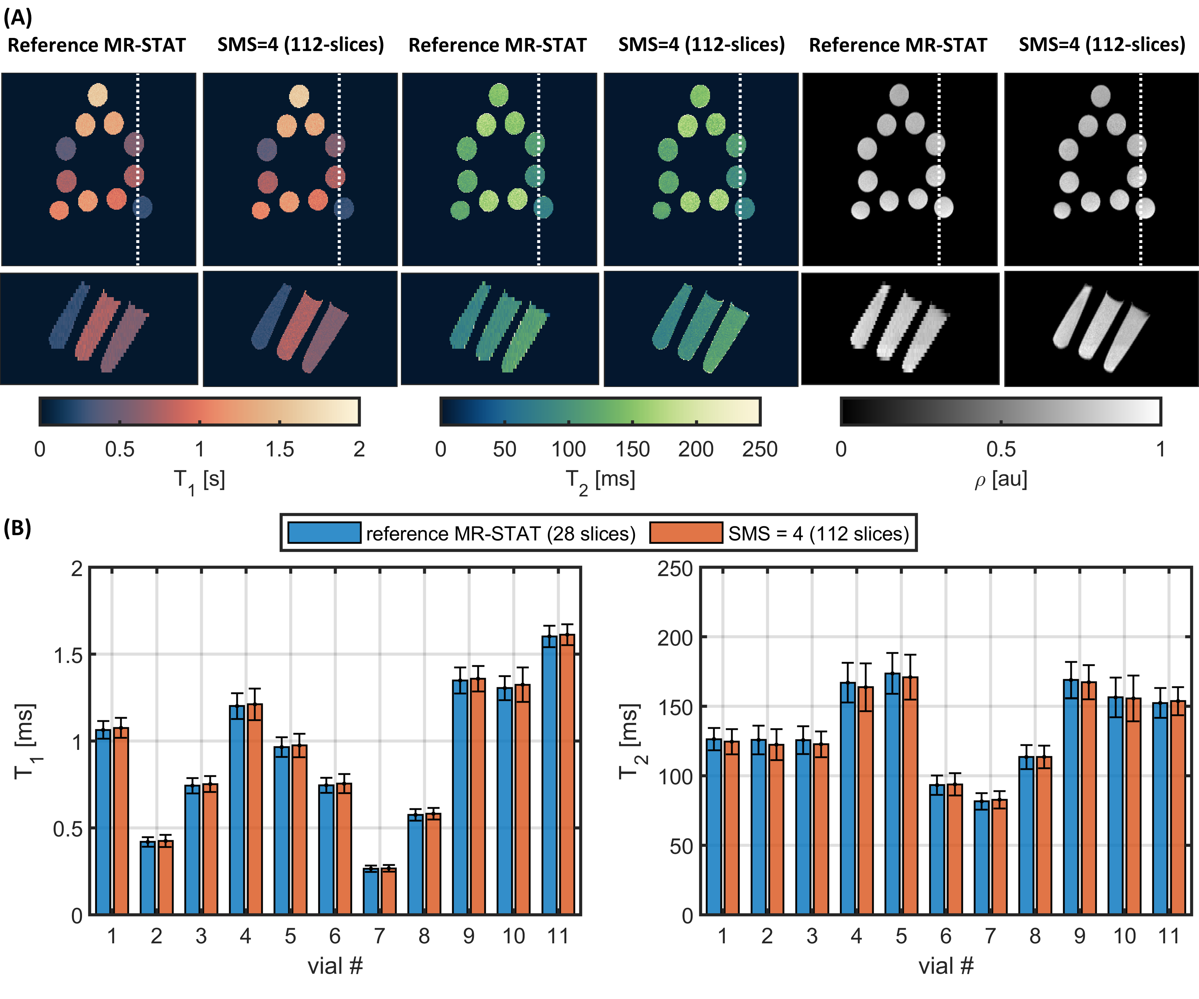
Figure 2 Results from the experiments on the gel-vials. (A) the results of the MR-STAT reconstructions for a transverse and sagittal slice. The white line shows the location of the sagittal slice. (B) Comparison of the estimated T1 and T2 in each vial.
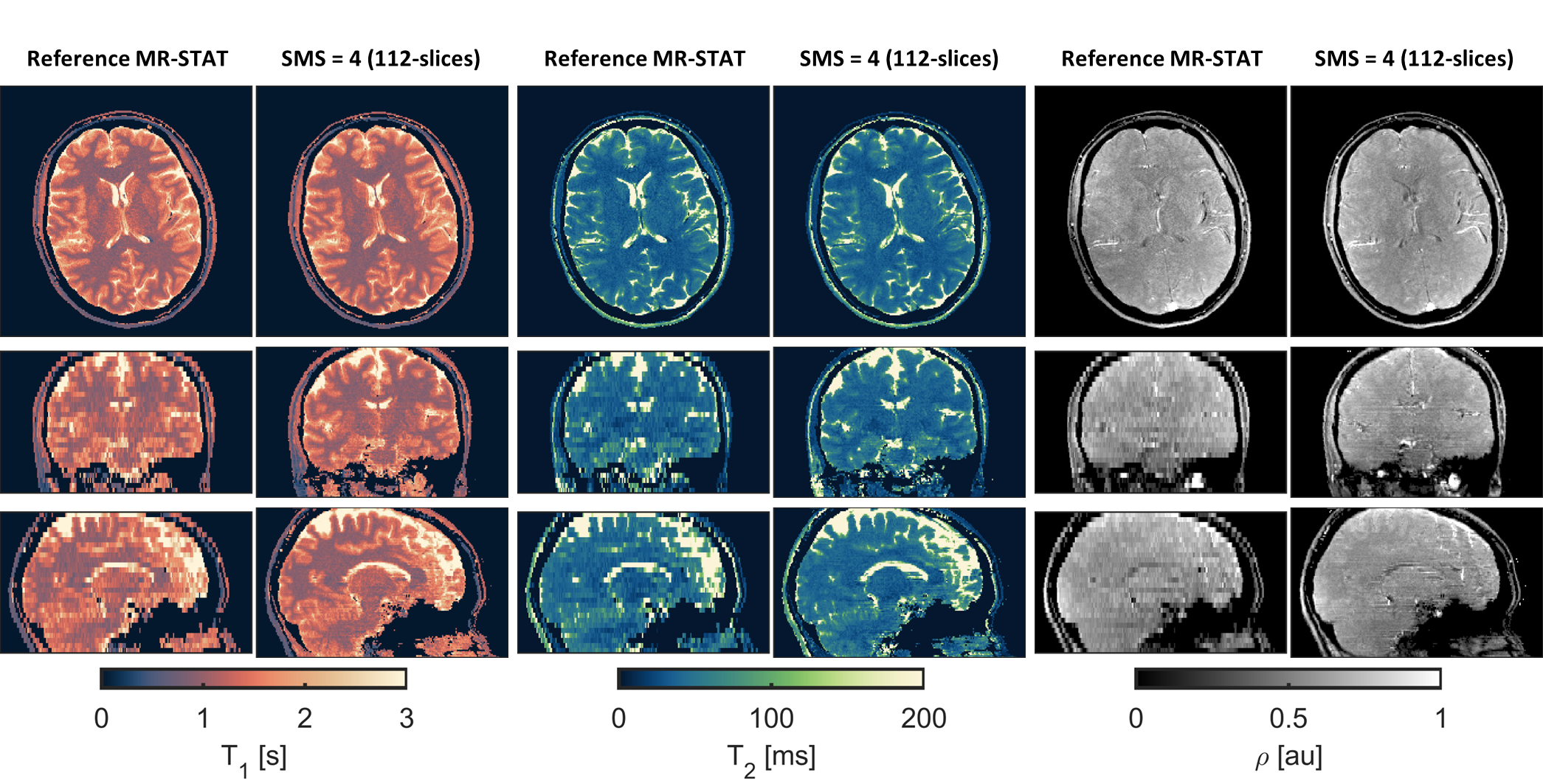
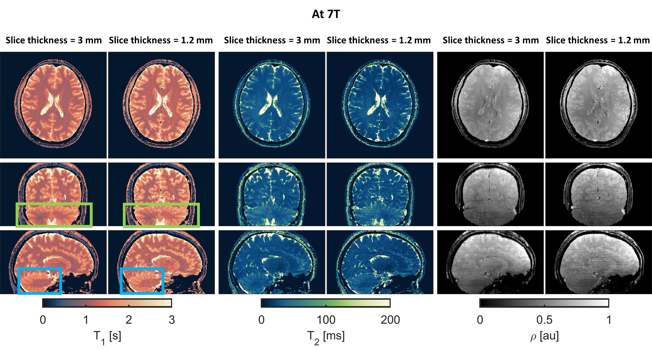
Figure 4 Results for the in-vivo scans at 7T Here, the highlighted sections of the T1-maps show the increase in detail observed when using 1.2 mm slice thickness instead of 3 mm slice thickness.