3822
Multi-contrast quantitative mapping with an unsupervised reconstruction method based on implicit neural representation1School of Biomedical Engineering, Shanghai Jiao Tong University, Shanghai, China, 2The National Engineering Research Center of Advanced Magnetic Resonance Technologies for Diagnosis and Therapy (NERC-AMRT), Shanghai Jiao Tong University, Shanghai, China
Synopsis
Keywords: Quantitative Imaging, Multi-Contrast
Motivation: Multi-contrast quantitative MRI usually requires multiple scans, leading to long acquisition time and potential inter-scan misalignment.
Goal(s): To achieve the multi-contrast quantitative MRI acquisition in a single scan and improve the accuracy of the quantitative mapping.
Approach: We developed a multi-contrast quantitative mapping sequence to simultaneously obtain T1, T2, T2* maps and subvoxel QSM. Reconstruction was conducted to directly estimate the underlying quantitative maps from the highly undersampled high-dimensional k-space data. The proposed framework was validated on the simulation, phantom and healthy volunteers.
Results: The results demonstrated that our proposed method exhibited a high correlation with references on the quantitative maps.
Impact: The proposed acquisition and reconstruction framework can simultaneously provide multi-contrast quantitative maps of the whole brain within a 5.8-minute scan. This new technique is clinically promising for tissue characterization and pathological research in neurosciences.
Introduction
Quantitative MRI offers diverse physical properties of tissue, reflecting the physiological information. Multi-contrast quantitative mapping can further provide comprehensive information, promising the clinical diagnosis and pathological research in neurosciences, such as Parkinson’s disease1. However, the conventional quantitative MRI, such as ME-GRE and ME-SE2, prolongs the acquisition time and may induce inter-scan misalignment between multiple scans. In this work, we proposed a new framework to simultaneously quantify T1, T2, T2* mapping and subvoxel QSM. An unsupervised reconstruction model based on implicit neural representation (INR)3 was proposed to reconstruct quantitative images.Methods
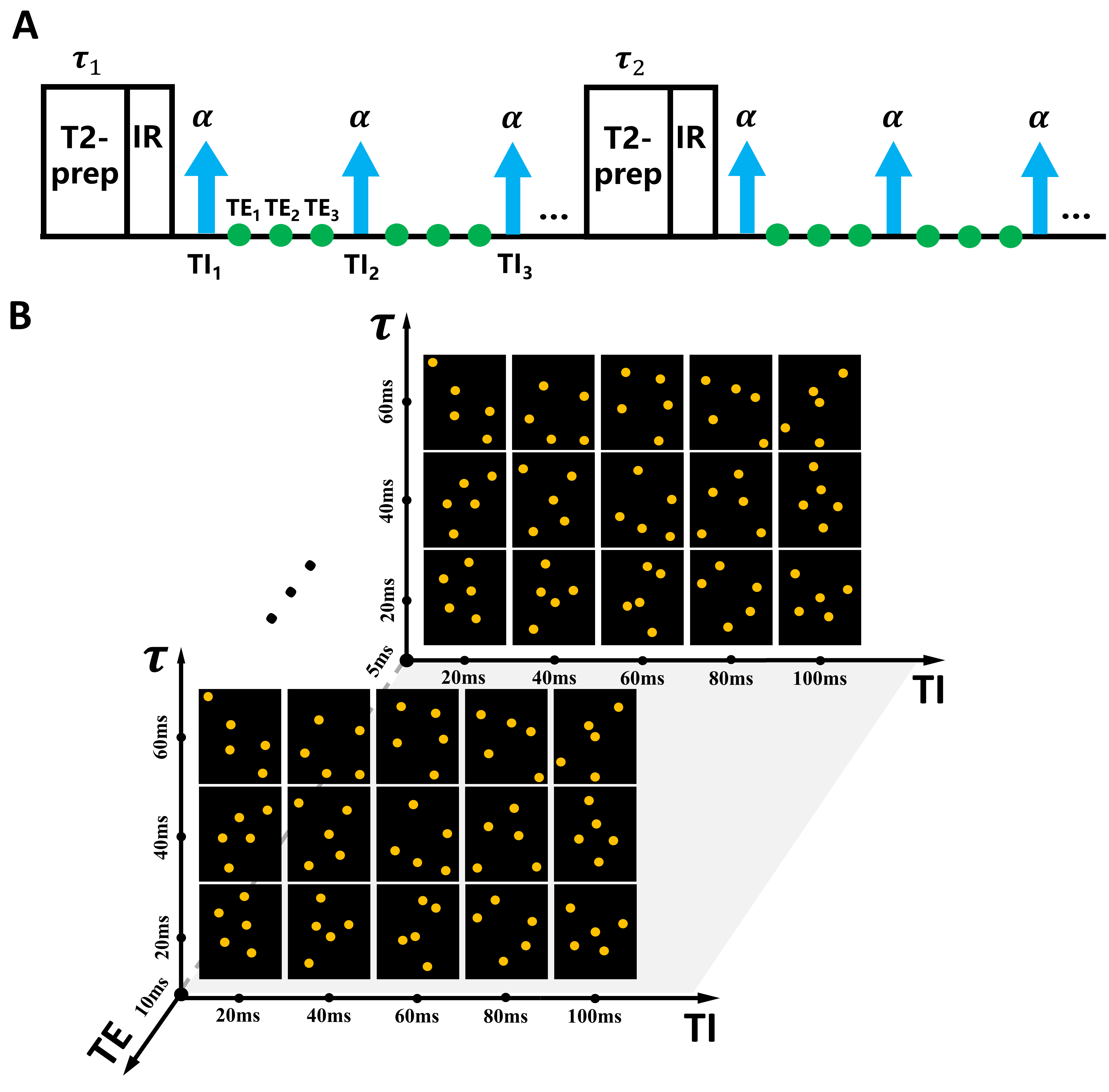
Sequence and data acquisition:The schematic diagram of the multi-contrast quantitative mapping sequence is shown in Fig. 1. The sequence is composed of T2-prep modules, IR modules, and ME-GRE readout. The IR module generates T1 weighting at each segment (TI) and the T2-prep module produces T2 weighting at each shot. The FLASH excitation is repeated throughout the shot followed by ME-GRE readout, generating T2* weighting. The high-dimensional k-space data is acquired following the Gaussian variable density sampling strategy along the ky and kz directions to enhance the spatial and temporal incoherence.
Image reconstruction:
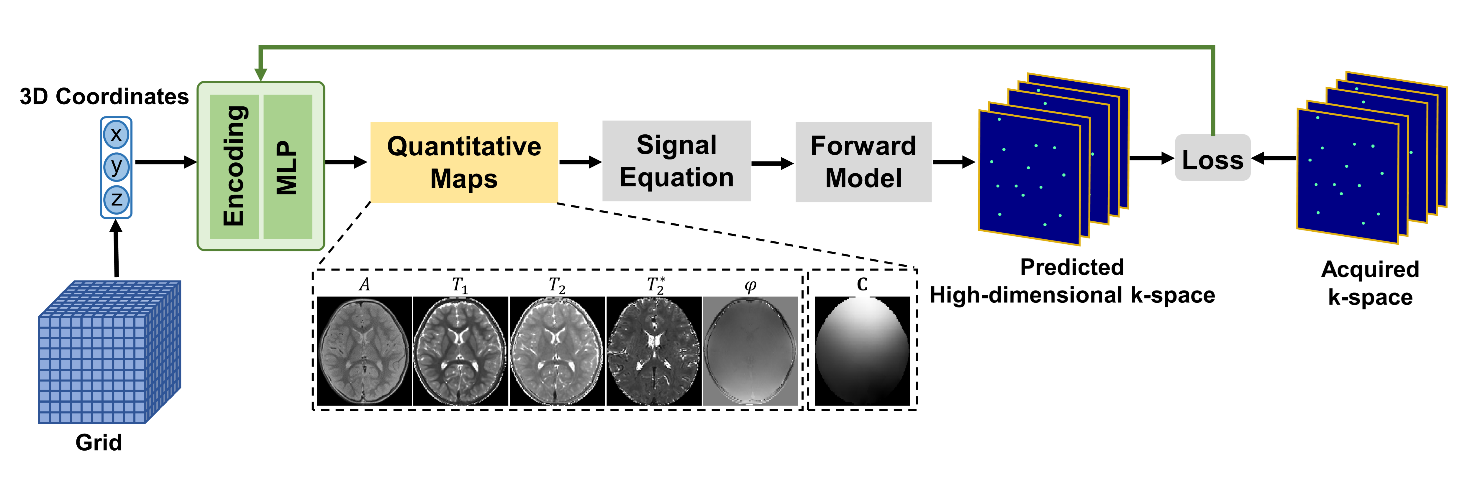
Fig. 2 displays the framework of the proposed multi-contrast mapping reconstruction. Inspired by the previous work4, the underlying quantitative maps, along with coil sensitivity maps are simultaneously estimated. Specifically, the quantitative image is generated by querying 3D spatial coordinates on the parameterized encoding module and $$$MLP_{\theta_{m}}(x,y,z)$$$, where $$$\theta_{m}$$$ represents the parameters of MLP to be optimized. The voxel-wise signal is calculated using the generated quantitative maps following Equation (1):
$$S\left(A,T_{1},T_{2},T_{2}^{*},\varphi_{e},B\right)=A\frac{1-e^{-\frac{T_{R}}{T_{1}}}}{1-e^{-\frac{T_{R}}{T_{1}}}cos\alpha}\left\lbrack 1+\left({Be^{-\frac{\tau}{T_{2}}}-1}\right)\left({e^{-\frac{T_{R}}{T_{1}}}cos\alpha}\right)^{n}\right\rbrack sin\alpha\cdot e^{-\frac{T_{E}}{T_{2}^{*}}}e^{j\varphi_{e}}\tag{1}$$
where $$$A$$$ denotes the proton density, $$$\varphi_{e}$$$ represents the phase on echo $$$e$$$, $$$B$$$ represents the inversion efficiency of the IR module, $$$n$$$ denotes the index of the FLASH excitation, and $$$\tau$$$ denotes the duration of the T2-prep module. The estimated high-dimensional k-space signal from the $$$j$$$th coil, i.e., $$$\hat{d_{j}}$$$, is predicted through the physical forward model:
$$\hat{d_{j}}=\mathbf{M}\mathbf{F}\mathbf{C}_{j}\left(\theta_{c}\right)S\tag{2}$$
where $$$\mathbf{C}_{j}\left(\theta_{c}\right)$$$ is the $$$j$$$th estimated coil sensitivity map, $$$\mathbf{F}$$$ denotes the Fourier transform and $$$\mathbf{M}$$$ represents the sampling mask. Then the parameters in MLP and encoding module can be optimized following the formulated problem:
$${\underset{\theta_{m},\theta_{c}}{argmin}{\sum\limits_{j=1}^{c}\left\|{d_{j}- \mathbf{M}\mathbf{F}\mathbf{C}_{j}S\left({A,T_{1},T_{2},T_{2}^{*},\varphi_{e},B}\right)}\right\|_{1}}} + \lambda\mathcal{R}\left({T_{1},T_{2},T_{2}^{*}}\right)\tag{3}$$
where $$$d_{j}$$$ is the acquired k-space signal of the $$$j$$$th coil, $$$\mathcal{R}( \cdot )$$$ denotes the regularizer that imposes the sharing features between the reconstructed quantitative images and $$$\lambda$$$ is a tunable parameter. After estimating T1, T2, T2* and phase maps, we applied a standard QSM reconstruction pipeline5-7 and utilized APART-QSM8 to reconstruct the subvoxel QSM.
Experiment:
The simulation was conducted with the parameters of FOV=224$$$\times$$$224$$$\times$$$64mm3, voxel size=2$$$\times$$$2$$$\times$$$4mm3, $$$T_{R}$$$=28ms, $$$\tau$$$=25,50,70,90ms, $$$T_{E}$$$=3.3,9.5,15.2,20.9ms, number of segments=56 and number of shots=16. The proposed method was compared with the low-rank tensor reconstruction method9. A standard ISMRM/NIST phantom10 was scanned for validation on a 3T uMR790 scanner with a 24-coil head array. The sequence parameters were: FOV=256$$$\times$$$256$$$\times$$$200mm3, voxel size=2$$$\times$$$2$$$\times$$$5mm3, $$$T_{R}$$$=30ms, $$$\tau$$$=0,30,60,90ms, $$$T_{E}$$$=5.2,10.9,16.6ms, number of segments=64 and scan time=5.3min. IR-SE, ME-SE and ME-GRE were used to respectively estimate T1, T2 and T2* maps as references. The in vivo experiment was performed on a healthy volunteer with the parameters of FOV=240$$$\times$$$240$$$\times$$$144mm3, voxel size=1$$$\times$$$1$$$\times$$$4mm3, $$$T_{R}$$$=20ms, $$$\tau$$$=25,50,70,90ms, $$$T_{E}$$$=3.9,9.6,15.3ms, number of segments=80 and scan time=5.8min. MTP, ME-SE and ME-GRE were used to estimate T1, T2, T2* maps and subvoxel QSM as references.
Results
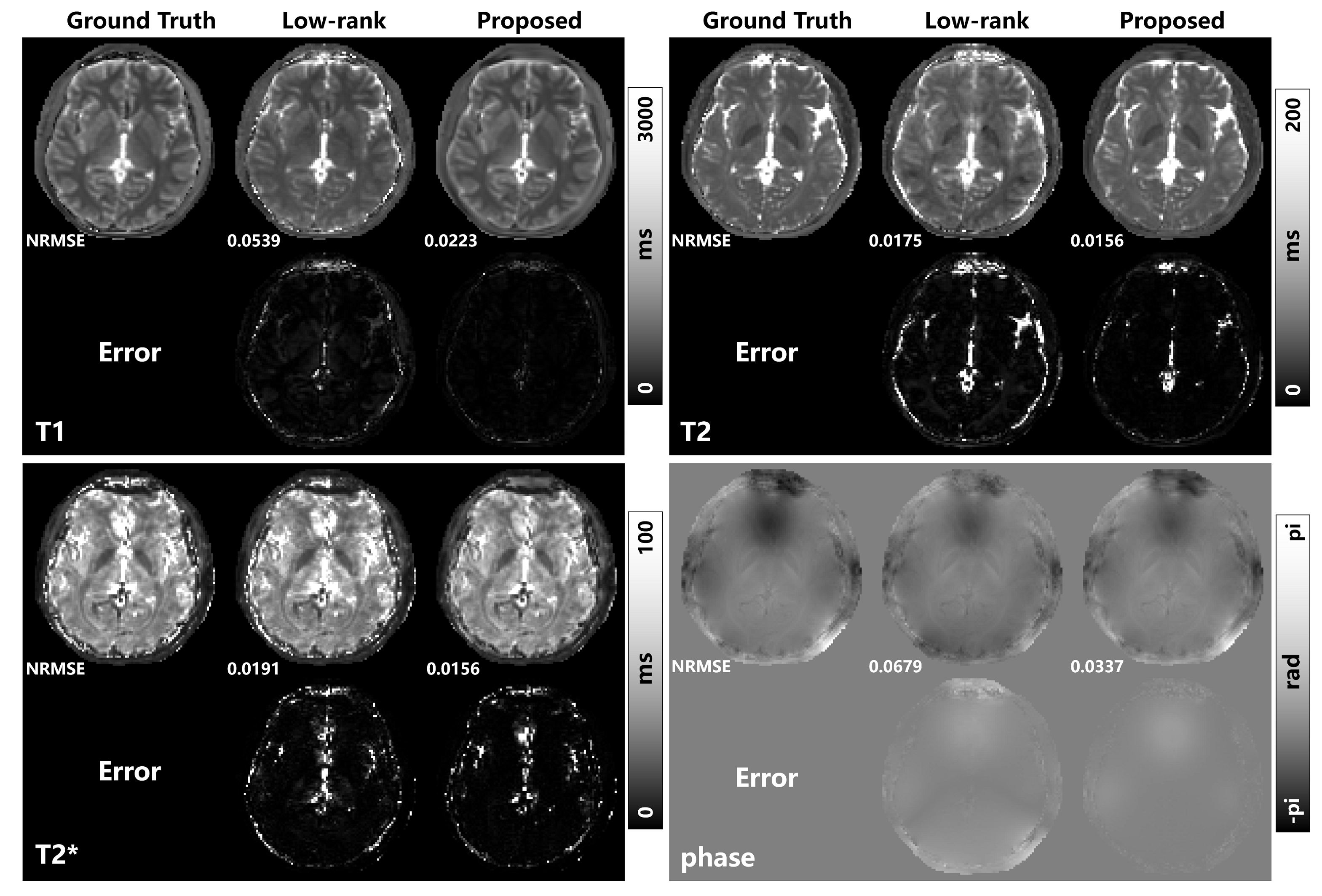
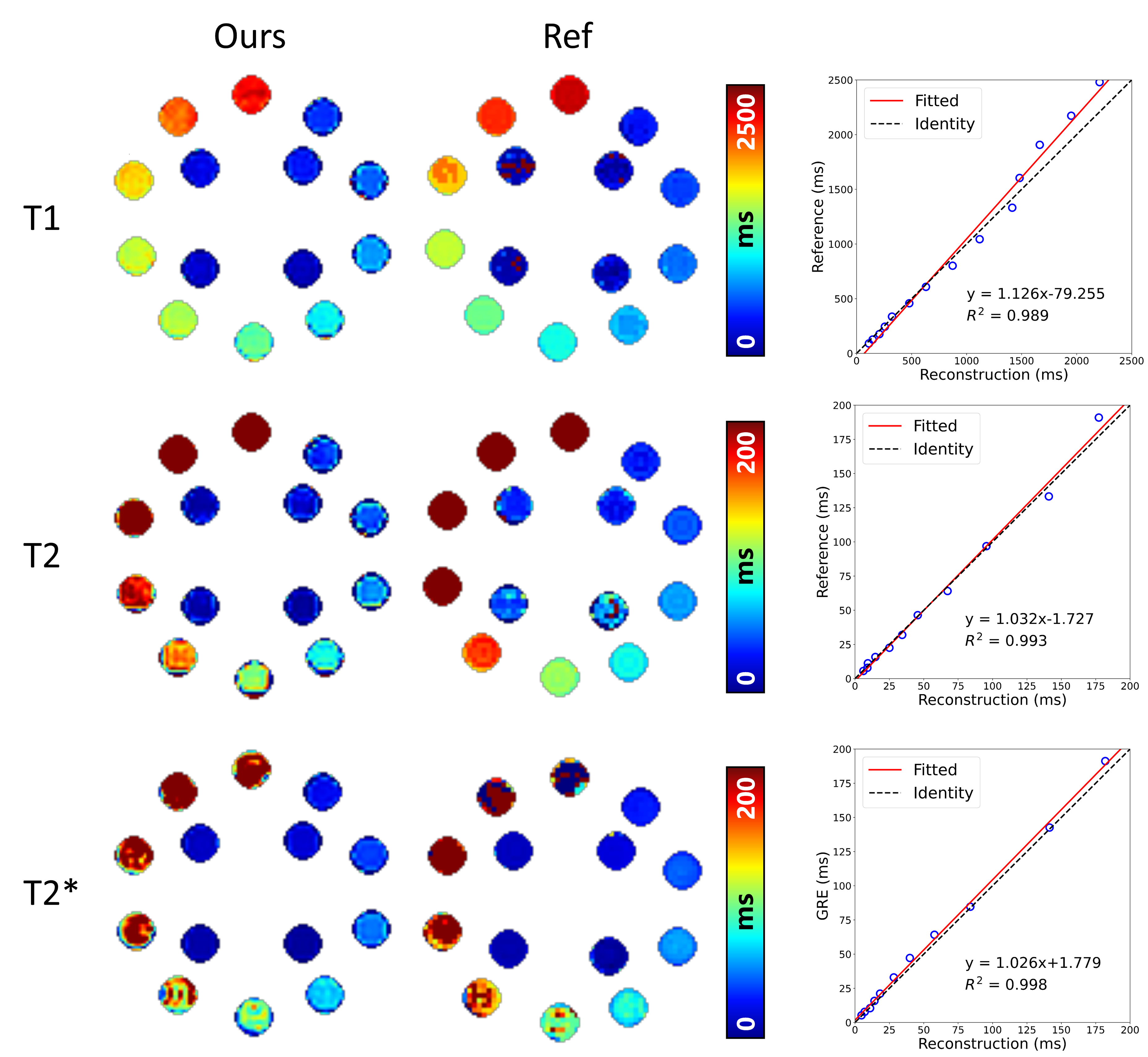
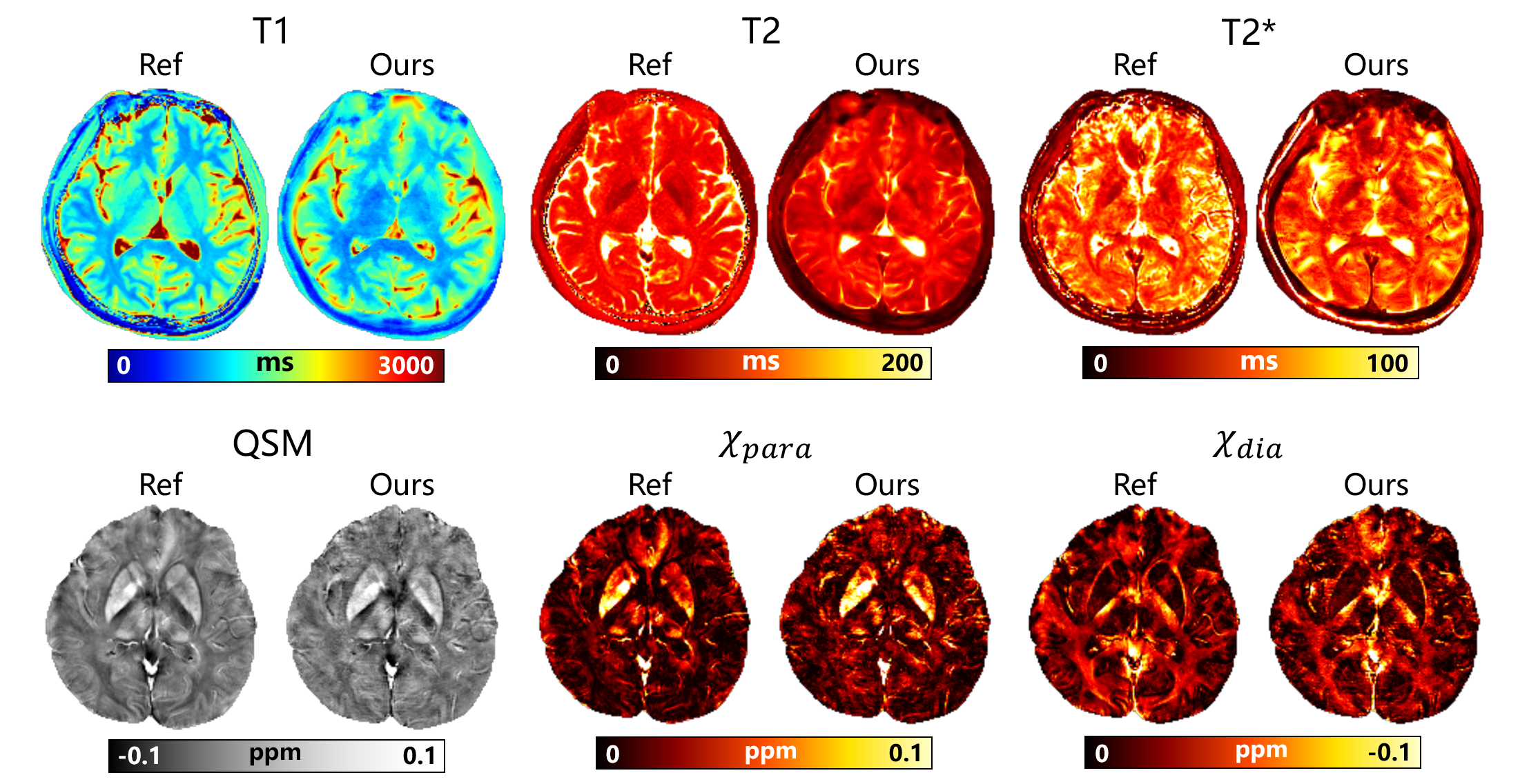
Fig. 3 shows the proposed method exhibits higher quantitative accuracy in terms of NRMSE and error maps on T1 and phase (the first echo) maps compared with the low-rank tensor reconstruction method on the simulation. Fig. 4 compares the multi-contrast quantitative maps and references on the phantom. The linear regression between the reconstruction values and the reference values exhibits a good consistency with a high $$$R^2$$$ and slope approaching 1. Fig. 5 exhibits the multi-contrast quantitative maps and references on a healthy volunteer. Our results show good image quality and agree well with the references.Discussion
The results demonstrate the superiority and fidelity of the proposed method, with reduced errors on the simulation and the consistency of quantitative maps on the phantom and in vivo results. This advantage comes from the fact that our method does not need to explicitly reconstruct the weighted images then subsequently fit the quantitative images, avoiding the accumulation of reconstruction errors. In addition, our proposed framework eliminates the need for navigator data to estimate the temporal subspace for the high-order tensor reconstruction11,12, consequently reducing scan times.Conclusion
In this study, we developed a new framework for multi-contrast quantitative mapping acquisition and reconstruction. Our method can provide whole-brain coverage with 1$$$\times$$$1$$$\times$$$4mm3 resolution within 5.8min and offer quantitative agreement with the reference scans.Acknowledgements
No acknowledgement found.References
1. S. Baudrexel et al., "Quantitative mapping of T1 and T2* discloses nigral and brainstem pathology in early Parkinson's disease," NeuroImage, vol. 51, no. 2, pp. 512-520, 2010, doi: 10.1016/j.neuroimage.2010.03.005.
2. N. Ben-Eliezer, D. K. Sodickson, and K. T. Block, "Rapid and accurate T2 mapping from multi-spin-echo data using Bloch-simulation-based reconstruction," Magn Reson Med, vol. 73, no. 2, pp. 809-817, 2015, doi: 10.1002/mrm.25156.
3. B. Mildenhall, P. P. Srinivasan, M. Tancik, J. T. Barron, R. Ramamoorthi, and R. Ng, "NeRF: Representing Scenes as Neural Radiance Fields for View Synthesis," Communications of the ACM, vol. 65, no. 1, pp. 99-106, 2021, doi: 10.1145/3503250.
4. R. Feng, Q. Wu, Y. Zhang, and H. Wei, "A scan-specific unsupervised method for parallel MRI reconstruction via implicit neural representation," in 2023 IEEE 20th International Symposium on Biomedical Imaging (ISBI), pp. 1-5, 2023, doi: 10.1109/ISBI53787.2023.10230813.
5. M. A. Schofield and Y. Zhu, "Fast and Robust Phase Unwrapping Algorithm for Electron Holography," Microscopy and Microanalysis, vol. 8, no. S02, pp. 532-533, 2002, doi: 10.1017/s1431927602105228.
6. W. Li, A. V. Avram, B. Wu, X. Xiao, and C. Liu, "Integrated Laplacian-based phase unwrapping and background phase removal for quantitative susceptibility mapping," NMR Biomed, vol. 27, no. 2, pp. 219-27, Feb 2014, doi: 10.1002/nbm.3056.
7. H. Wei et al., "Streaking artifact reduction for quantitative susceptibility mapping of sources with large dynamic range," NMR Biomed, vol. 28, no. 10, pp. 1294-303, Oct 2015, doi: 10.1002/nbm.3383.
8. Z. Li et al., "APART-QSM: An improved sub-voxel quantitative susceptibility mapping for susceptibility source separation using an iterative data fitting method," NeuroImage, vol. 274, p. 120148, 2023, doi: 10.1016/j.neuroimage.2023.120148.
9. J. He, Q. Liu, A. G. Christodoulou, C. Ma, F. Lam, and Z. P. Liang, "Accelerated High-Dimensional MR Imaging With Sparse Sampling Using Low-Rank Tensors," IEEE Trans Med Imaging, vol. 35, no. 9, pp. 2119-29, Sep 2016, doi: 10.1109/TMI.2016.2550204.
10. K. F. Stupic et al., "A standard system phantom for magnetic resonance imaging," Magn Reson Med, vol. 86, no. 3, pp. 1194-1211, 2021, doi: 10.1002/mrm.28779.
11. T. Cao et al., "Three-dimensional simultaneous brain mapping of T1, T2, T 2 * and magnetic susceptibility with MR Multitasking," Magn Reson Med, vol. 87, no. 3, pp. 1375-1389, Mar 2022, doi: 10.1002/mrm.29059.
12. S. Ma et al., "Three-dimensional whole-brain simultaneous T1, T2, and T1rho quantification using MR Multitasking: Method and initial clinical experience in tissue characterization of multiple sclerosis," Magn Reson Med, vol. 85, no. 4, pp. 1938-1952, Apr 2021, doi: 10.1002/mrm.28553.
Figures