3821
Accelerated Low-rank MPnRAGE Denoising and T1 Reconstruction using Jointly Trained U-Net Regularizers1Waisman Center, University of Wisconsin-Madison, Madison, WI, United States, 2Medical Physics, University of Wisconsin-Madison, Madison, WI, United States, 3Psychiatry, University of Wisconsin-Madison, Madison, WI, United States, 4Pediatrics, University of Wisconsin-Madison, Madison, WI, United States, 5Radiology, University of Wisconsin-Madison, Madison, WI, United States
Synopsis
Keywords: Quantitative Imaging, Image Reconstruction
Motivation: Quantitative T1 (qT1) using MPnRAGE is a promising brain biomarker. However, reliable qT1 measurements at 1 mm isotropic resolution across the brain may require long scan times (>8min).
Goal(s): We aim to develop a low-rank denoising strategy providing reliable qT1 estimations from accelerated scans in about 2 minutes.
Approach: We developed a novel strategy using two U-Net models, the denoiser and T1-estimator, trained together to jointly convert accelerated low-rank scans into accurate qT1 maps.
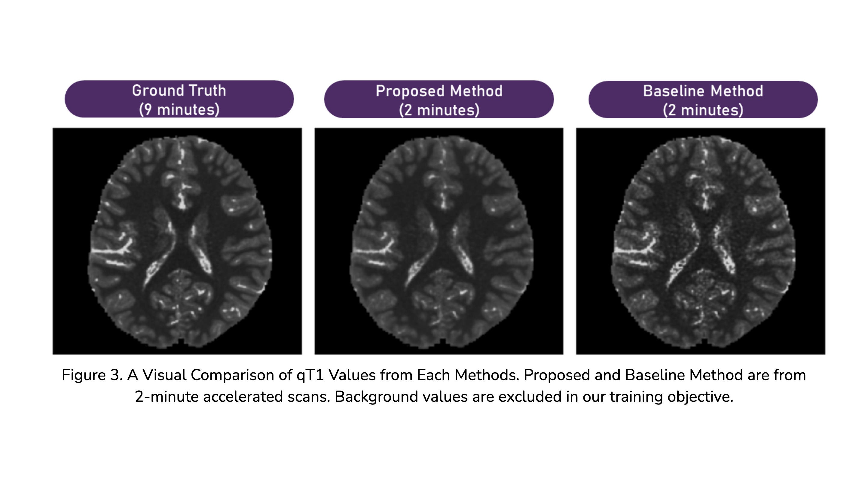
Results: Our method exhibits good bias correction with low errors in both gray matter and white matter (<3%) with high image acceleration.
Impact: Our method provides fast and accurate whole-brain high-resolution qT1 estimation from MPnRAGE scans in about 2 minutes.
Introduction
Quantitative T1 (qT1) relaxometry with magnetic resonance imaging (MRI) is a promising marker of myelination and maturation in the brain1, proving to be integral for the assessment of human neurological development and degeneration2. Recently, a non-invasive 3D radial MPnRAGE method was developed to obtain highly reliable and repeatable qT1 measures with 1 mm isotropic spatial resolution in about 9 minutes3,4. A faster acquisition method would be imperative to facilitate wider clinical adoption and reduce patient burden.Method
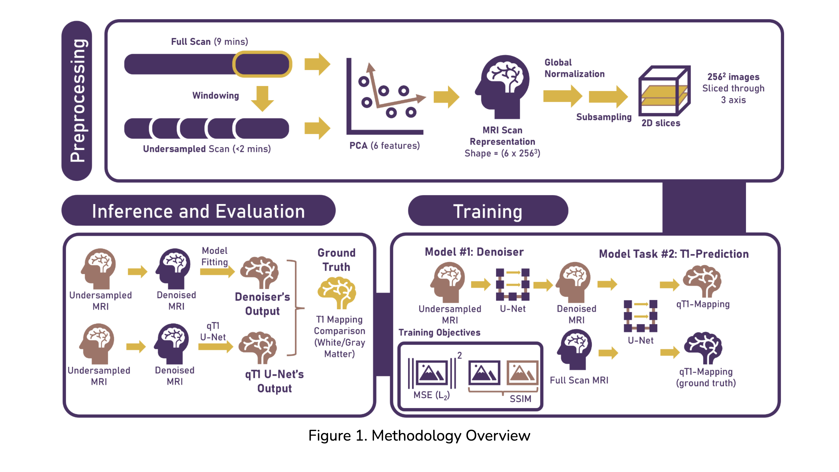
The study included 60 MPnRAGE datasets for training and evaluation. The image reconstruction utilized dimensionality reduction from 400 inversion recovery images into 6 principal components images. To evaluate the effects of undersampling, the raw 9-minute k-space data for each subject were retrospectively undersampled to synthesize acquisition times of 2, 4, and 7 minutes. The undersampled data sets, created by removing parts of the radially scanned data, were deemed input, and the 9-minute model images served as intermediate ground truth. The qT1 mappings generated from Y were used as labels. Each volumetric triplet was sliced orthogonal to each axis into images of 256x256 pixels.Two U-Net5 models labeled the denoiser and T1-predictor are used in the pipeline. U-Net is a powerful neural network architecture that has achieved remarkable success in medical image processing5, including MRI6. The denoiser aims to improve the quality of the principal components image with a mapping from undersampled to fully scanned low-rank images. The T1-predictor takes a principal component image (full scan, or the denoised undersampled images) and creates an estimation with respect to the ground truth qT1 values.
The denoiser was trained to preserve the structural similarity (SSIM) between the full and denoised scans. The T1-predictor minimizes the L2 norm between ground truth and the predicted qT1 values. The T1-predictor’s gradients are propagated through the denoiser to regularize both networks. We evaluated our models using five-fold cross-validation.
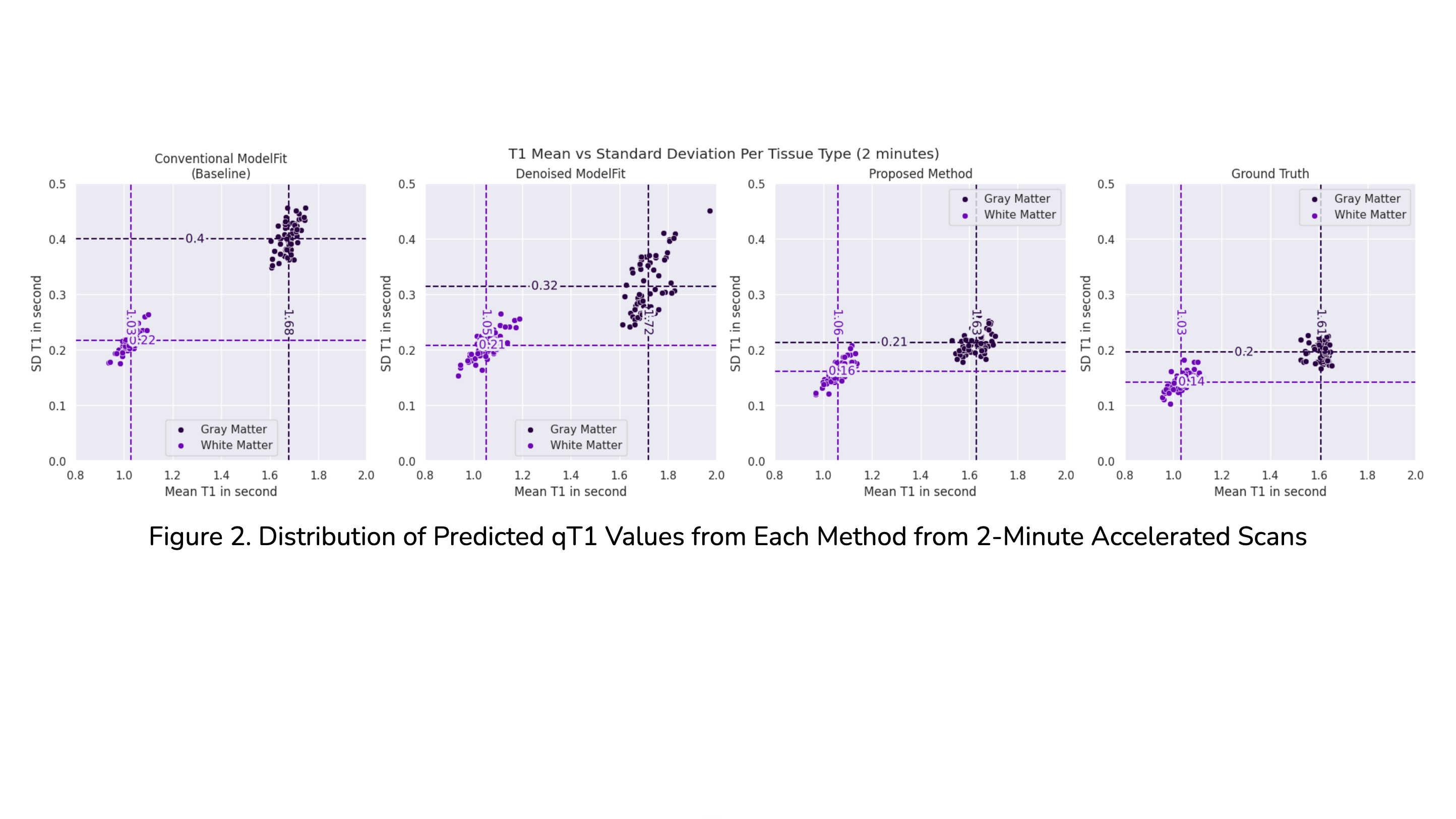
The trained denoiser-T1-predictor pair is compared to the conventional qT1 model fitting technique of the source images. We also compared the conventional model fitting7 result from the intermediate output from the denoiser to investigate the individual effects of the denoiser and T1-predictor. We evaluated the mean and standard deviations of qT1 values in a sample-wise, tissue-specific manner on a set of test datasets.
Discussion
As the denoiser was trained on optimizing the SSIM between the PCA images, it aims to improve the image quality8 while neglecting biases, thus the results demonstrated positive biases. Our T1-predictor, on the other hand, minimizes this bias via the L2 training objective. The larger white matter error is hypothesized to arise from the tendency of L2-based regressors to predict values close to the mean, failing to estimate the higher qT1 of white matter tissues correctly. As such, applying different loss weights to each tissue type may circumvent the issue. In addition, the processing time of our proposed method has a fast processing time of 1-2 minutes.Conclusion
We proposed and implemented a machine learning algorithm capable of producing reliable qT1 measures from undersampled scans as short as 2 minutes. Compared to the baseline, our algorithm produces smaller errors and bias on gray matter tissues with respectable errors on white matter. Moreover, our algorithm is faster compared to the baseline. This greatly reduces the acquisition time of MPnRAGE scans, opening the door to more convenient and affordable MRI scans.Acknowledgements
No acknowledgement found.References
- Heath, F., Hurley, S. A., Johansen‐Berg, H., & Sampaio‐Baptista, C. (2018). Advances in noninvasive myelin imaging. Developmental neurobiology, 78(2), 136-151.
- Ganzetti, M., Wenderoth, N., & Mantini, D. (2014). Whole brain myelin mapping using T1-and T2-weighted MR imaging data. Frontiers in human neuroscience, 8, 671.
- Kecskemeti, S., & Alexander, A. L. (2020). Three‐dimensional motion‐corrected T1 relaxometry with MPnRAGE. Magnetic resonance in medicine, 84(5), 2400-2411.
- Kecskemeti, S., Freeman, A., Travers, B. G., & Alexander, A. L. (2021). FreeSurfer based cortical mapping and T1-relaxometry with MPnRAGE: test-retest reliability with and without retrospective motion correction. Neuroimage, 242, 118447.
- Ronneberger, O., Fischer, P., & Brox, T. (2015). U-net: Convolutional networks for biomedical image segmentation. In Medical Image Computing and Computer-Assisted Intervention–MICCAI 2015: 18th International Conference, Munich, Germany, October 5-9, 2015, Proceedings, Part III 18 (pp. 234-241). Springer International Publishing.
- Mehta, D., Padalia, D., Vora, K., & Mehendale, N. (2022, December). MRI image denoising using U-Net and Image Processing Techniques. In 2022 5th International Conference on Advances in Science and Technology (ICAST) (pp. 306-313). IEEE.
- Maier, O., Schoormans, J., Schloegl, M., Strijkers, G. J., Lesch, A., Benkert, T., ... & Stollberger, R. (2019). Rapid T1 quantification from high resolution 3D data with model‐based reconstruction. Magnetic resonance in medicine, 81(3), 2072-2089.
- Nilsson, J., & Akenine-Möller, T. (2020). Understanding SSIM. arXiv preprint arXiv:2006.13846.
Figures