3816
Cross-frequencies Coil Sensitivity Profile Prediction Using Convolutional Neural Networks: Explorative study1UMC Utrecht, Utrecht, Netherlands, 2Tesla Dynamic Coils B.V., Zaltbommel, Netherlands, 3Eindhoven University of Technology, Eindhoven, Netherlands
Synopsis
Keywords: AI/ML Image Reconstruction, Image Reconstruction, cross-frequency B1 prediction
Motivation: Coil sensitivity profiles are essential for multi-channel MRI/MRSI data processing, yet not acquirable within reasonable scan time for X-nuclei with low natural abundance.
Goal(s): Predict sensitivity patterns of lowly abundant X-nuclear species based on sensitivities of highly abundant nuclei acquired by the same multi-tuned coil array.
Approach: We scanned 8 subjects at 1.5T and 3T using similar commercial head arrays. A 3D patch-based convolutional neural network is used to predict 3T sensitivity patterns from 1.5T sensitivities.
Results: Predicted 3T sensitivity patterns show high similarity to the ground truth. 3T signal-combination is feasible using the 1.5T-based predicted sensitivities, despite subject repositioning and hardware deviation.
Impact: An adequate prediction of coil sensitivity profiles at 128MHz based on 64MHz sensitivity profiles using highly similar receiver arrays was achieved. It opens up new possibilities for combining multi-channel signals acquired by multi-tuned (e.g., 31P-23Na, 19F-1H, etc.) receiver arrays.
Introduction
X-nuclear MR techniques have the unique capacity to non-invasively probe the underlying chemistry of physiological processes. Depiction of chemical information is valuable for metabolism study, cancer diagnosis and prognosis, treatment efficacy tracking, etc.1-7 The main hurdle of X-nuclear MR is the low SNR due to low natural abundance of most X-nuclei. Higher magnetic fields and parallel imaging improved the SNR efficiency of X-nuclear signal acquisition. However, the SNR of most X-nuclei is not sufficient to efficiently measure the coil sensitivities. The commonly used methods are using the averages of the first few FID points of the acquired data as the coil sensitivity, and the WSVD method8. Here we propose a new method for sensitivity approximation, taking advantage of the rising machine learning techniques. Most X-nuclear coils are multi-tuned (i.e., X-nuclear and 1H). When both frequencies share the same coil geometry, we hypothesize that the coil sensitivities at one frequency can be predicted with the coil sensitivities of the other. Therefore, we aim to predict the coil sensitivities (i.e., B1- distributions) of lowly abundant X-nuclei based on the easily accessible B1- of the highly abundant nuclei, that is acquired with the same receiver array. This abstract presents an explorative study to verify the feasibility of this method. Eight subjects were scanned at 1.5T and 3T using commercial head coil arrays with similar configurations. The Larmor frequency of 1H at 1.5T and at 3T are 64MHz and 128MHz respectively, similar to that of 23Na (high natural abundance) and 31P (low natural abundance) at 7T. For sensitivity prediction, a 3D patch-based convolutional neural network architecture is used. The network is trained to predict the coil array sensitivity distributions at 3T from the sensitivity distributions of the same subject at 1.5T. Despite the inter-scanner relocation of subjects, as well as small differences between these two coil arrays, the predicted 3T sensitivity distributions show high similarity to the ground truth. The combined 3T images using the predicted sensitivity distributions from 1.5T show very good image quality.Methods
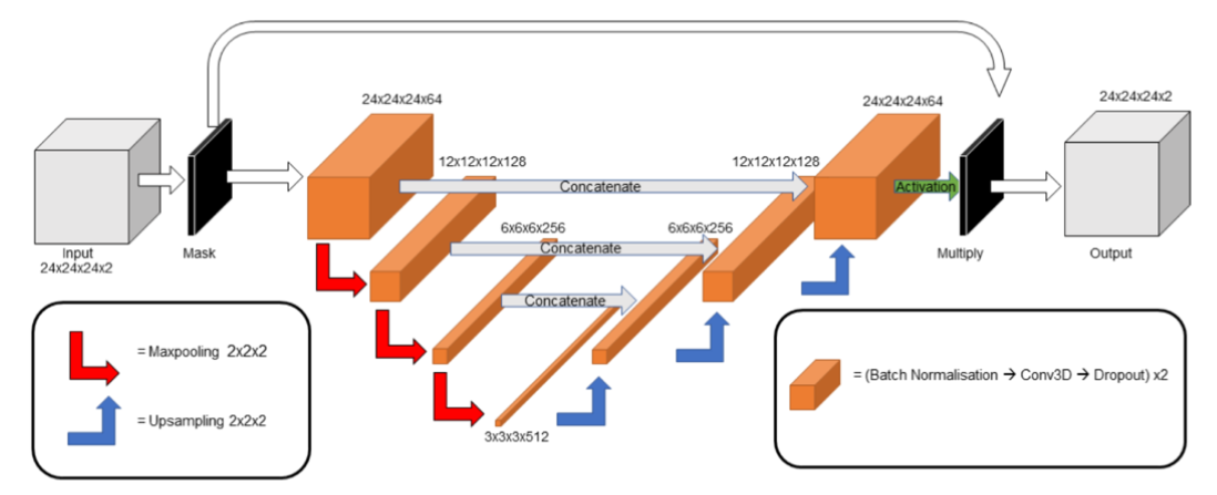
MR experiments: The MR data of 1.5T and 3T were acquired on Philips Ingenia systems (Philips b.v., Best) using 15-ch dStream head arrays (Philips Healthcare, Best, NL). Scans of the same subject are performed minutes apart. The volunteers were asked to try to reproduce the same position relative to the coil after moving to the other scanner. The positioning consistency was reviewed during survey scans. A so-called SenseRefScan is used for sensitivity profile acquisition (128x128x128 voxels, 4x4x4 mm3 voxel size). Anatomy information is acquired using a TFE sequence.Neural Network Architecture: A three-dimensional convolutional neural network inspired by the U-Net architecture9 was built using TensorFlow10. The model uses convolutional layers, batch normalization, drop-out, up- sampling, down-sampling, scaled exponential linear units (SELU) activation11 and a custom lambda layer. The complete architecture of the model is illustrated in Figure 1. Drop-out layers were used to further increase regularization and to prevent over-fitting12. The loss function for training is the mean absolute percentage error: $$L(y,\hat{y})=100\times\frac{1}{n}\sum_{i=1}^{n}|\frac{y_{i}-\hat{y_{i}}}{y_{i}+\epsilon}|,$$ where $$$y$$$ is the ground truth, $$$\hat{y}$$$ is the prediction. $$$\epsilon$$$ is added to the denominator to avoid division by zero. This cost function is chosen over the absolute difference or the mean square error to balance the impact of regions with low sensitivity and high sensitivity.
Image reconstruction: The 3T signal combination is done by Roemer’s commonly used uniform noise combination method13.
Results
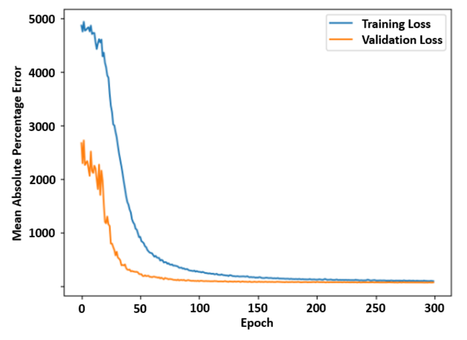
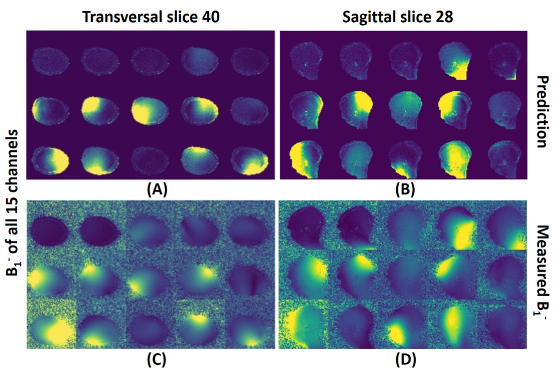
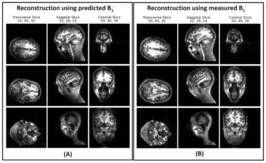
The CNN converged after 150 epochs (Figure 2). The pattern of the predicted coil sensitivities matches well with the ground truths (Figure 3). However, there is a scaling mismatch. Despite this mismatch, the data reconstruction using the predicted and measured coil sensitivity maps is comparable as is shown in Figure 4.Conclusion and discussion
This study has explored the idea of cross-frequency coil sensitivity prediction for multi-tuned coil arrays. It is limited by a small training pool, the repositioning of subjects, and small differences between the coil arrays. Despite these limitations, the training history shows no sign of overfitting, and the predicted sensitivity patterns match the ground truth adequately. Most importantly, they are able to reconstruct 3T data without significant artifacts. Extensive studies on sensitivity prediction for 1H-19F (297MHz, 280MHz) and 23Na-31P (79MHz, 121MHz) using a 7T quintuple-tuned coil14 will follow. Note that in these foreseen scenarios, the subjects will be scanned for two different nuclei in the same session using the same receiver array, thus the limitations caused by switching system and RF coils will be excluded.Acknowledgements
No acknowledgement found.References
1. Presant CA, Wolf W, Waluch V, et al. Association of intratumoral pharmacokinetics of fluorouracil with clinical response. The Lancet. 1994;343(8907):1184-1187. doi:https://doi.org/10.1016/S0140-6736(94)92399-X
2. Stevens AN, Morris PG, Iles RA, Sheldon PW, Griffiths JR. 5-fluorouracil metabolism monitored in vivo by 19F NMR. Br J Cancer. 1984;50(1):113-117. doi:10.1038/bjc.1984.146
3. Klomp D, van Laarhoven H, Scheenen T, Kamm Y, Heerschap A. Quantitative 19F MR spectroscopy at 3 T to detect heterogeneous capecitabine metabolism in human liver. NMR in Biomedicine: An International Journal Devoted to the Development and Application of Magnetic Resonance In vivo. 2007;20(5):485-492.
4. Rothman DL, de Feyter HM, de Graaf RA, Mason GF, Behar KL. 13C MRS studies of neuroenergetics and neurotransmitter cycling in humans. NMR Biomed. 2011;24(8):943-957.
5. Ianniello C, Moy L, Fogarty J, et al. Multinuclear MRI to disentangle intracellular sodium concentration and extracellular volume fraction in breast cancer. Sci Rep. 2021;11(1):5156. doi:10.1038/s41598-021-84616-9
6. Chen C, Stephenson MC, Peters A, Morris PG, Francis ST, Gowland PA. 31P magnetization transfer magnetic resonance spectroscopy: assessing the activation induced change in cerebral ATP metabolic rates at 3 T. Magn Reson Med. 2018;79(1):22-30.
7. van der Kemp WJM, Stehouwer BL, Luijten PR, van den Bosch MAAJ, Klomp DWJ. Detection of alterations in membrane metabolism during neoadjuvant chemotherapy in patients with breast cancer using phosphorus magnetic resonance spectroscopy at 7 Tesla. Springerplus. 2014;3(1):1-7.
8. Rodgers CT, Robson MD. Receive array magnetic resonance spectroscopy: whitened singular value decomposition (WSVD) gives optimal Bayesian solution. Magnetic Resonance in Medicine: An Official Journal of the International Society for Magnetic Resonance in Medicine. 2010;63(4):881-891.
9. O. Ronneberger, P. Fischer, and T. Brox, “U-net: Convolutional networks for biomedical image segmentation,” in Lecture Notes in Computer Science (including subseries Lecture Notes in Artificial Intelligence and Lecture Notes in Bioinformatics), 2015.
10. F. Marcham, “Tensorflow: LArge-scale Machine Learning on Heterogeneous Distributed Systems,” The Library, vol. s4-X, no. 3, p. 339, 2015.
11. V. Nair and G. E. Hinton, “Rectified linear units improve Restricted Boltzmann machines,” in ICML 2010 - Proceedings, 27th International Conference on Machine Learning, 2010.
12. G. E. Hinton, N. Srivastava, A. Krizhevsky, I. Sutskever, and R. Salakhutdinov, “Improving neural networks by preventing coadaptation of feature detectors,” CoRR, vol. abs/1207.0, 2012. [Online]. Available: http://arxiv.org/abs/1207.0580
13. Roemer, P.B., Edelstein, W.A., Hayes, C.E., Souza, S.P. and Mueller, O.M. (1990), The NMR phased array. Magn Reson Med, 16: 192-225. https://doi.org/10.1002/mrm.1910160203
14. Dai J, Gosselink M, van der Velden TA, Meliadò EF, Raaijmakers AJE, Klomp DWJ. An RF coil design to enable quintuple nuclear whole-brain MRI. Magn Reson Med. 2023 May;89(5):2131-2141. doi: 10.1002/mrm.29577. Epub 2023 Feb 5. PMID: 36740899.
Figures