3809
Neural network inversion in transversely isotropic materials1Mayo Clinic, Rochester, MN, United States
Synopsis
Keywords: Diagnosis/Prediction, Elastography, Anisotropy, Stiffness, Shear, Tensile, Inversion
Motivation: Most Magnetic Resonance Elastography (MRE) inversion algorithms assume isotropic materials. However, in tissues with a preferred fiber direction, the effective mechanical properties computed under this assumption will reflect a mixture of the true underlying elastic moduli.
Goal(s): In this study, we extend neural network inversion (NNI) to include transversely isotropic (TI) materials. Assumptions are progressively relaxed and the TI inversion in each case is compared against isotropic inversion.
Approach: Data was generated to train and test multiple TI inversions.
Results: An NNI trained to handle TI material can more accurately estimate shear moduli in anisotropic materials and can predict the amount of anisotropy.
Impact: This research expands on currently used MRE to allow for more accurate property estimation in highly organized tissues such as brain and muscle. Moreover, it opens new paths of investigation into pathological changes of the highly organized tissues.
Introduction
Most inversion methods for MRE assume that materials possess isotropic properties. When this assumption does not hold, the resulting estimates yield an effective modulus that mixes various parameters in proportions that depend on the boundary conditions. Anisotropic materials, which exhibit different mechanical responses depending on the direction of strain, may benefit from an anisotropic inversion technique. For this reason, several groups have begun investigating anisotropic inversion for use in highly organized tissues such as brain and muscle1,2,3,4,5. In this study, we test the hypothesis that a neural network inversion (NNI) trained on transversely isotropic materials can improve accuracy in these anisotropic materials. To this end, we progressively increased model complexity and compared accuracy against isotropic inversion in independent test sets.Methods
A previously described finite difference model was modified to model harmonic motion in transversely isotropic materials6,7. Training data were generated at 3-mm resolution under a homogeneity assumption within a 96-mm field of view. Three datasets were generated: isotropic, TI with only shear anisotropy, TI with only tensile anisotropy. A separate NNI was trained using each dataset.To create datasets with shear anisotropy, first one shear modulus was randomly selected between 1kPa and 5kPa for each dataset, and this was assigned at random to either $$$C_{44}$$$ or $$$C_{66}$$$. The remaining shear modulus was assigned by applying a randomly selected scalar to the first shear modulus in the range of ½ to 2. The damping ratios for the shear moduli were independently selected with ranges from 0–0.5.
To create datasets with tensile anisotropy both shear moduli, ($$$C_{44}$$$ and $$$C_{66}$$$), were assigned the same randomly selected value. Then a randomly selected ratio of tensile anisotropy (TA) was selected. At random either the longitudinal or transverse Young’s modulus was assigned using the relationship $$$E_1=2*G_1*(1+ν_1)$$$ where $$$G_1$$$ and $$$ν_1$$$ are the corresponding shear modulus and Poisson’s ratio. The other Young’s modulus was computed as $$$E_2=(-1*E_1)*\frac{(TA/2)+1}{(TA/2)-1}$$$.
Two random rotations were applied to each training example such that all possible fiber directions were equally likely. Motion was introduced through randomly assigned, smoothly varying, Dirichlet boundary conditions. These boundary conditions create multiple directions of wave propagation and polarization, which is required to estimate transversely isotropic properties. Training examples were further augmented with randomized phase, a random noise field, and through the selection of a smaller 33-mm field of view. An inception-like neural network was trained. The network’s inputs were the gradients of the displacements and a normalized vector map pointing in the fiber direction. The vector map had noise added to mimic the estimation of the first eigenvector from diffusion imaging8. Angle noise was simulated as normally distributed rotations with a standard deviation of 10 degrees around the two relevant axes. The net was trained using an Adam optimizer at two learning rates. The network used a patch of 11x11x11 voxels (at 3mm voxel size) to estimate the real and complex components of $$$C_{44}$$$ and $$$C_{66}$$$ at the center of each patch. For tensile ratio prediction, a network was trained with the same described inputs, but the target was the ratio between the fiber and non-fiber Young’s moduli.
Independent test sets of each type were generated for inversion evaluation.
Results
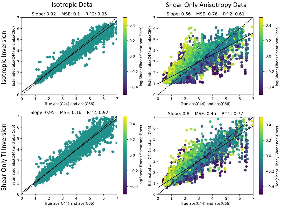
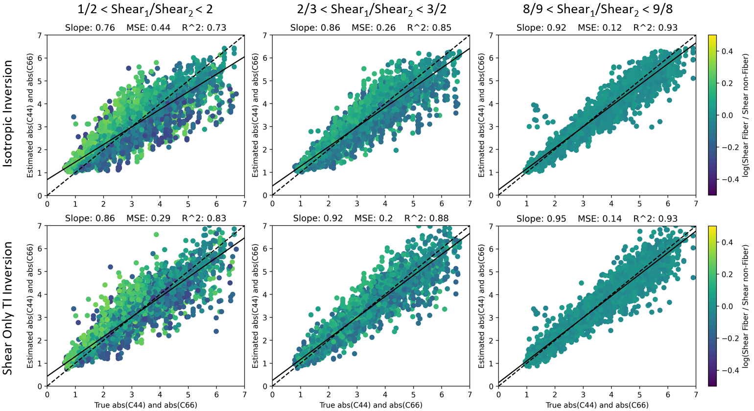
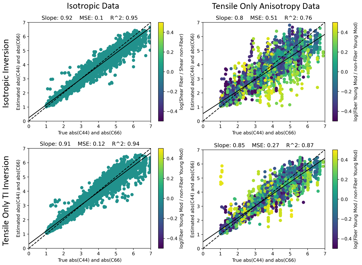
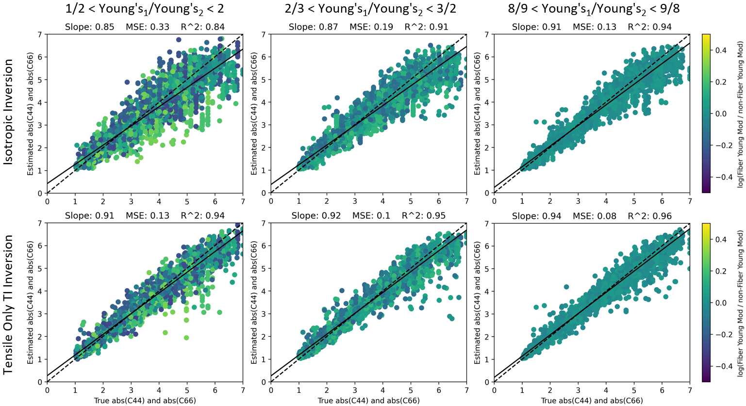
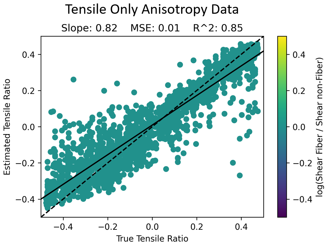
Figure 1 shows the performance of an isotropic and a Shear TI inversion in both isotropic and shear anisotropy only TI test data. The Shear TI network performs slightly worse on isotropic data but outperforms the isotropic NNI on data with shear anisotropy present. Figure 2 examines the effect of the degree of anisotropy on the relative performance of the two inversion algorithms. The Shear TI inversion outperforms an isotropic inversion on data with various amounts of shear anisotropy; the benefit decreases as shear anisotropy decreases. Analogous results, under a tensile anisotropy only assumption, are presented in Figures 3 and 4. Figure 5 examines the capability to estimate the ratio between Young’s moduli in data with tensile anisotropy.Conclusion
Tissues that present as tracts or with fibrous structures, or tissues with non-linear elastic properties that have been preferentially stretched in one direction, are governed by two different shear moduli. MRE approaches that assume isotropic properties will be less accurate when imaging these materials, potentially reducing their diagnostic utility. This research shows that an NNI trained to handle transversely isotropic material can more accurately estimate shear moduli in materials with anisotropy and can predict the amount of anisotropy. Further work is needed to implement a full TI NNI including simultaneous shear and tensile anisotropy, and relaxation of the homogeneity assumption both with respect to elastic moduli and fiber direction.Acknowledgements
Funding: (R37EB001981, R01AG076636)References
[1] A. Romano, M. Scheel, S. Hirsch, J. Braun, and I. Sack, “In vivo waveguide elastography of white matter tracts in the human brain,” Magn Reson Med, vol. 68, no. 5, pp. 1410–1422, Nov. 2012, doi: 10.1002/mrm.24141.
[2] A. Romano et al., “In vivo waveguide elastography: effects of neurodegeneration in patients with amyotrophic lateral sclerosis,” Magn Reson Med, vol. 72, no. 6, pp. 1755–1761, Dec. 2014, doi: 10.1002/mrm.25067.
[3] D. J. Tweten, R. J. Okamoto, J. L. Schmidt, J. R. Garbow, and P. V. Bayly, “Estimation of material parameters from slow and fast shear waves in an incompressible, transversely isotropic material,” J Biomech, vol. 48, no. 15, pp. 4002–4009, Nov. 2015, doi: 10.1016/j.jbiomech.2015.09.009.
[4] J. Guo, S. Hirsch, M. Scheel, J. Braun, and I. Sack, “Three-parameter shear wave inversion in MR elastography of incompressible transverse isotropic media: Application to in vivo lower leg muscles,” Magn Reson Med, vol. 75, no. 4, pp. 1537–1545, Apr. 2016, doi: 10.1002/mrm.25740.
[5] M. McGarry et al., “A heterogenous, time harmonic, nearly incompressible transverse isotropic finite element brain simulation platform for MR elastography,” Physics in Medicine and Biology, vol. 66, p. 055029, Mar. 2021, doi: 10.1088/1361-6560/ab9a84.
[6] J. M. Scott et al., “Artificial neural networks for magnetic resonance elastography stiffness estimation in inhomogeneous materials,” Medical Image Analysis, vol. 63, p. 101710, Jul. 2020, doi: 10.1016/j.media.2020.101710.
[7] J. Trevathan et al., “Initial Evaluation of a Transverse Isotropic Finite Difference Model for Training Learned Inversion,” p. 4076, doi: 10.58530/2022/4076.
[8] K. G. Schilling, V. Janve, Y. Gao, I. Stepniewska, B. A. Landman, and A. W. Anderson, “Histological validation of diffusion MRI fiber orientation distributions and dispersion,” NeuroImage, vol. 165, pp. 200–221, Jan. 2018, doi: 10.1016/j.neuroimage.2017.10.046.
Figures