3807
Model-based deep learning reconstruction by SmartSpeed AI for head and neck contrast-enhanced 3D-T1 weighted imaging1Hokkaido University Hospital, Sapporo, Japan, 2Philips Japan Ltd, Tokyo, Japan, 3Faculty of Medicine, Graduate School of Medicine, Hokkaido University, Sapporo, Japan
Synopsis
Keywords: Head & Neck/ENT, Head & Neck/ENT
Motivation: Fat-suppressed (Fs) contrast-enhanced (CE) three-dimensional (3D) T1-weighted imaging (T1WI) enables the clear visualization of head and neck structures; however, it requires a long scanning time to obtain high quality images.
Goal(s): To demonstrate the utility of model-based deep learning (DL) reconstruction, named SmartSpeed AI, for the acquisition of Fs-CE-3D T1WI of the head and neck.
Approach: Three reconstruction techniques were compared for head and neck Fs-CE-3D T1WI: 1) conventional compressed-sensing sensitivity-encoding (CS), 2) CS followed by end-to-end DL reconstruction, and 3) SmartSpeed AI.
Results: SmartSpeed AI provided the superior image quality than other two reconstruction techniques.
Impact: SmartSpeed AI, a model-based deep learning deep learning reconstruction technique, demonstrated improved image quality in head and neck Fs-CE-3D T1WI, even with a high reduction factor of 12, compared to conventional CS and CS followed by end-to-end deep learning reconstruction.
Introduction
Fat-suppressed (Fs) contrast-enhanced (CE) three-dimensional (3D) T1-weighted imaging (T1WI) enables clear visualization of head and neck structures. However, this sequence sometimes needs long scanning times which can cause the patient discomfort and even pain. Recently, deep learning (DL)-based image reconstruction techniques, particularly using convolutional neural networks (CNNs), have shown promising results for accelerating the data acquisition process of MRI. Particularly, the advanced DL reconstruction technique of model-based reconstruction using Adaptive-CS-Net, named SmartSpeed AI, have been introduced effective for denoising by combining the CNN and the image reconstruction process of the compressed-sensing sensitivity-encoding (CS)-based denoising cycle1. The aim of this study was to assess the utility of SmartSpeed AI, by comparing among three reconstruction technique of 1) conventional CS, 2) CS followed by end-to-end DL reconstruction, and 3) model-based DL reconstruction (i.e., SmartSpeed AI) in Fs-CE-3D T1WI of the head and neck.Methods
The protocol of this retrospective study was approved by our institutional review board.Patients: Twenty-four patients with head and neck tumor were included in this study. All patients underwent MR scanning by a 3.0-Tesla MR unit (Ingenia Elition X; Philips Healthcare, Best, Netherlands) with a 16-channel neurovascular coil.
Image acquisition: The Fs-CE-3D T1WI of the head and neck were acquired as follows: a 3D-T1 fast-field echo (T1-FFE) sequence, TR 5.9 ms, TE 1.2 ms, flip angle 8 degree, acquired matrix 300 × 300 (reconstructed matrix, 640 × 640), field of view (FOV) 240 × 240 mm, pixel size 0.375 × 0.375 mm, slice thickness 0.8 mm, slice pitch 0.4 mm (total 500 slices), reduction factor 12, scanning time 1 min 36 s. By using the same raw-image dataset, three types of images were reconstructed: 1) conventional CS reconstruction, 2) conventional CS followed by end-to-end DL reconstruction, and 3) SmartSpeed AI. End-to-end DL reconstruction mainly consists of the denoising convolutional neural network (DnCNN) for image processing2. SmartSpeed AI utilized a reconstruction model incorporating the DL architecture of Adaptive-CS-Net into the CS-based iterative denoising cycle, described as model-based type DL reconstruction1,3,4.
Image analysis: Qualitative image assessment was performed by the board-certified radiologists who specialized the head and neck imaging in blinded fashion. The overall image quality, visualizations of anatomical structures, and degree of artifacts were visually evaluated based-on the following five-point (1-5; 1 very poor, 2 poor, 3 moderate, 4 good, 5 excellent) grading system. Quantitative assessment was also performed by placing the region of interests (ROIs) on the tumor and posterior neck muscle to measure the value of signal to noise ratio (SNR). The SNRs were calculated as the mean signal in the ROI divided by the standard deviation (SD) of the signal in the ROI.
Statistical analysis: Statistical comparisons were performed using the Wilcoxon signed-rank test for qualitative analysis and the paired t-test for quantitative analysis. A p value <0.05 was considered significant, with Bonferroni correction of p-value for multiple comparisons among three types of reconstruction techniques.
Results
Twenty-four patients had primary head and neck tumors as follows: pharyngeal squamous cell carcinoma (SCC) (n=10), oral cavity SCC (n=4), nasal or sinonasal cavity SCC (n=4), and parotid gland tumor (n=6).In the qualitative analysis, scores in SmartSpeed AI-based images were significantly higher than other two types of reconstruction techniques in all evaluation items (p<0.05, respectively). In addition, scores in CS followed by end-to-end DL reconstruction-based images were significantly higher than CS reconstruction in all evaluation items (p<0.05, respectively), except the lesion edge sharpness. Details of qualitative analysis are presented in Fig 1.
In the quantitative analysis, all SNRs in SmartSpeed AI-based images were significantly higher than other two types of reconstruction techniques (p<0.05, respectively). In addition, all SNRs in CS followed by end-to-end DL reconstruction-based images were significantly higher than CS reconstruction (p<0.05, respectively). Details of qualitative analysis are presented in Fig 2.
The representative case is presented in Fig 3.
Discussion
Our results indicated that model-based DL reconstruction (i.e., SmartSpeed AI) consists of the CNN architecture embedded into the CS denoising cycle successfully demonstrated improved image quality in both qualitative and quantitative assessments of the Fs-CE-3D T1WI for evaluation of head and neck compared to the conventional CS and CS followed by end-to-end DL reconstruction technique. We speculated that the algorithm in SmartSpeed AI successfully demonstrated image processing with large amounts of processed data in iterative process through the Adaptive-CS-Net for more accurate denoising3,4.Conclusion
Smartspeed AI has the potential to achieve clinical usefulness by providing high image quality within a short scanning time for the detailed evaluation of the head and neck.Acknowledgements
NoneReferences
1. Pezzotti N, Yousefi S, Elmahdy MS, et al. An Adaptive Intelligence Algorithm for Undersampled Knee MRI Reconstruction. IEEE Access 2020;8:204825–38.
2. Kaye EA, Aherne EA, Duzgol C, et al. Accelerating Prostate Diffusion-weighted MRI Using a Guided Denoising Convolutional Neural Network: Retrospective Feasibility Study. Radiol Artif Intell. 2020;2:e200007.
3. Foreman SC, Neumann J, Han J, et al. Deep learning-based acceleration of Compressed Sense MR imaging of the ankle. Eur Radiol 2022;32:8376–85.
4. Wu X, Tang L, Li W, et al. Feasibility of accelerated non-contrast-enhanced whole-heart bSSFP coronary MR angiography by deep learning-constrained compressed sensing. Eur Radiol 2023. https://doi.org/10.1007/s00330-023-09740-8.
Figures
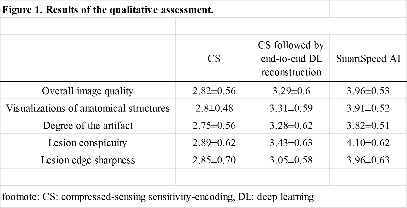
Fig 1. Results of the qualitative assessment.
All data are presented as mean ± standard deviation (1 very poor, 2 poor, 3 moderate, 4 good, 5 excellent). In all evaluation items, SmartSpeed AI showed significantly higher scores than other two types of reconstruction techniques. In addition, CS followed by end-to-end DL reconstruction were significantly higher than CS reconstruction in all evaluation items except the lesion edge sharpness.
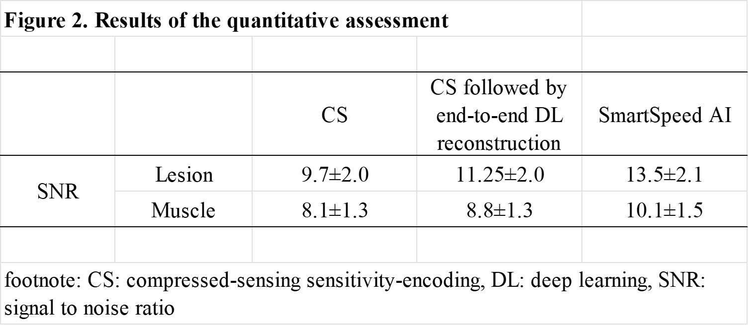
Fig 2. Results of the quantitative assessment.
All values of SNR are presented as mean ± standard deviation. SmartSpeed AI-based images showed significantly higher tumor and muscle SNRs than other two types of reconstruction techniques. CS followed by end-to-end DL reconstruction-based images showed significantly higher tumor and muscle SNRs than CS reconstruction-based images.
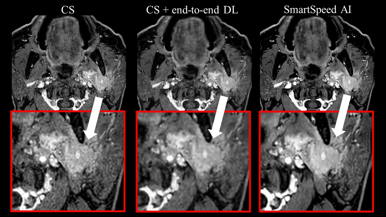
Fig 3. Representative images obtained with three reconstruction techniques.
The case with a left parotid gland tumor was presented using conventional CS-based reconstruction (left image), CS followed by end-to-end DL-based reconstruction (center image), and SmartSpeed AI-based reconstruction (right image), respectively. Red boxes indicate zoomed-in images of the tumor and surrounding structures. The image with SmartSpeed AI-based reconstruction showed marked improvement in image quality.