3804
MOdel-free Diffusion-wEighted MRI (MODEM) with Machine Learning for Accurate Tissue Characterization1Center for Magnetic Resonance Research, University of Illinois Chicago, Chicago, IL, United States, 2Department of Biomedical Engineering, University of Illinois Chicago, Chicago, IL, United States, 3Department of Radiology, Tongji Hospital, Wuhan, China, 4Departments of Radiology and Neurosurgery, University of Illinois Chicago, Chicago, IL, United States
Synopsis
Keywords: AI Diffusion Models, Pelvis
Motivation: Mathematical, biophysical, and/or statistical models are typically used to analyze diffusion-weighted imaging signals, yielding quantitative biomarkers. Those model-based approaches, however, often suffer from limited model capability, fitting instability, and degeneracy.
Goal(s): To use a MOdel-free Diffusion-wEighted MRI technique (MODEM) to differentiate underlying tissues based on diffusion signal intensities.
Approach: We developed a machine-learning-based approach which we call MOdel-free Diffusion-wEighted MRI technique(MODEM) and assess its performance by using synthetic DWI data from Monte Carlo simulations and cervical staging dataset.
Results: MODEM exhibited superior diagnostic performance to the model-based approach in both Monte Carlo simulations and cervical cancer staging data.
Impact: A model-free machine-learning-based approach provides superior performance to the conventional diffusion-model-based approach for differentiating the underlying tissue properties.
Introduction
Over the past two decades, diffusion-weighted imaging (DWI) has been increasingly used in routine clinical MRI examinations for cancer detection due to its ability to identify lesions often unnoticed by conventional MRI sequences.1-4 The decay pattern of the diffusion-weighted signal contains valuable information regarding the underlying tissue microstructures and microenvironment. Traditionally, probing the tissue microstructure and microenvironment is accomplished by fitting the signal attenuation to mathematical, biophysical, and/or statistical diffusion models.5-9 However, fitting diffusion models to experimental data is subject to the assumptions that deviate from the actual dynamics, leading to oversimplification or overfitting. Moreover, several studies indicated that the conventional model-fitting approach is prone to instability and degeneracy even when diffusion data is vastly oversampled.9,10 The goal of this study is two-fold: 1) to develop a machine-learning-based approach which we call MOdel-free Diffusion-wEighted MRI technique (MODEM) and assess its performance by using synthetic DWI data from Monte Carlo simulations; and 2) to apply MODEM for staging cervical carcinomas and compare its performance with conventional model-based methods.Materials and Methods
Data from Monte Carlo Simulation: Camino Diffusion MRI Toolkit (UCL)11 was employed to generate a set of diffusion-weighted signals via Monte Carlo simulations. Four pairs of substrates were constructed to simulate varying cell size (5µm vs. 10µm), cell density (46.3% vs. 80.7%), cell size distribution (Gamma vs. uniform distribution), and cell membrane permeability (0% vs. 0.2%), respectively, as illustrated in Figure 1. In each substrate, 100,000 random walkers were randomly initialized, each taking 20,000 time-steps toward the echo time with an intrinsic diffusivity of 2.0×10⁻³mm2/s. DWI signals were synthetized at 13 b-values from 0 to 3000 s/mm2. Seven Gaussian noise levels (0%, 1%, 5%, 10%, 15%, 20%, 25%) were added to the DWI signals separately, each repeated 1000 times.Cervical Cancer Data: DWI was performed on 54 cervical cancer patients with 17 b-values from 0 to 4500 s/mm2 on a 3T scanner (Discovery MR750, GE Healthcare). Among the patients, 26 were in the early International Federation of Obstetrics and Gynecology (FIGO) stage and 28 in the late stage. Lesion ROIs were determined along the border of the largest tumor on the diffusion-weighted images with b = 1000 s/mm2, yielding 3,169 early-stage tumor voxels and 6,666 late-stage tumor voxels.
MODEM Analysis: Feed-forward backward-propagation deep neural networks were built using MLPClassifier in Python Scikit-learn (version 1.2.2, scikit-learn.org) to differentiate different substrates in the simulation dataset and different stages in the cervical cancer dataset. Normalized signal intensities at each b-value served as input to the neural network. Data were split into 80% and 20% for training and testing, respectively. A receiver operating characteristic (ROC) analysis was performed on the testing data. To avoid sampling bias, the data split, classification, and evaluation steps were repeated 100 times.
Diffusion Model Analysis: Along with each MODEM iteration, a model-based approach was also employed using each of the following models: mono-exponential, fractional order calculus (FROC)5, diffusion kurtosis imaging (DKI)8, and continuous-time random-walk (CTRW) models6 on both Monte Carlo simulation and cervical cancer datasets. Additionally, an intravoxel incoherent motion (IVIM) model7 was also applied to the cervical cancer dataset. The diffusion parameters from each model were combined using multivariable logistic regression, where the regression coefficients were estimated using the training dataset. The testing dataset underwent ROC analysis, followed by comparisons between the model-based approaches and MODEM.
Results
Figures 2 and 3 respectively display the AUC and accuracy values obtained from MODEM and diffusion models in differentiating substrates in four simulations over 100 iterations at different noise levels. The AUC and accuracy values are close to 100% in both MODEM and diffusion models at low noise levels (≤1%) while decreasing as the noise level increases. MODEM provided a higher AUC and accuracy over any of the investigated diffusion models at all noise levels >1%. In addition, the AUC and accuracy of MODEM exhibited excellent stability with standard deviations within 1% in all simulations. Figure 4 displays the boxplots of AUC and accuracy of MODEM and the diffusion models in differentiating early-stage vs. late-stage cervical cancer voxels over 100 iterations. MODEM outperformed all diffusion models with the highest mean AUC (0.770) and accuracy (68.6%).Discussion and Conclusion
We have developed a model-free, machine-learning-based approach – MODEM – for analyzing diffusion-weighted signals. MODEM outperformed the diffusion models we have investigated as measured by AUC and accuracy for differentiating tissue structures in simulation and early-state vs. late-stage cervical cancer. The perspective of using model-free machine-learning-based approach to substitute conventional model-based methods will likely improve the robustness of diffusion MRI for tissue characterizations, particularly in cancer detection and treatment monitoring.Acknowledgements
No acknowledgement found.References
[1] Murata T, Shiga Y, Higano S, et al. Conspicuity and evolution of lesions in Creutzfeldt-Jakob disease at diffusion-weighted imaging. Am. J. Neuroradiol. 2002;23(7):1164-72.
[2] Eiber M, Holzapfel K, Ganter C, et al. Whole‐body MRI including diffusion‐weighted imaging (DWI) for patients with recurring prostate cancer: technical feasibility and assessment of lesion conspicuity in DWI. J. Magn. Reason. Imaging. 2011;33(5):1160-70.
[3] Padhani AR, Liu G, Koh DM, et al. Diffusion-weighted magnetic resonance imaging as a cancer biomarker: consensus and recommendations. Neoplasia. 2009;11(2):102–125.
[4] Tang L, Zhou XJ. Diffusion MRI of cancer: From low to high b‐values. Journal of Magnetic Resonance Imaging. 2019;49(1):23-40.
[5] Zhou XJ, Gao Q, Abdullah O, et al. Studies of anomalous diffusion in the human brain using fractional order calculus. Magn Reson Med. 2010;63(3):562-569.
[6] Karaman MM, Sui Y, Wang H, et al. Differentiating low‐and high‐grade pediatric brain tumors using a continuous‐time random‐walk diffusion model at high b‐values. Magn Reson Med. 2016;76(4):1149-1157.
[7] Le Bihan D, Breton E, Lallemand D, et al. Separation of diffusion and perfusion in intravoxel incoherent motion MR imaging. Radiology. 1988;168(2):497-505.
[8] Jensen JH, Helpern JA, Ramani A, et al. Diffusional kurtosis imaging: the quantification of non‐gaussian water diffusion by means of magnetic resonance imaging. Magn Reson Med. 2005;53(6):1432-1440.
[9] Novikov DS, Kiselev VG, Jespersen SN. On modeling. Magn Reson Med. 2018;79(6):3172-3193.
[10] Jelescu IO, Veraart J, Fieremans E, et al. Degeneracy in model parameter estimation for multi‐compartmental diffusion in neuronal tissue. NMR Biomed. 2016;29(1):33-47.
[11] Hall MG, Alexander DC. Convergence and parameter choice for Monte-Carlo Simulations of Diffusion MRI. IEEE Trans. Med. Imaging. 2009;28(9):1354-1364.
Figures
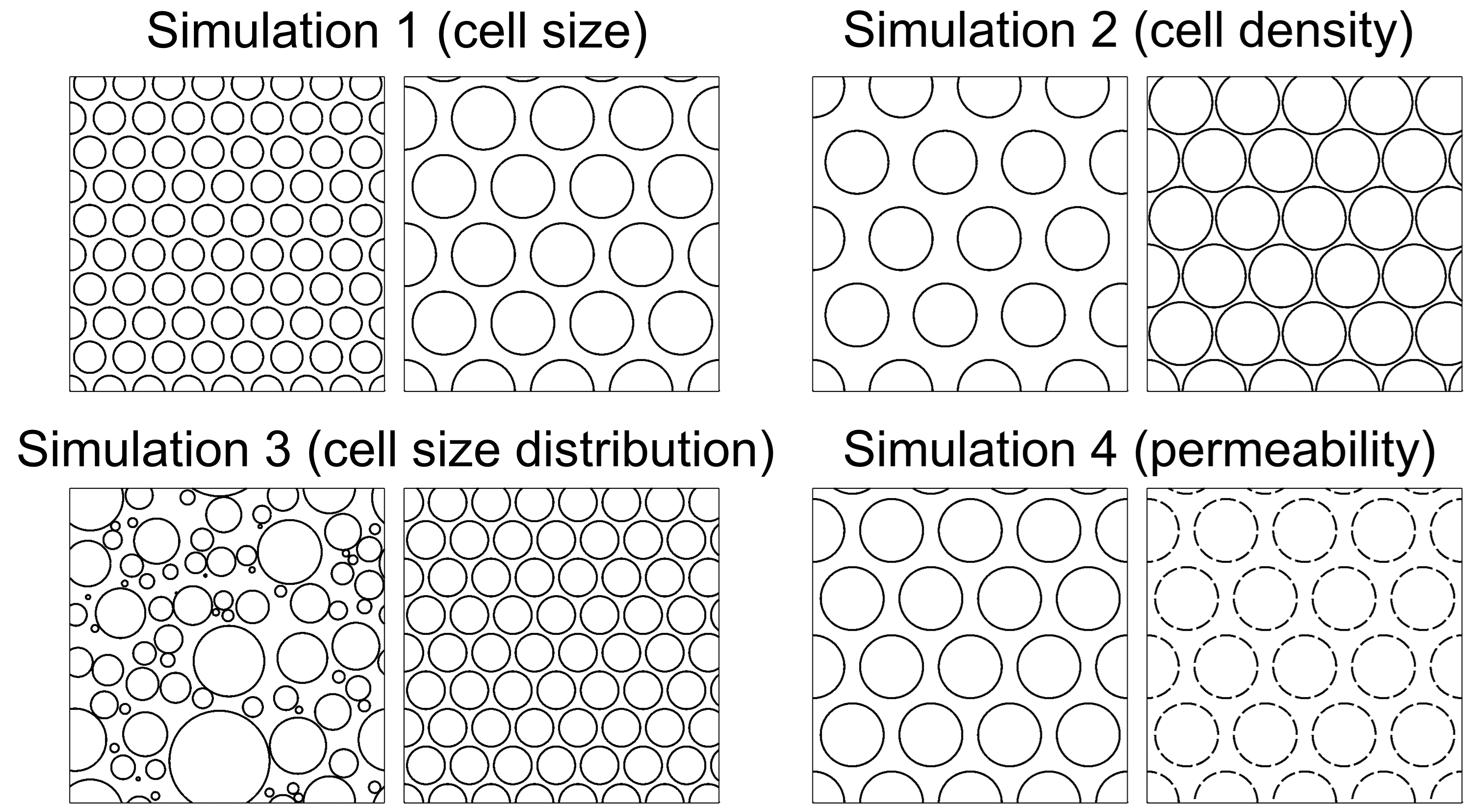
Figure 1. Substrates of the Monte Carlo simulations. Simulation 1: varying cell size (5µm vs. 10µm); simulation 2: varying cell density (46.3% vs. 80.7%); simulation 3: varying cell size distribution (Gamma vs. uniform distribution); simulation 4: varying cell membrane permeability (0% vs. 0.2%).
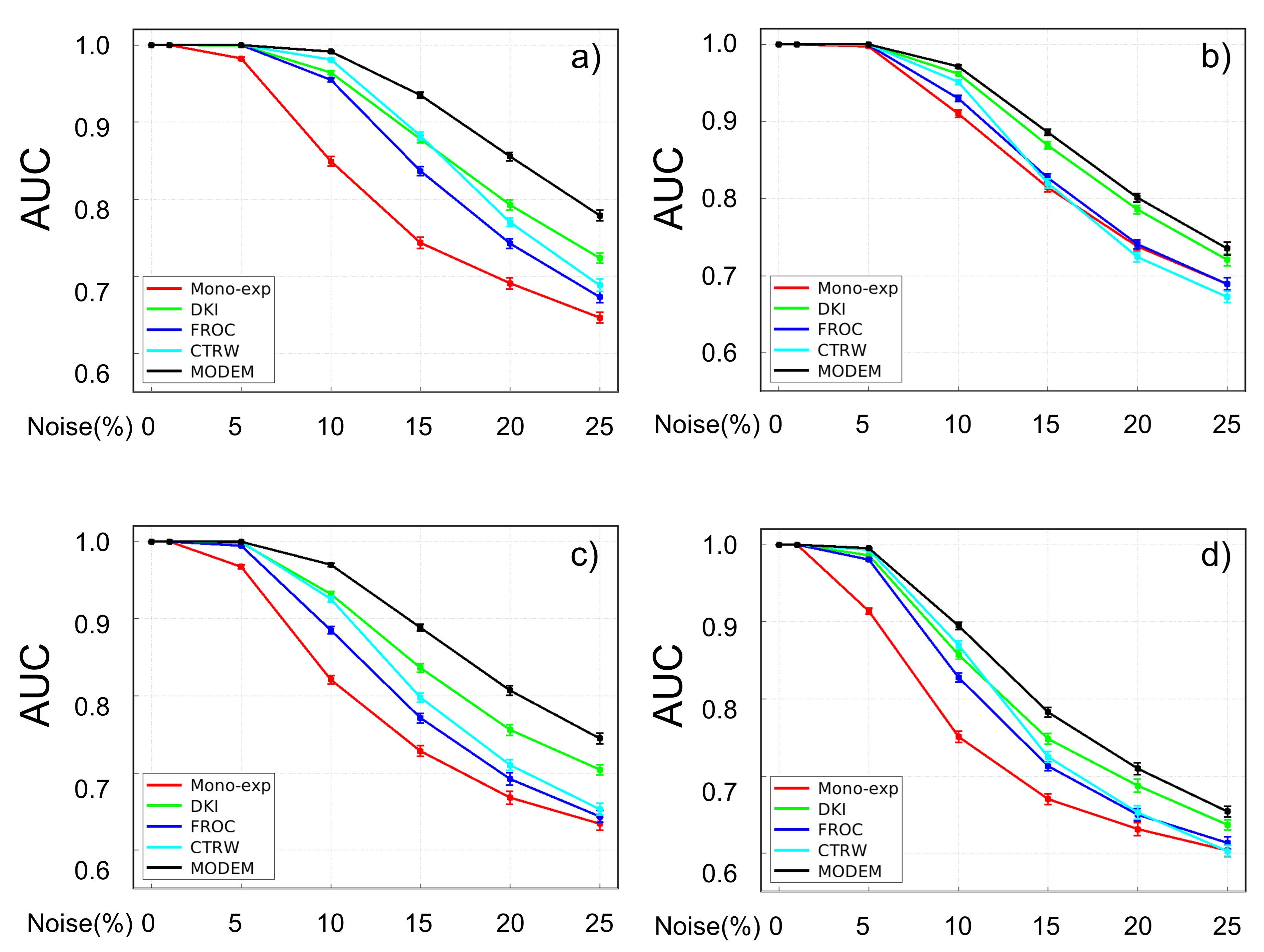
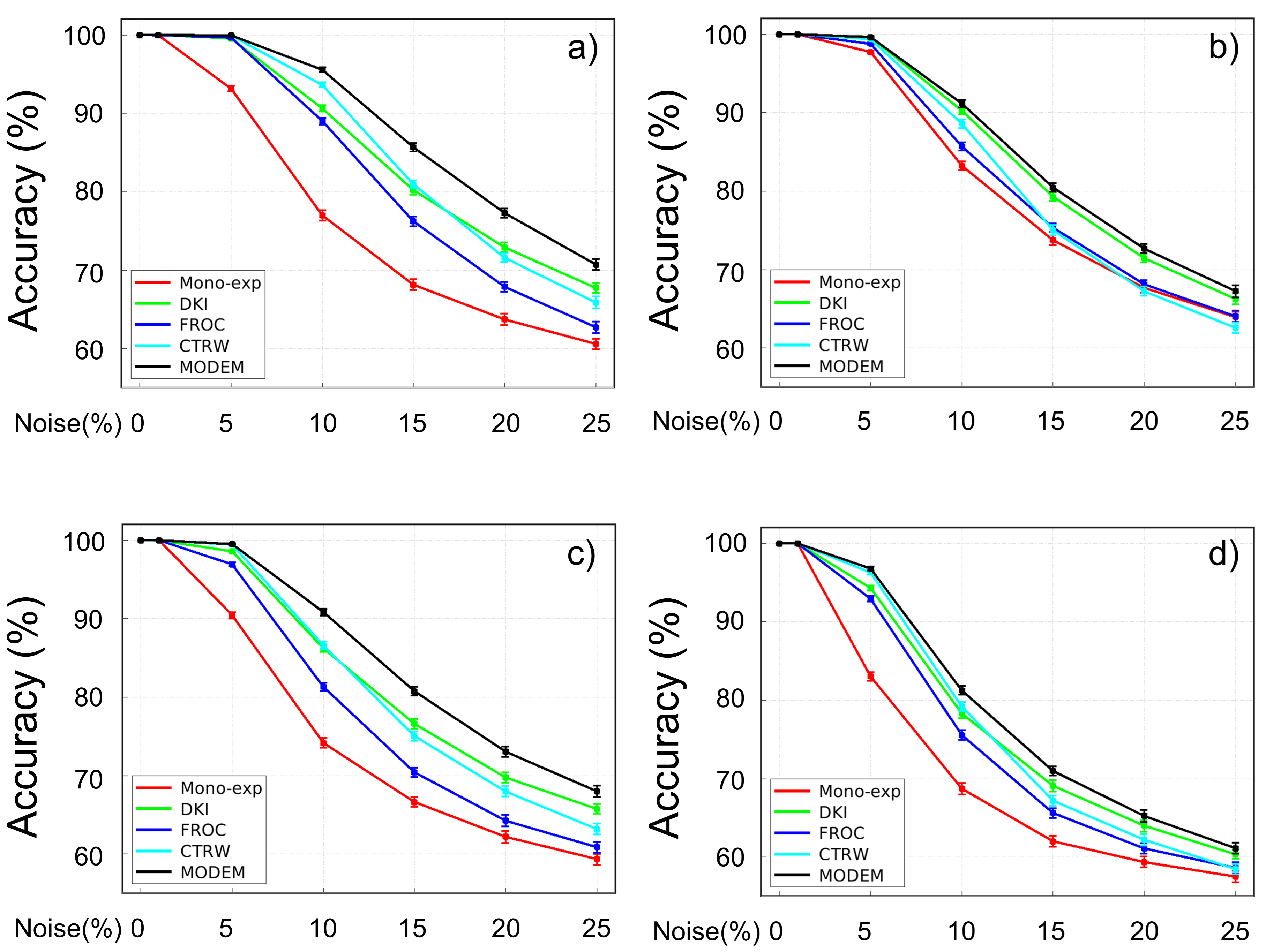
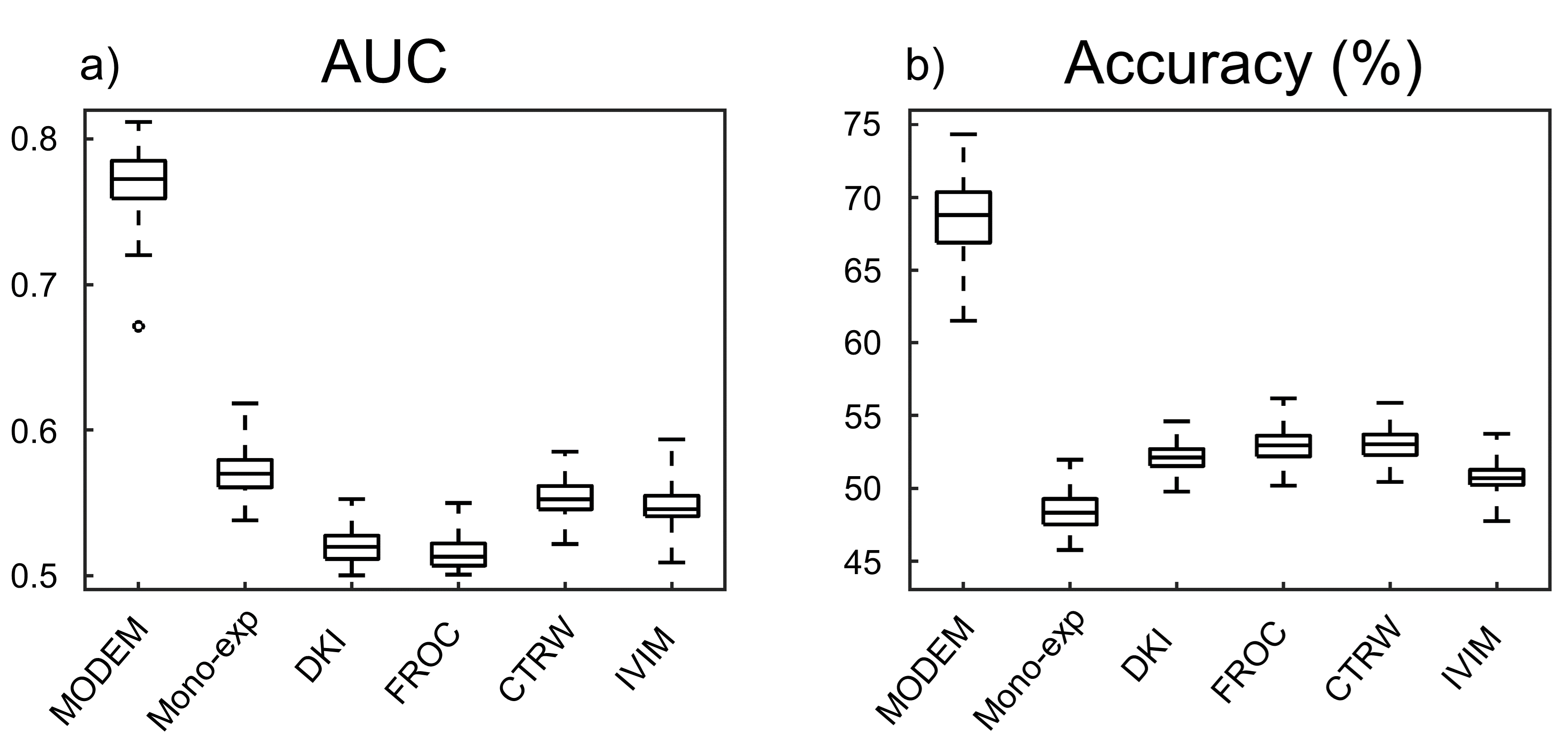
Figure 4. Boxplots of AUC (a) and accuracy (b) of MODEM and the diffusion models in differentiating early-stage vs. late-stage cervical cancer over 100 iterations.