3803
Quantitative Assessment of Synthetic MR with Deep Learning Reconstruction in Clinical Diagnosis of Nasopharyngeal Carcinoma1Radiology Department, Sun Yat-sen University Cancer Center, Guangzhou, China, 2GE Healthcare, MR Research, Beijing, China
Synopsis
Keywords: AI/ML Software, Cancer
Motivation: To enhance nasopharyngeal carcinoma (NPC) diagnostics, this study aims to assess the accuracy and image quality of relaxometry maps using fast synthetic MRI with deep learning reconstruction.
Goal(s): The primary goal is to evaluate the potential of DL Recon for NPC diagnosis, focusing on reduce scan time, improve image quality and quantitative accuracy to enable early lesion detection.
Approach: Two protocols (Trad: lower acceleration rate without DL Recon, DLR: higher acceleration rate with DL Recon) was performed on twenty-four NPC patients to evaluated T1/T2/PD measurements and image quality.
Results: Fast MAGiC acquisition with DL Recon can retain accuracy and improve image quality.
Impact: With DL Recon, the MAGiC acquisition can achieve in shorter scan time, with enhanced image quality and maintained quantitative accuracy in NPC diagnosis use.
Introduction
Nasopharyngeal carcinoma (NPC) ranks as a frequent malignancy in the field of otorhinolaryngology, demanding precise diagnostic measures across all stages1. The utilization of MRI in NPC diagnostics has been pivotal, with quantitative tissue information (T1/T2/PD) proving indispensable for lesion identification and pretreatment prognosis2-4.Recent breakthroughs in deep learning-based MRI reconstruction have presented a giant shift of image quality by offering higher Signal-to-Noise Ratio (SNR) and sharpness in a significantly reduced scan time. As for the relaxometry maps acquired from synthetic MR, images required more than quality but accuracy and stability to represent the authentic information of tissues, especially for lesion distinguishment. So far, only few investigations of synthetic MR with deep learning reconstruction showing positive reproducibility on different weighted images or pediatric neuroimaging5, 6.
This study is aimed to evaluate the accuracy and image quality of the lesion relaxometry maps in NPC patients acquired from a fast synthetic MR with deep learning reconstruction method, comparing to the traditional clinical-use approach.
Method
From September to October 2023, twenty-four patients diagnosed with NPC underwent MRI examinations using a 3.0T MR scanner (SIGNA Premier, GE Healthcare) following an IRB-approved protocol with written informed consent. Each patient underwent two sets of synthetic MR protocols known as magnetic resonance image compilation (MAGiC), with distinct different acceleration and reconstruction methods. The shared parameters are as follow: TR = 4000 ms; TE = 15.5/93.1 ms; ETL = 16; FOV = 24 cm; slice thickness = 5 mm with 1 mm spacing; image matrix = 320x256; NEX = 1. The traditional reconstruction method (Trad) employed an acceleration rate of 2.5, while AIR Recon DL method (DLR) used a rate of 3, resulting in total scan time of 4 min and 3min28s, respectively.All quantitative T1/T2/PD maps were reconstructed from MAGiC workstation in scanner. The T1/T2/PD values were calculated within region-of-interest (ROI) designated areas to assess accuracy and stability performance using AIR Recon DL. All ROIs were manually placed on lesions based on contrast-enhanced images by an experienced radiologist. The evaluation of overall image quality was executed through a standardized 5-point Likert scale assessment7 by two independent oncologists. Statistical analyses involved two-tailed paired t-test for T1/T2/PD value within lesion, with statistically significant set at p<0.001.
Results
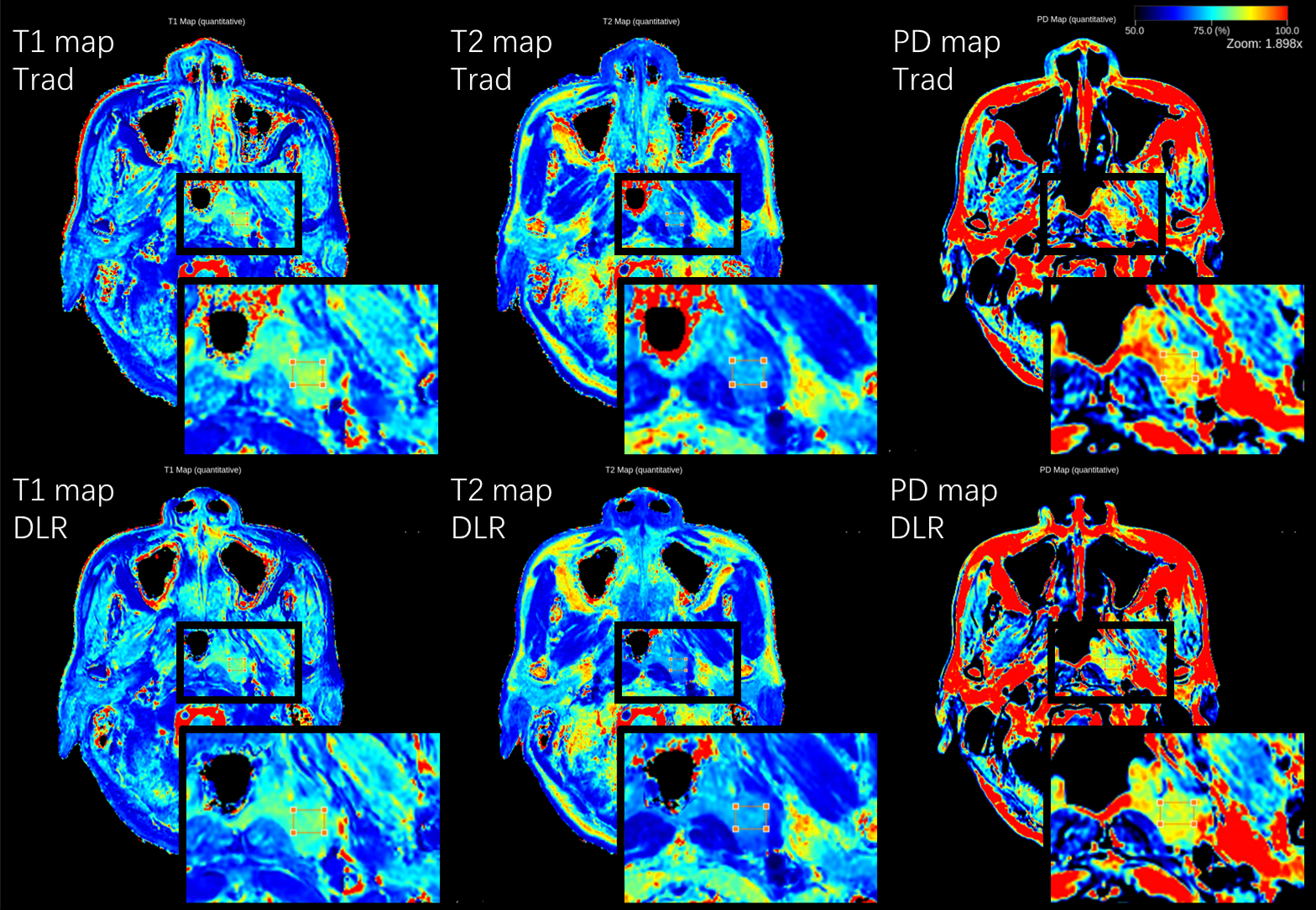
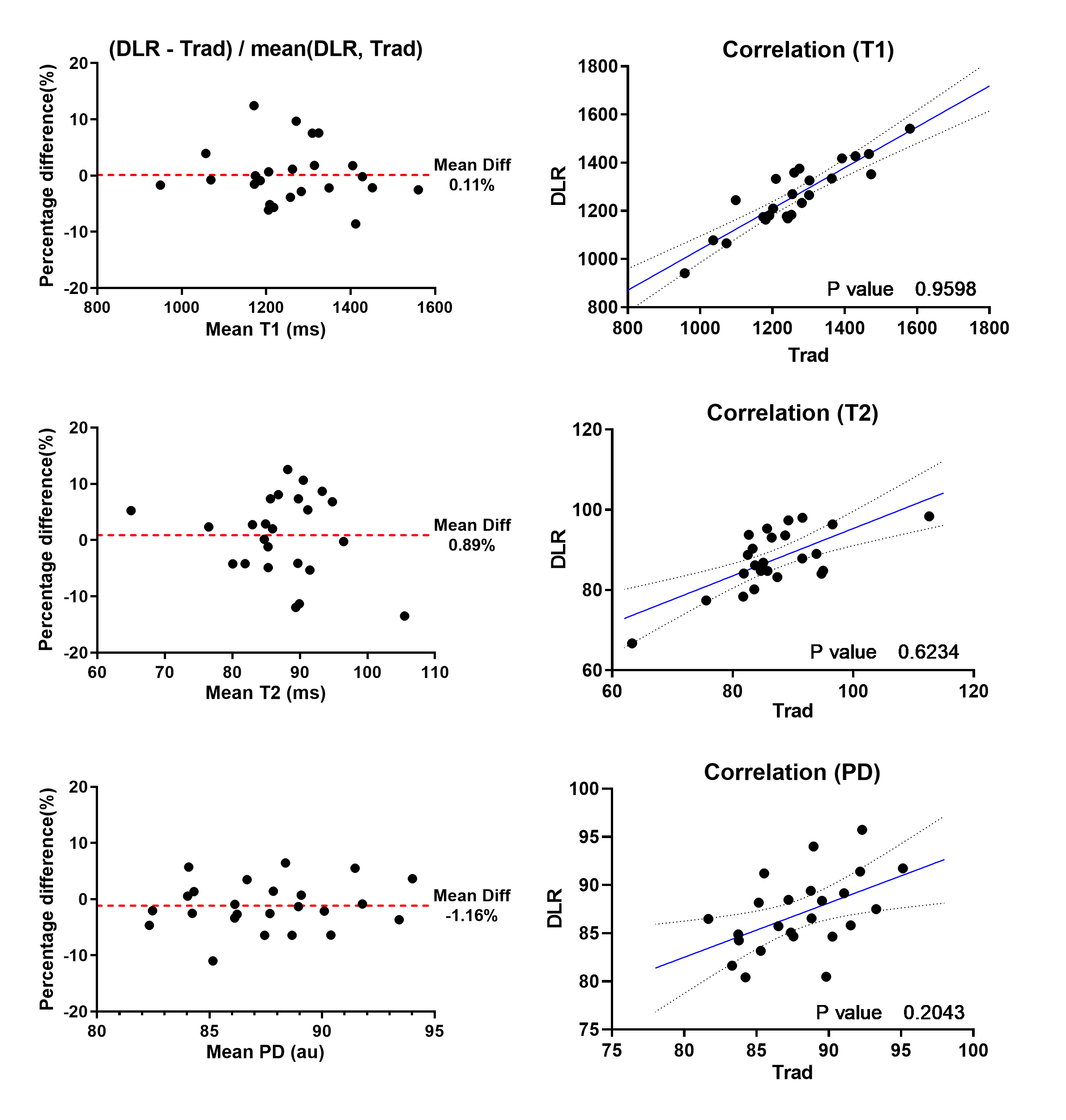
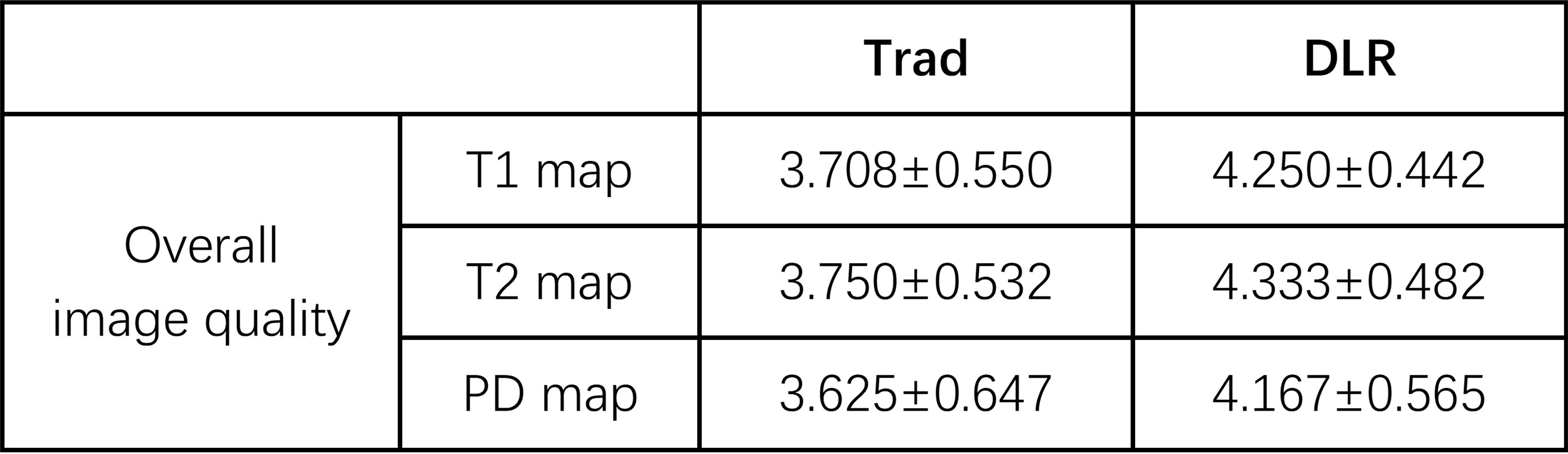
Figure 1 displayed the T1/T2/PD maps from a typical NPC patient with a zoomed-in view of the lesion, emphasizing the improved sharpness around the lesion edges with DLR compared to the Trad method. Table 1 showed the average quantitative image quality scores assigned by the oncologists. The higher average score for the DLR method suggests enhanced lesion conspicuity in the quantitative maps. Figure 2 presented the percentage differences and correlation regression diagrams, supported by paired t-test results of T1/T2/PD values within NPC lesions obtained through both methods. The detailed statistical outcomes are cataloged in Table 2, revealing no significant differences in quantitative values between the two methods.Discussion
As a synthetic MR method, MAGiC with Multi-Dynamic Multi-Echo (MDME) acquisition is capable of producing T1/T2/PD maps within a single sequence, which significantly reduces clinical scan time while offering superior diagnostic confirmation in oncology cases. However, for patients with NPC, to stay still in a 4-minute scan duration may prove to be intolerable. Moreover, a higher acceleration rate can lead to lower signal intensity during reconstruction.As this study demonstrated, the combination of AIR Recon DL with a higher acceleration rate can reduce the scan time by more than half a minute for a full brain scan. Figure 2 illustrates a minor overall difference percentage in quantitative values within the lesion, reflecting this proposed rapid DLR method consistently provides stable and accurate quantitative measurements, aligning with the performance of traditional methods.
As a preliminary study, this study involved a relatively small patient amount, which limited the depth of statistical analysis. A relatively large standard deviation arises in Table 2 due to the patients' inability to maintain stillness between the two protocols. In further investigations, a condensed 2-minute, lesion-focused protocol would be designed to minimize quantitative deviations attributable to patient movement.
Conclusion
When integrated with AIR Recon DL, the expedited MAGiC acquisition proves proficient in delivering precise quantitative maps and impressive image quality for NPC diagnosis.Acknowledgements
No acknowledgement found.References
- Lai, Vincent, and Pek Lan Khong. "Updates on MR imaging and 18F-FDG PET/CT imaging in nasopharyngeal carcinoma." Oral oncology 50.6 (2014): 539-548.
- Meng, T., et al. "Investigation of the feasibility of synthetic MRI in the differential diagnosis of non-keratinising nasopharyngeal carcinoma and benign hyperplasia using different contoured methods for delineation of the region of interest." Clinical Radiology 76.3 (2021): 238-e9.
- Wang, Peng, et al. "Synthetic MRI in differentiating benign from metastatic retropharyngeal lymph node: combination with diffusion-weighted imaging." European Radiology 33.1 (2023): 152-161.
- Yang, Fan, et al. "Pretreatment synthetic magnetic resonance imaging predicts disease progression in nonmetastatic nasopharyngeal carcinoma after intensity modulation radiation therapy." Insights into Imaging 14.1 (2023): 1-12.
- Tanenbaum, L. N., et al. "Deep Learning–Generated Synthetic MR Imaging STIR Spine Images Are Superior in Image Quality and Diagnostically Equivalent to Conventional STIR: A Multicenter, Multireader Trial." American Journal of Neuroradiology 44.8 (2023): 987-993.
- Kim, E., et al. "Accelerated Synthetic MRI with Deep Learning–Based Reconstruction for Pediatric Neuroimaging." American Journal of Neuroradiology 43.11 (2022): 1653-1659.
- Joshi, Ankur, et al. "Likert scale: Explored and explained." British journal of applied science & technology 7.4 (2015): 396.
Figures