3801
Test-Retest analysis of atTRACTive with a dissimilarity uncertainty sampling scheme on the Corpus Callosum of mice1German Cancer Research Center (DKFZ), Division of Medical Image Computing, Heidelberg, Germany, 2Medical Faculty Heidelberg, Heidelberg University, Heidelberg, Germany, 3NCT Heidelberg, National Center for Tumor Diseases (NCT), Heidelberg, Germany, 4Faculty of Bioscience, Heidelberg University, Heidelberg, Germany, 5HIDSS4Health - Helmholtz Information and Data Science School for Health,, Karlsruhe/Heidelberg, Germany, 6Faculty of Mathematics and Computer Science, Heidelberg University, Heidelberg, Germany, 7German Cancer Research Center (DKFZ), Division of Radiology, Heidelberg, Germany, 8Neurology Clinic and National Center for Tumor Diseases, Heidelberg University Hospital, Heidelberg, Germany, 9Department of Neurosurgery, Heidelberg University Hospital, Heidelberg, Germany, 10IU, International University of Applied Sciences, Erfurt, Germany, 11Department of Psychiatry and Psychotherapy, Central Institute of Mental Health, Medical Faculty Mannheim, University of Heidelberg, Mannheim, Germany, 12Department of Psychiatry and Psychotherapy, Central Institute of Mental Health, Mannheim, Germany, 13National Center for Tumor Diseases (NCT), Heidelberg, Germany, 14Pattern Analysis and Learning Group,, Heidelberg University Hospital, Heidelberg, Germany, 15German Cancer Consortium (DKTK), partner site Heidelberg, Germany
Synopsis
Keywords: AI/ML Software, Machine Learning/Artificial Intelligence, Active Learning, Tractography, White Matter
Motivation: Accurate tractography-based segmentation of white matter tracts is crucial for tasks such as pre-surgical planning. Fully automated methods are limited to predefined tracts and struggle with anatomical deviations, e.g. caused by tumors.
Goal(s): Our goal is to enhance the manual segmentation process through a novel and intuitive approach.
Approach: We recently developed atTRACTive, a tool for semi-automatic fiber dissection relying on entropy-based active learning. In this work, we have improved atTRACTive and conducted an initial evaluation of its test-retest reliability in comparison to traditional ROI-based tract segmentation methods.
Results: atTRACTive has demonstrated superior test-retest reliability compared to traditional ROI-based segmentation approaches.
Impact: The method offers guidance to researchers in the intuitive and efficient segmentation of arbitrary white matter tracts. Instead of drawing challenging-to-reproduce ROIs, users can simply annotate meaningful streamlines, which are then used to train a classifier.
Introduction
Delineation of individual white matter pathways based on fiber tractography is a widely used technique, e.g. for pre-surgical planning1, brain development studies2 or the characterization of psychiatric disesases3,4. To address the limitations of manually extracting tracts from whole-brain tractograms (known as virtual fiber dissection), which predominantly depends on defining regions of interest (ROI) and is very time-consuming and hard to reproduce, fully automated approaches based on supervised machine learning have been developed5,6. However, these methods are limited to pre-defined tracts, mostly relying on healthy adult data, require large amounts of pre-annotated data for training and can struggle with large anatomical deviations, e.g. caused by tumors. For all cases not covered by fully automatic methods, we recently introduced atTRACTive, an entropy-based active learning method, where we redefined the concept of manual fiber dissection as a semi-automatic approach in which a classifier iteratively enhances its performance by proposing ambiguous streamlines to a human expert for annotation7. The method is implemented as a graphical user-based tool into MITK Diffusion (https://github.com/MIC-DKFZ/MITK-Diffusion)8. In this work, we propose an improved uncertainty sampling scheme based on dissimilarity. Moreover, we have performed initial test-retest experiments to evaluate the reliability of the GUI-based tool.Method
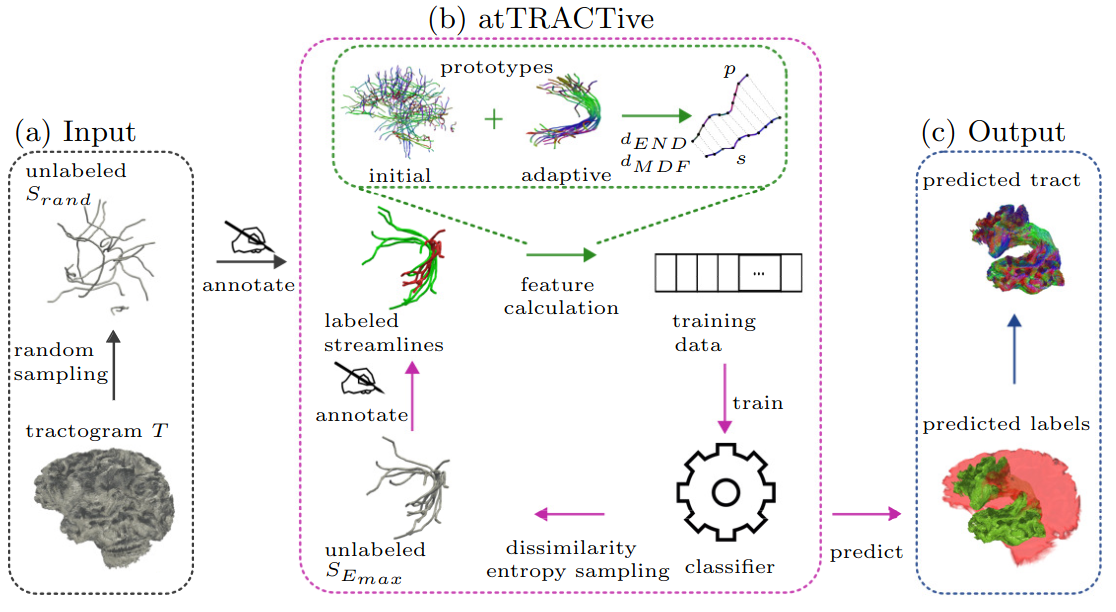
We have considered the fiber dissection problem as a binary classification task, where streamlines are classified based on whether they belong to a particular tract. In an active learning setting, a classifier iteratively proposes streamlines to a human expert for annotation and learns from its decision. Instead of randomly choosing samples for annotation, the samples with the highest uncertainty of the classifier represented by the entropy are proposed9. Considering that similar streamlines lead to equal uncertainty, this sampling approach may have the drawback of providing redundant streamlines due to similar high entropy scores. Consequently, less information and sample variance are presented to the model, which hypothetically reduces reproducibility and increases annotation effort. To address this issue, we use farthest first traversal of a subset of the samples with the highest entropy scores according to the method proposed by Olivetti and colleagues10.The schematic workflow of atTRACTive is visualized in Figure 1. As this method relies heavily on the visual representation of data and a user-friendly workflow, involving user interaction, we have implemented it within MITK-Diffusion8. This offers a GUI-based tool allowing users to annotate streamlines effortlessly by simply hovering over them in either a 3D or 2D render window using structural information, e.g. from T1 weighted images, for orientation.
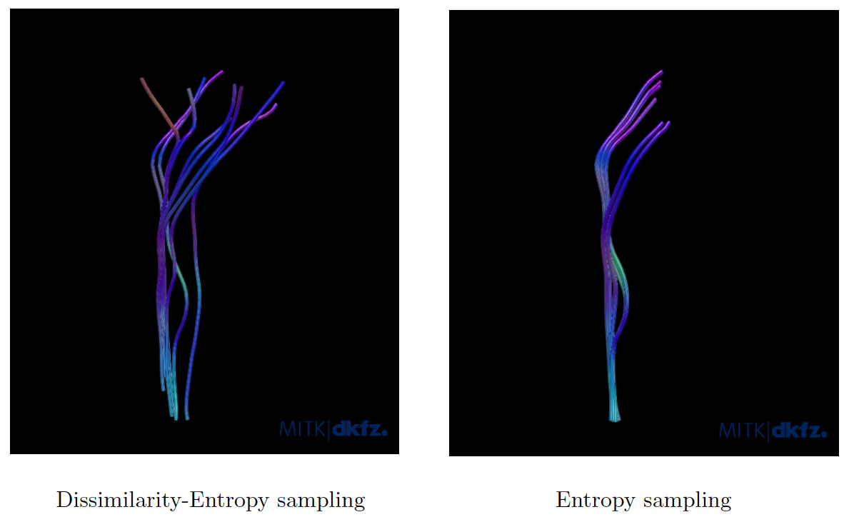
We qualitatively compared the novel sampling scheme to pure entropy-based sampling on the corticospinal tract on the ISMRM dataset11.
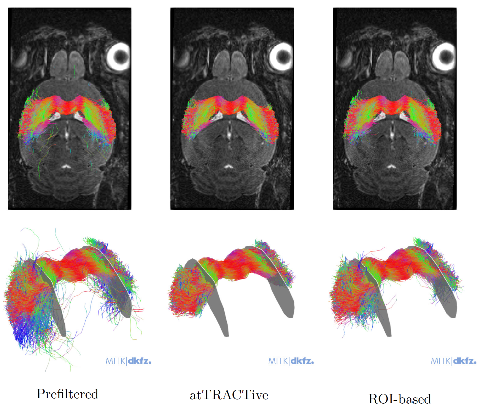
To investigate the reproducibility characteristics of atTRACTive compared to traditional ROI-based approaches, we performed initial experiments for analyzing test-retest reliability. The experiment was performed by a medical expert on five subjects of a mouse brain dataset on the task of extracting the Corpus Callosum. Since no fully automated methods for tract segmentation on mouse data are available so far, this task represents an ideal use-case for atTRACTive. Whole-brain tractography was conducted for all subjects. Two regions of interest were placed to prefilter the corpus callosum fibers from whole brain tractography that consisted of more than one million streamlines. Subsequently, either atTRACTive was applied or additional ROIs were added until the expert was satisfied with the results. The experiments were repeated after two weeks, and tract overlap was assessed using the Dice score to evaluate reliability.
Results
Figure 2 contrasts dissimilarity entropy sampling with pure entropy sampling. Both images show ten fibers suggested by the classifier for labeling by the human expert. Notably, the latter results in fibers with remarkably similar shapes, whereas dissimilarity introduces increased variability among the streamlines, thereby incorporating more information into the model.The initial reliability experiments with atTRACTive revealed its superior robustness compared to pure ROI-based tract dissection, measured with a Dice score of 0.92 versus 0.88 (p=0.377). Qualitative results in Figure 2 display the ability of atTRACTive to provide a more compact representation of the Corpus Callosum, minimizing implausible and false positive fibers.
Conclusion
By introducing dissimilarity into entropy-based sampling, we enhance the capacity of the model to capture a broader range of structural variations among tract fibers. In the future, this technique will be compared quantitatively with respect to efficiency and reproducibility to pure-based entropy sampling.atTRACTive demonstrates promising results in terms of test-retest reliability when compared to traditional manual methods that rely on ROIs. Future experiments will further explore atTRACTive's reproducibility across diverse cases. We are also planning to investigate the generalization capabilities of atTRACTive across different subjects.
Acknowledgements
This work was supported by the German Research Foundation (DFG) grant NE 2069/2-1 as well as by the Helmholtz Association under the jointresearch school "HIDSS4Health – Helmholtz Information and Data Science School for Health.References
1 Berman, J. (2009). Diffusion MR tractography as a tool for surgical planning. Magnetic resonance imaging clinics of North America, 17(2), 205-214.
2 Mukherjee, P., & McKinstry, R. C. (2006). Diffusion tensor imaging and tractography of human brain development. Neuroimaging Clinics, 16(1), 19-43.
3 Wasserthal, J., Maier-Hein, K. H., Neher, P. F., Northoff, G., Kubera, K. M., Fritze, S., ... & Hirjak, D. (2020). Multiparametric mapping of white matter microstructure in catatonia. Neuropsychopharmacology, 45(10), 1750-1757.
4 McIntosh, A. M., Maniega, S. M., Lymer, G. K. S., McKirdy, J., Hall, J., Sussmann, J. E., ... & Lawrie, S. M. (2008). White matter tractography in bipolar disorder and schizophrenia. Biological psychiatry, 64(12), 1088-1092.
5 Bertò, G., Bullock, D., Astolfi, P., Hayashi, S., Zigiotto, L., Annicchiarico, L., ... & Olivetti, E. (2021). Classifyber, a robust streamline-based linear classifier for white matter bundle segmentation. NeuroImage, 224, 117402.
6 Wasserthal, J., Neher, P., & Maier-Hein, K. H. (2018). TractSeg-Fast and accurate white matter tract segmentation. NeuroImage, 183, 239-253.
7 Peretzke, R., Maier-Hein, K. H., Bohn,
J., Kirchhoff, Y., Roy, S., Oberli-Palma, S., ... & Neher, P.
(2023, October). atTRACTive: Semi-automatic white matter tract
segmentation using active learning. In International Conference on Medical Image Computing and Computer-Assisted Intervention (pp. 237-246). Cham: Springer Nature Switzerland.
8 Fritzsche, K. H., Neher, P. F., Reicht, I., van Bruggen, T., Goch, C., Reisert, M., ... & Stieltjes, B. (2012). MITK diffusion imaging. Methods of information in medicine, 51(05), 441-448.
9 Holub, A., Perona, P., & Burl, M. C. (2008, June). Entropy-based active learning for object recognition. In 2008 IEEE Computer Society Conference on Computer Vision and Pattern Recognition Workshops (pp. 1-8). IEEE.
10 Olivetti, E., Nguyen, T. B., & Garyfallidis, E. (2012, July). The approximation of the dissimilarity projection. In 2012 Second International Workshop on Pattern Recognition in NeuroImaging (pp. 85-88). IEEE.
11 Maier-Hein, K. H., Neher, P. F.,
Houde, J. C., Côté, M. A., Garyfallidis, E., Zhong, J., ... &
Descoteaux, M. (2017). The challenge of mapping the human connectome
based on diffusion tractography. Nature communications, 8(1), 1349.
Figures