3798
RegGAN-CBAM based virtual LGE and its application on hypertrophic cardiomyopathy patients1Human Phenome Institute, Fudan university, Shanghai, China, 2Digital Medical Research Center, School of Basic Medical Sciences, Fudan University, Shanghai, China, 3Philips Healthcare, Shanghai, China
Synopsis
Keywords: Diagnosis/Prediction, Cardiomyopathy, Cine, LGE, Hypertrophic cardiomyopathy, RegGAN, CBAM
Motivation: LGE CMR, the standard clinical non-invasive characterization, is limited by its reliance on intravenous contrast agents and long waiting time. Therefore, developing a contrast agent-free technology is essential for achieving fast and cost-effective CMR scans.
Goal(s): To evaluate the reproducibility and reliability of the virtual LGE based on Cine, and compare it with native LGE to assess its efficiency in diagnosing HCM in clinical context.
Approach: RegGAN was employed to forecast LGE imaging and rectify the outcomes. And CBAM was utilized to quantify the influence of diverse components in LGE.
Results: RegGAN-CBAM demonstrates favorable performance in both image and enhancement prediction of LGE.
Impact: The performance of virtual LGE based on Cine exhibits a notable level of diagnostic efficiency and reliability. This approach serves as a non-invasive myocardial tissue characterization method with practical applicability in the clinical assessment of HCM.
Introduction
Late gadolinium enhancement (LGE) imaging stands as the established gold standard for non-invasive myocardial tissue characterization [1]. However, this technique necessitates the administration of intravenous contrast agents and requires long waiting time [2]. Therefore, it is highly desired to develop a contrast agent-free technology as an alternative to LGE, enabling faster and more cost-effective cardiovascular magnetic resonance (CMR) scans [3].Cine, the most widely used sequence for CMR, with its fast imaging speed, shows promise as a contrast agent-free technique for detecting intrinsic imaging signals associated with myocardial abnormalities observed in LGE.
In this regard, we propose a registration-based generative adversarial network (RegGAN-CBAM) [4,5] to assess the reproducibility and reliability of the Cine-based LGE approach, and compare its diagnostic performance for hypertrophic cardiomyopathy with LGE in clinical setting.
Methods
Study PopulationThe local institutional review board approved this retrospective study. Informed consent was obtained from all subjects. In total, 149 patients were enrolled between July 2017 and December 2019. Hypertrophic Cardiomyopathy (HCM) patients were diagnosed according to the guidelines [6]. The end-diastolic left ventricular (LV) wall thickness ≥15mm in one or more segments, not solely explained by loading conditions, or a lesser LV wall thickness of 13-14mm combined with a relevant family history.
Image Acquisition
CMR was performed using a 3 T MRI machine (Ingenia, Philips Healthcare, Best, The Netherlands) using a dS torso coil anterior to the chest. Cine imaging parameters were as follows: repetition time (TR) = 2.8ms, echo time (TE) = 1.4ms, section thickness = 7mm, section gap = 3mm, field of view (FOV) = 300mm×300mm, acquired resolution = 1.2mm×1.2mm. LGE imaging parameters were as follows: TR = 6.1ms, TE = 3ms, FOV = 300mm × 300mm, acquired resolution = 1.6mm × 1.9mm, section thickness = 7mm, section gap = 3mm [7]. The injection plan was 0.15 mmol/kg of gadolinium-DTPA with 15 ml saline flushing.
RegGAN-CBAM Model
The flowchart can be seen in Figure.1. The datasets were randomized into 2 independent groups for method development (70%) and testing (30%). The development group was further divided into training (60%) and validation (10%) datasets. Deep learning models were blinded to the test group. The model training parameters were set with epoch = 200 and learning rate = 0.0001. Additionally, the registration module functions as a label noise model to refine the generated results. And CBAM outputs weights for the multi-channels (corresponding to the LGE different components), enabling the calculation of the influence of different components in LGE.
Model Evaluation
The virtual LGE imaging is evaluated using three metrics: mean absolute error (MAE), peak signal-to-noise ratio (PSNR), and structural similarity index (SSIM). Statistical analysis was conducted to measure myocardium enhancements using native LGE and virtual LGE (Figure.4). Spearman's correlation test was applied to assess the correlation of liver regions (Figure.4a). A Bland-Altman plot was constructed to assess the consistency of myocardium enhancements between native and virtual LGE (Figure.4b). Moreover, in order to provide a visual representation of the myocardium enhancements, box plots were constructed (Figure.4c).
Results
Intermediate results during trainingFigure.2 illustrates the generation of virtual LGE during the training process of RegGAN-CBAM at epochs 10, 80, 150, and 200. It can be observed that with an increasing number of iterations, the virtual LGE becomes progressively clearer.
Image comparison
Figure.3 presents the comparative results of Cine, virtual LGE (epoch = 200), and native LGE. The colorbar visualizes the numerical range corresponding to virtual and native LGE. It is evident that virtual and native LGE exhibit a remarkable level of concordance in terms of their enhancement values.
Model score
The comprehensive scores are presented in Figure 5, including training set, validation set, and test set. Particularly, in the testing set, the MAE was 0.076, the PSNR reached 10.142, and the SSIM reached 0.393.
Statistical analysis
In Figure.3a, a strong positive correlation was observed between native and virtual MRE liver stiffness (r = 0.908, p < 0.01). In Figure.4b, the mean difference line represented an average deviation of -3.391 between the myocardium enhancements. The upper and lower limit lines, corresponding to the 96% limits of agreement, were 184.113 and -190.894, indicating a strong agreement between the two modalities.
Discussion
This study demonstrates that RegGAN-CBAM performs well in the prediction of LGE. The virtual LGE displays notable similarity to native LGE in the images, and their myocardium enhancement values exhibit high correlation and consistency.Conclusion
The LGE approach based on Cine demonstrates high diagnostic efficiency and reliability, serving as a non-invasive method for diagnosing HCM with practical application value in clinical setting.Acknowledgements
This study was supported in part by the National Natural Science Foundation of China (No. 62001120, 62331021), The Royal Society (IEC\NSFC\211235) and the Shanghai Sailing Program (No. 20YF1402400, 22YF1409300).
The correspondence should be sent to Prof. Chengyan Wang (Email: wangcy@fudan.edu.cn)
References
[1] Becker M A J, Cornel J H, Van de Ven P M, et al. The prognostic value of late gadolinium-enhanced cardiac magnetic resonance imaging in nonischemic dilated cardiomyopathy: a review and meta-analysis[J]. JACC: Cardiovascular Imaging, 2018, 11(9): 1274-1284.
[2]Zhang Q, Burrage M K, Lukaschuk E, et al. Toward replacing late gadolinium enhancement with artificial intelligence virtual native enhancement for gadolinium-free cardiovascular magnetic resonance tissue characterization in hypertrophic cardiomyopathy[J]. Circulation, 2021, 144(8): 589-599.
[3] Zhang Q, Burrage M K, Shanmuganathan M, et al. Artificial intelligence for contrast-free MRI: Scar assessment in myocardial infarction using deep learning–based virtual native enhancement[J]. Circulation, 2022, 146(20): 1492-1503.
[4] Kong L, Lian C, Huang D, et al. Breaking the dilemma of medical image-to-image translation[J]. Advances in Neural Information Processing Systems, 2021, 34: 1964-1978.
[5] Woo S, Park J, Lee J Y, et al. Cbam: Convolutional block attention module[C]//Proceedings of the European conference on computer vision (ECCV). 2018: 3-19.
[6]Elliott PM, Anastasakis A, Borger MA, et al. ESC guidelines on diagnosis and management of hypertrophic cardiomyopathy: the task force for the diagnosis and management of hypertrophic cardiomyopathy of the European society of cardiology (ESC). Eur Heart J 2014;35(39):2733-79.
[7] Shi R Y, Wu R, An D A L, et al. Texture analysis applied in T1 maps and extracellular volume obtained using cardiac MRI in the diagnosis of hypertrophic cardiomyopathy and hypertensive heart disease compared with normal controls[J]. Clinical Radiology, 2021, 76(3): 236. e9-236. e19.
Figures
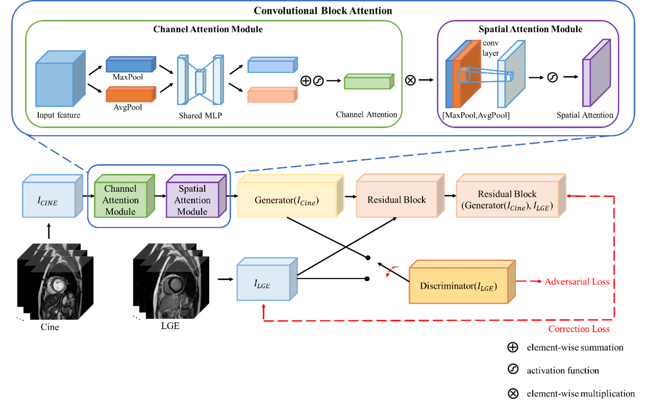
Figure 1. Flowchart of the RegGAN-CBAM model.
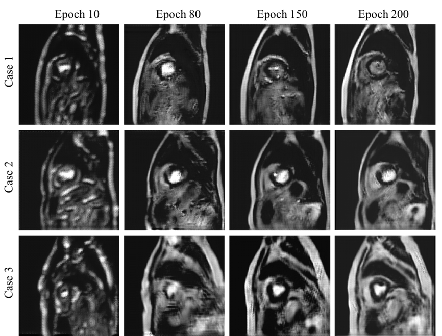
Figure 2. Intermediate 3 cases of virtual LGE during RegGAN-CBAM training.
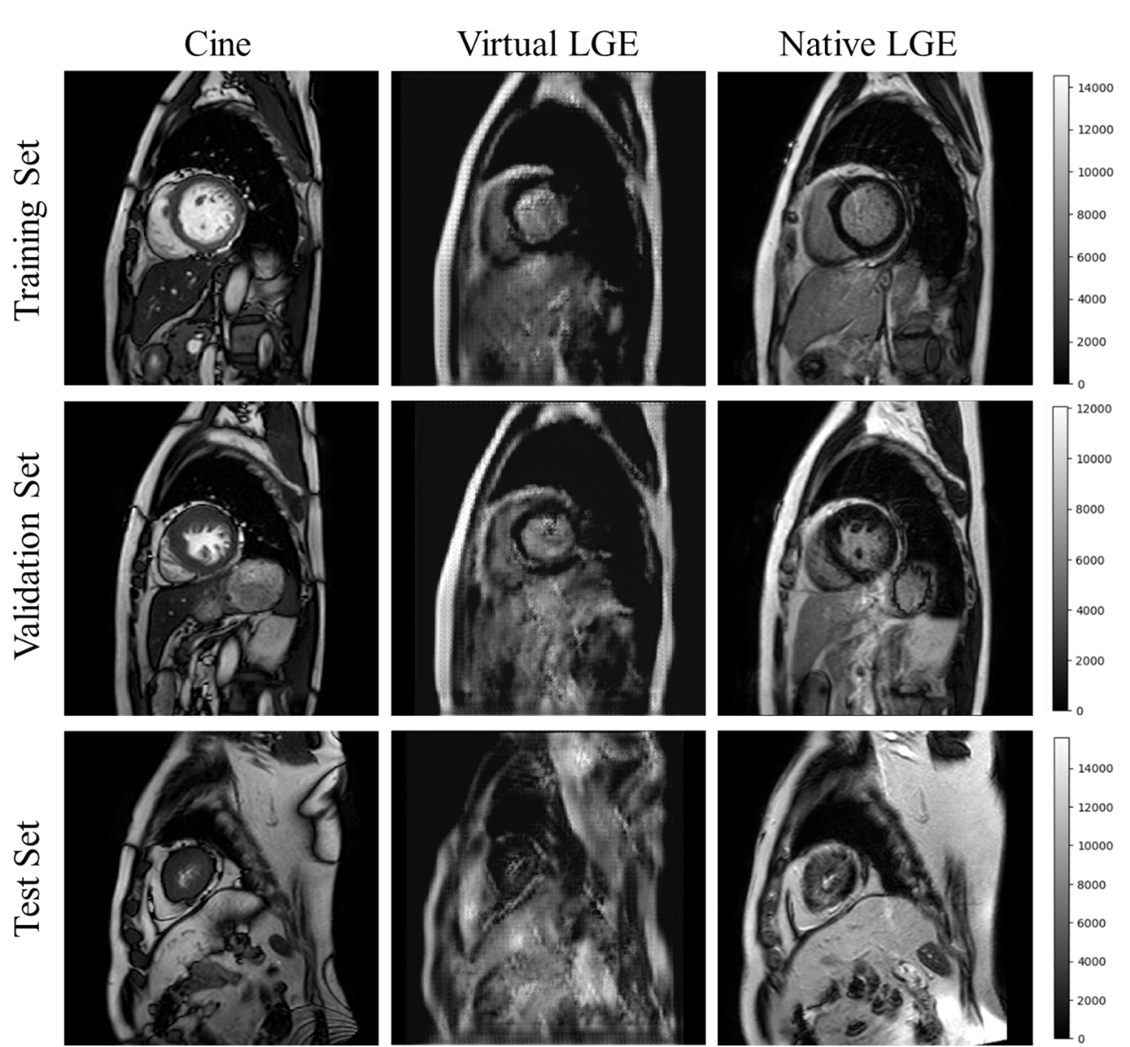
Figure 3. Representative cases of Cine, virtual LGE (epoch = 200), and native LGE images in the training set, validation set, and test set. The colorbar is positioned on the right side of the image to represent the enhancement range of virtual and native LGE. It is evident that virtual and native LGE demonstrate a high level of consistency in terms of their enhancement values.
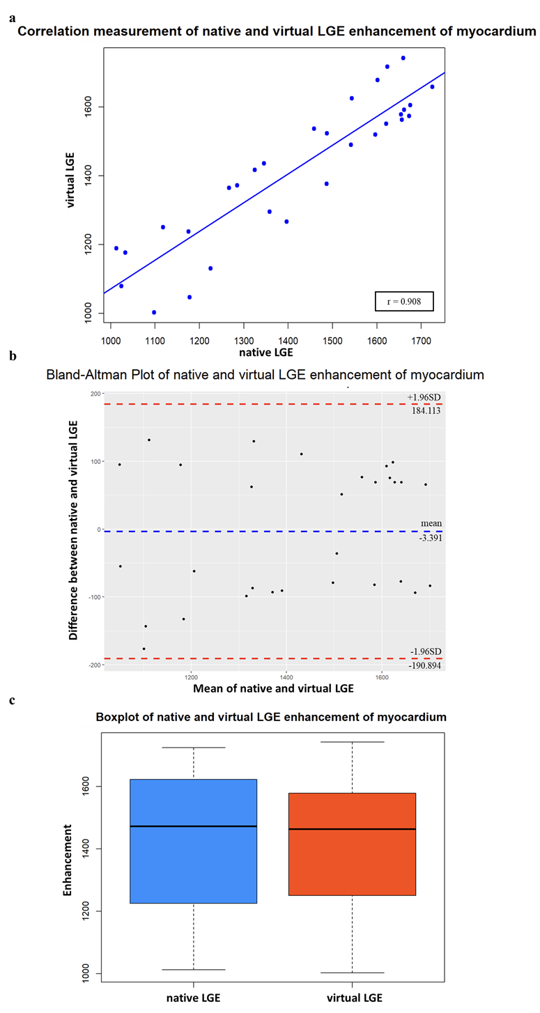
Figure 4. Statistical analysis, including (a) Correlation analysis of native and virtual LGE enhancement of myocardium, (b) Bland-Altman Plot of native and virtual LGE enhancement of myocardium and (c) Boxplot of native and virtual LGE enhancement of myocardium.

Figure 5. Scores of training set, validation set, and test set on RegGAN-CBAM.