3795
Automatic classification of Cine MRI images using CNN: Apical-to-Basal vs Extreme slices1Centre for Biomedical Engineering, Indian Institute of Technology, Delhi, New Delhi, India, 2Department of Radiology, Fortis Memorial Research Institute, Gurugram, India, 3Department of Biomedical Engineering, AIIMS, New Delhi, New Delhi, India, 4Yardi School for Artificial Intelligence, Indian Institute of Technology, Delhi, New Delhi, India
Synopsis
Keywords: Diagnosis/Prediction, Machine Learning/Artificial Intelligence
Motivation: Manual segmentation of cardiac MRI images is a time-consuming and laborious task prone to observer bias. Automatic segmentation approaches provide poor results in extreme slices. A slice classification step applied before automatic segmentation will lead to better results and reduced variability.
Goal(s): To develop a classifier model with high classification performance on short-axis(SA) cine MRI images for slice selection.
Approach: We trained and compared 2 CNN models for classifying SA cine MRI images into Apical-to-Basal vs Extreme slices.
Results: Xception model had better classification accuracy (0.90) and F1- score (0.93) when compared to InceptionV3 (0.87 and 0.89, respectively).
Impact: The proposed model will provide automatic, fast and accurate classification of MRI cine images, which will improve the accuracy of automatic segmentation of myocardium and its assessment.
Introduction
Cardiac MRI is considered the gold standard for quantitative analysis of heart for objective disease assessment 1. However, this requires segmentation of the left ventricular (LV) myocardium, which is time-consuming, laborious, and prone to inter-observer variability if done manually. The utilization of convolutional-neural-networks(CNNs) have shown great potential in the automatic segmentation of LV myocardium. Several studies have shown that a prior slice classification step in addition to segmentation models improves the LV segmentation accuracy2,3 This study aims to develop a DL-based classifier for categorizing short-axis(SA) cine MRI stack into apical-to-basal(A2B) and extreme(EX) classes, which will enhance the efficacy of the subsequent segmentation process.Materials and Methods
MRI Data:This study uses an open-source dataset (M&Ms dataset) 4 containing multi-center, multi-vendor, and multi-disease cine MRI data of 285 patients with approximately 11 slices and 30 time-frames each.
Methodology:
The cine MRI dataset of 285 patient was split into training(n=200), validation(n=50) and testing(n=35). All the images were normalized and resized to 224×224 and labelled as A2B or EX slices depending on the presence or absence of the LV myocardium in 2 chamber view. Training, validation and testing set contained 60162 images, 14865 images, and 10255 images, respectively. A data pipeline to convert single channel 224×224 images to 299×299×3 output was used to feed two pre-trained CNN classifiers, namely, InceptionV35 and Xception6 (pre-trained on the ImageNet dataset7). The models were trained with Adam8 optimizer and binary cross-entropy loss function with a learning rate of 1×10⁻⁴. Fig.1 represents the methodology of the study. Performance of the models were evaluated on the testing dataset using accuracy, F1-score, precision, recall, and area under ROC curve (AUROC) metrics.
Results
The slice classification accuracy achieved by the InceptionV3 model on test dataset was 87%, while for the Xception model was 90%. Other metrics like F1 score, precision and recall for both models are presented in Table 1. A comparison between confusion matrix and ROC curves of the two models is shown in Fig.2 and Fig.3, respectively. The inference time of Xception model was 3.53ms /image (as computed on GPU with configuration: NVIDIA RTX A2000 12GB).Discussion
This study uses CNN based classifier prior to the segmentation to improve segmentation accuracy on SA cine images. This research contributes to the growing body of evidence supporting the integration of DL-based methodologies in the field of cardiac imaging and diagnosis. The promising results indicate the feasibility of leveraging CNN-based slice classification for enhanced segmentation performance and further quantitative analysis. However, a future study on the effects of the proposed slice classification model on the LV segmentation accuracy will be carried out.Conclusion
This study proposed a fully automated method for slice classification on SA cine MRI data with an accuracy of 90%.Acknowledgements
Authors, Sandeep Kumar and Raufiya Jafari share equal authorship. The authors acknowledges dedication and collaboration of MedImg lab(IIT Delhi) members, whose hard work has been invaluable to the study.References
1. Piersson, A. D. Essentials of cardiac MRI in clinical practice. Journal of Cardiovascular Magnetic Resonance 18, 1 (2016).
2. Ho, N. & Kim, Y. C. Evaluation of transfer learning in deep convolutional neural network models for cardiac short axis slice classification. Scientific Reports 2021 11:1 11, 1–11 (2021).
3. Bartoli, A. et al. Deep Learning–based Automated Segmentation of Left Ventricular Trabeculations and Myocardium on Cardiac MR Images: A Feasibility Study. Radiol Artif Intell 3, (2021).
4. Campello, V. M. et al. Multi-Centre, Multi-Vendor and Multi-Disease Cardiac Segmentation: The M&Ms Challenge. IEEE Trans Med Imaging 40, 3543–3554 (2021).
5. Szegedy, C., Vanhoucke, V., Ioffe, S., Shlens, J. & Wojna, Z. Rethinking the Inception Architecture for Computer Vision. Proceedings of the IEEE Computer Society Conference on Computer Vision and Pattern Recognition , 2818–2826 (2016).
6. Chollet, F. Xception: Deep Learning with Depthwise Separable Convolutions. Proceedings - 30th IEEE Conference on Computer Vision and Pattern Recognition, CVPR, 1800–1807 (2016).
7. Russakovsky, O. et al. ImageNet Large Scale Visual Recognition Challenge. Int J Comput Vis 115, 211–252 (2014).
8. Kingma, D. P. & Ba, J. L. Adam: A Method for Stochastic Optimization. 3rd International Conference on Learning Representations, ICLR- Conference Track Proceedings (2014).
Figures
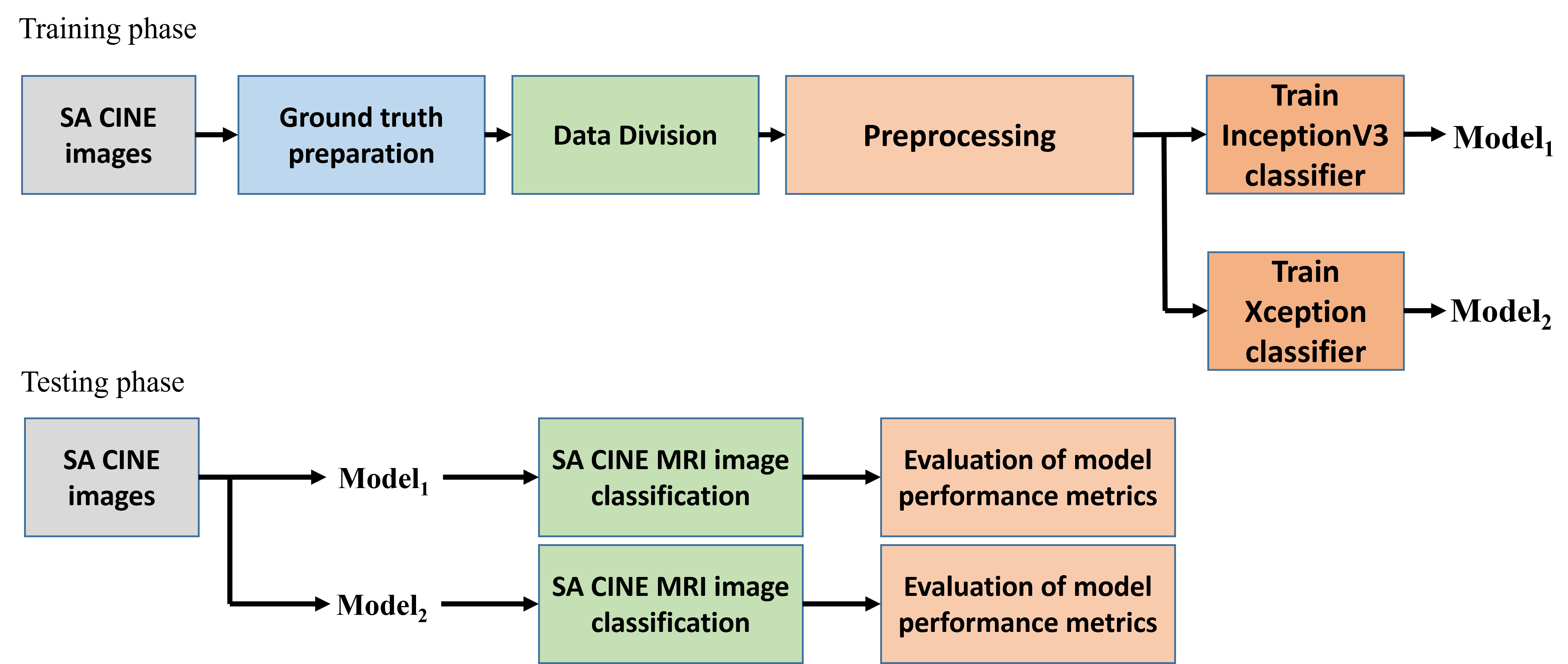
Figure 1: Flowchart of proposed methodology
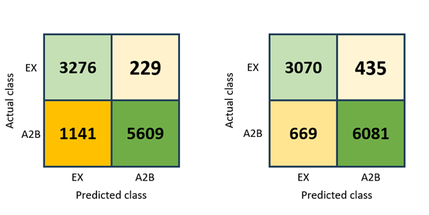
Figure 2: Confusion matrix of InceptionV3 (left) and Xception (right) showing the number of slices classified and the true labels for test dataset.
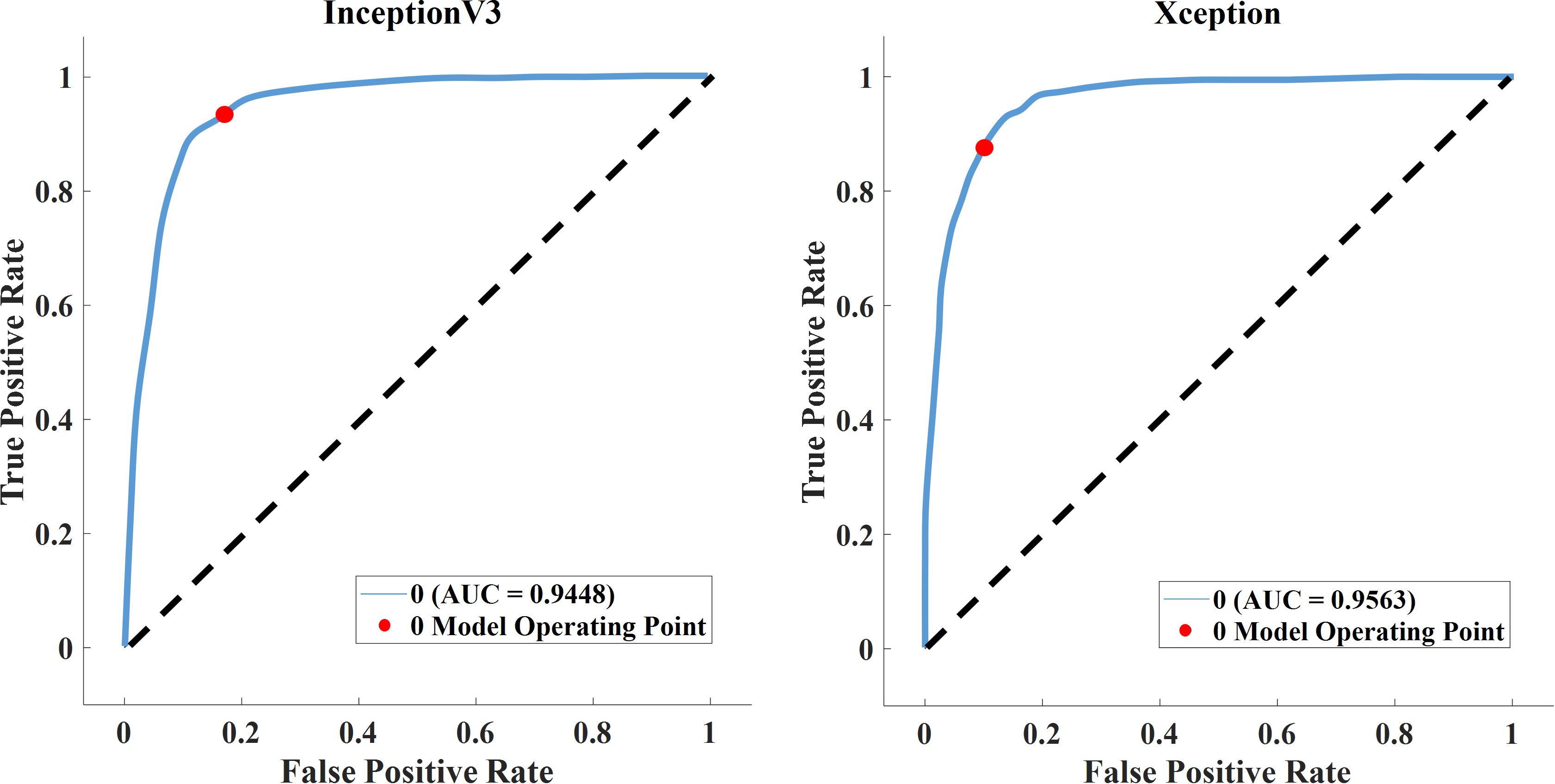
Figure 3: ROC Curve analysis of InceptionV3 (left) and Xception (right) for test dataset.
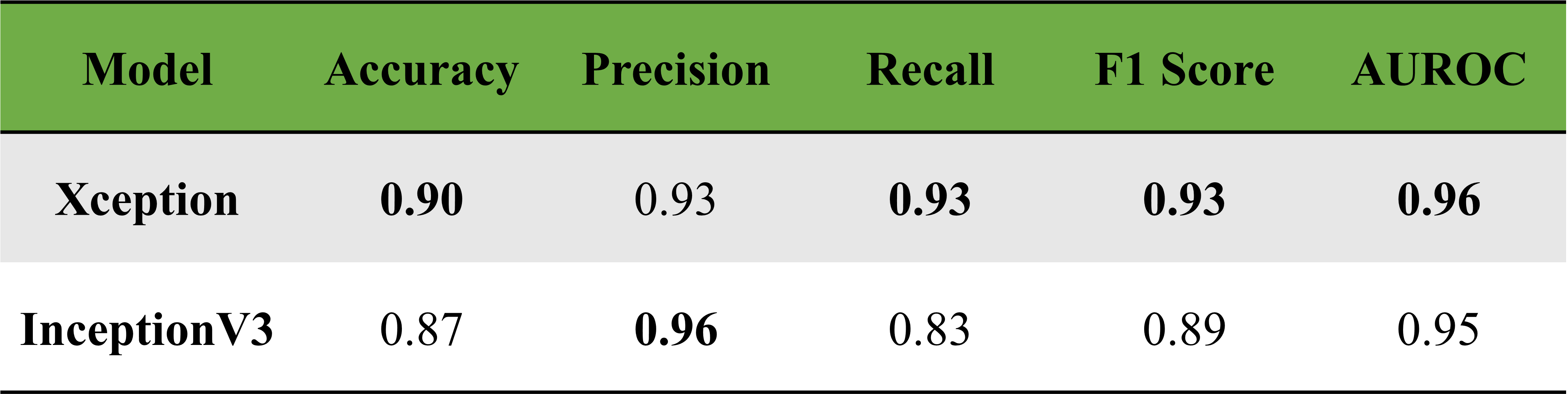
Table. 1: Comparison of performance metrics of InceptionV3 and Xception on test dataset.