3794
The Clinical Utility of Radiomics Models Based on Non-Contrast T1 Mapping in CMR for Discriminating Acute and Chronic Myocardial Infarction1The First Hospital of China Medical University, Shenyang, China, 2University of Electronic Science and Technology of China, Chengdu, China, 3MR Research Collaboration Team,Siemens Healthineers Ltd., Shanghai, China
Synopsis
Keywords: Diagnosis/Prediction, Radiomics, cardiac magnetic resonance
Motivation: The complexity of differentiation between acute and chronic myocardial infarctions(MI) not only complicates the choice of treatment plans but also poses challenges for post-treatment follow-up.
Goal(s): This study aimed to determine whether radiomics model based on T1 mapping can be applied in differential diagnosis of acute and chronic MI.
Approach: Images of 61 patients were included for feature extraction, and the statistically significant features were selected to establish the radiomics model.
Results: This radiomics model demonstrates significant efficacy in distinguishing between acute and chronic myocardial infarction lesions in patients, providing valuable support for clinical diagnosis and follow-up treatment.
Impact: The development of radiomics model to differentiate acute from chronic myocardial infarction lesions holds promise for facilitating prompt clinical decision-making. This advancement enables early medical intervention for MI patients, reducing the risk of adverse cardiovascular events and enhancing patient prognosis.
Introduction
Cardiovascular disease currently stands as the leading global cause of mortality [1]. Among its manifestations, acute myocardial infarction represents a critical and perilous condition, with cardiomyocyte necrosis stemming from myocardial infarction significantly contributing to the unfavorable prognosis of cardiovascular patients. Research has shown that many MI patients present with both acute and chronic infarctions at their initial diagnosis, posing difficulties in treatment planning and follow-up [2]. Electrocardiograms and coronary angiography have limitations in pinpointing new lesions. In certain cases, especially when clinical information and medical history are limited or when the two conditions manifest similarly, distinguishing between new and old MI lesions can be more complex. Therefore, from a practical clinical perspective, differentiation between acute and chronic MI holds diagnostic value[3]. Due to the intricate nature of cardiac structure and dynamics, as well as the high observer variability in widely used visual analyses and qualitative assessments in conventional clinical diagnostics, there is a clinical need for more objective data assessment to enhance diagnostic reliability while reducing interpretation and phenotype description errors. Cardiac magnetic resonance (CMR) imaging has gained widespread recognition for its non-invasive and versatile nature in assessing cardiac structure and function[4].Methods
This retrospective analysis encompassed clinical data from 61 myocardial infarction (MI) patients, featuring 29 images acquired during the acute phase and 32 during the chronic phase. Imaging was performed on a 3T scanner (MAGNETOM Prisma, Siemens Healthcare, Erlangen, Germany), involving the acquisition of black blood and bright blood sequences, as well as MOLLI-T1 mapping scan with 4 chambers, 2 chambers and all the short axis. The global myocardial image was segmented using a Simens self-developed segmentation algorithm as ROI (Figure 1). A specialized MATLAB program was employed for global myocardial segmentation from plain T1 mapping images, and FAE 0.5.8 open-source software (https://github.com/salan668/FAE) was used to extract radiomics features.To standardize the radiomics feature data, an independent sample T-test was applied for feature selection. Subsequently, 49 patients were selected as the training dataset, while an additional 12 cases served as an independent testing dataset to establish the radiomics model based on statistically significant parameters. The model's performance was evaluated using receiver operating characteristic (ROC) curve analysis, and the area under the ROC curve (AUC) was calculated to quantify its discriminative predictive power.
Results and Discussion
Features: A total of 14 shape features and 20 first-order statistical features were extracted from the T1 mapping images. Seven of the first-order statistical features were found to be statistically significant (P < 0.05, Figure 2). ROC curves were generated for these statistically significant characteristics. Notably, the AUC for first-order statistical features, including Interquartile Range, Mean Absolute Deviation, and Robust Mean Absolute Deviation, in the differential diagnosis, all exceeded 0.8. The AUC for Robust Mean Absolute Deviation reached a maximum of 0.832 (Figure 3).Model: Cross-validation with 5-fold on the training dataset revealed that the model based on six features achieved the highest AUC on the validation dataset. At this point, the AUC and accuracy of the model reached 0.889 and 0.917 on the testing dataset. Clinical statistics in the diagnosis and the selected features are presented in Table 1 and Table 2. The ROC curve is illustrated in Figure 4.
Limitation: In this study, only shape features and first-order statistical features were extracted. Further research is needed to explore higher-order features in future investigations.
Conclusion
Significant differences in the imaging characteristics of myocardial infarction were observed, although some imaging findings between the two groups of cases overlapped.The establishment of radiomics model based on T1Mapping technology can enhance diagnostic efficacy and hold high clinical value for identifying lesions in patients with myocardial infarction during different stages.
Acknowledgements
No acknowledgement found.References
[1] Santulli G (2013) Epidemiology of cardiovascular disease in the 21st century: updated updated numbers and updated facts. J Cardiovasc Dis Res 1:1–2
[2] Differentiation of Acute Myocardial Infarction from Chronic Myocardial Scar with MRI[J]: 1-3.
[3] Byoung Wook Choi, MD,Department of Radiology, Yonsei University, College of Medicine, Severance Hospital, Seoul 120-752, Korea.
[4] Karamitsos TD, Francis JM, Myerson S, Selvanayagam JB,Neubauer S (2009) The role of cardiovascular magnetic resonance imaging in heart failure. J Am Coll Cardiol 54:1407–14242321 European Radiology (2023) 33:2312–2323
Figures
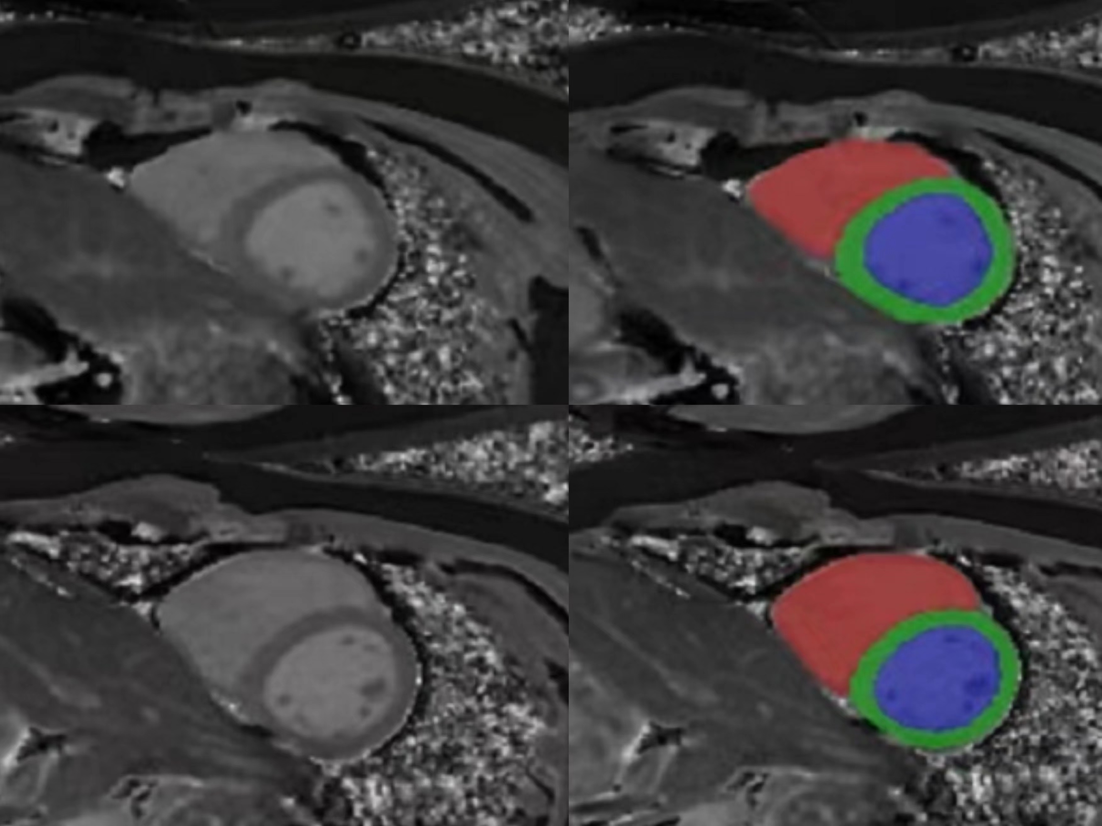
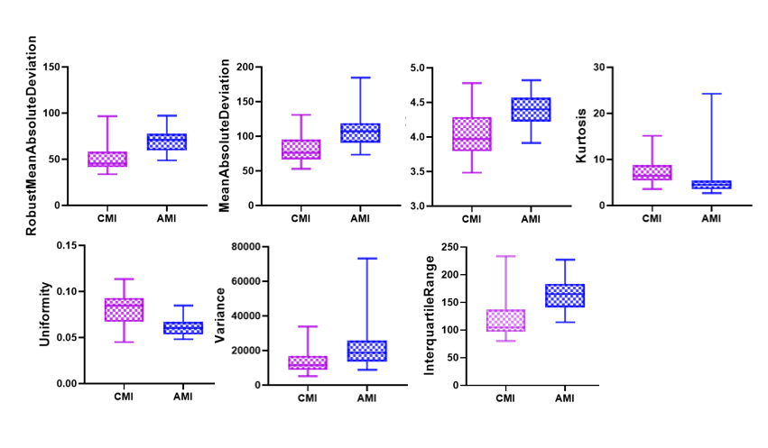
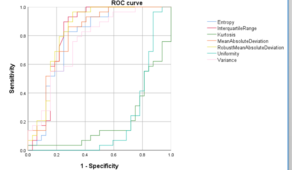
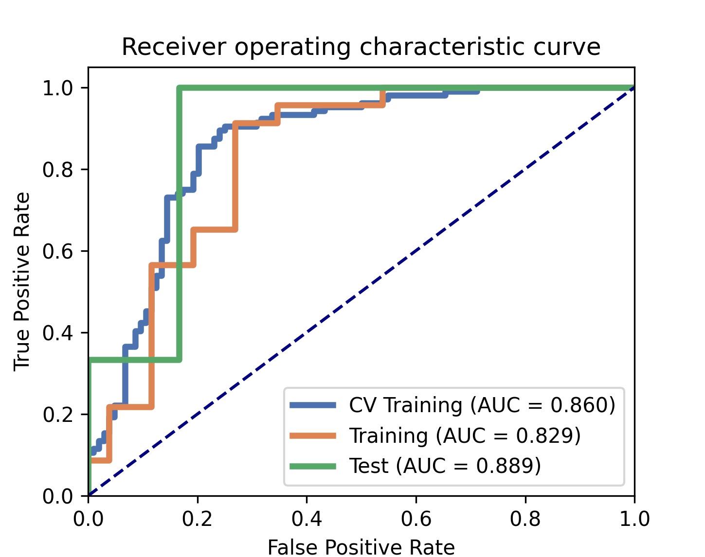
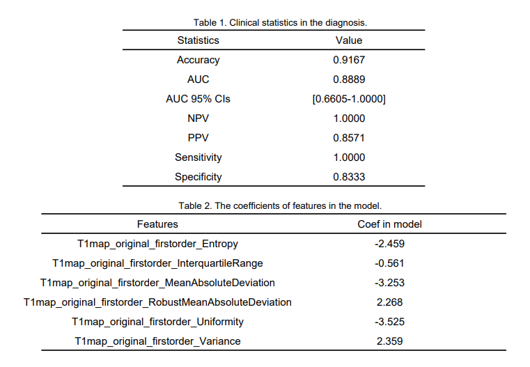
Table 1. Clinical statistics in the diagnosis.
Table 2. The coefficients of features in the model.