3786
Differentiation Magnetic Resonance Images of Tuberculous and Brucellar Spondylitis Using Convolutional Neural Network Based on VGG191Department of Radiology, Shandong Provincial Qianfoshan Hospital, Shandong University, Jinan, China, 2Beijing United Imaging Research Institute of Intelligent Imaging, Yongteng North Road, Haidian District, Beijing 100089, Beijing, China, 3Clinical Medical College of Jining Medical University, Jining, China, 4Department of Radiology, The First Affiliated Hospital of Shandong First Medical University & Shandong Provincial Qianfoshan Hospital, Jinan, China
Synopsis
Keywords: Diagnosis/Prediction, Infection, spondylitis
Motivation: Tuberculous spondylitis (TS) and brucellosis spondylitis (BS) are two common infectious diseases in spinal surgery, and the differential diagnosis of these diseases is challenging but important to ensure appropriate treatment.
Goal(s): The aim of this study was to evaluate the performance of a convolutional neural network CNN) based on VGG19 in distinguishing between TS and BS on different parameter magnetic resonance imaging (MRI) and to compare it with three radiologists.
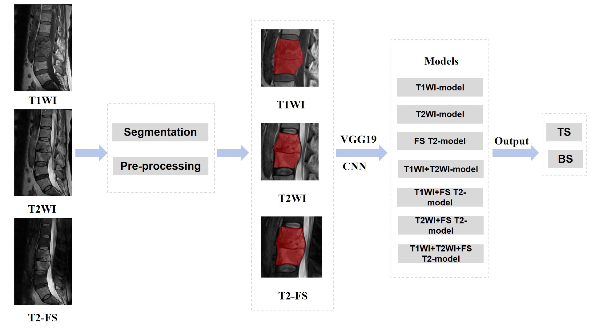
Approach: MRIs of 383 patients were randomly divided into training (n = 307) and validation (n = 76) groups.
Results: VGG19-based CNN outperforms radiologist assessment in distinguishing TS from BS.
Impact: The proposed CNN based on VGG19 is effective in diagnosing TS and BS on MRI, which could not only help in clinical decision making, but also improve efficiency and reduce medical costs.
Purpose
The aim of this study was to evaluate the performance of a convolutional neural network CNN) based on VGG19 in distinguishing between Tuberculous spondylitis (TS) and brucellosis spondylitis (BS) on magnetic resonance imaging (MRI) with different parameters and to compare it with the performance of three skilled radiologists.Methods
A total of 383 patients with TS (n = 182) and BS (n = 201) were randomly divided into training (n = 307) and validation (n = 76) groups. Various MRI sequences, including sagittal T1-weighted (T1WI),T2-weighted (T2WI), and fat-suppressed T2-weighted imaging (FS T2), were used to construct three discrimination models using VGG19 architecture. To enhance the differentiating ability for single MRI sequence we conducted a fusion strategy by averaging the predicted probabilities from the aforementioned three discrimination models. The fusion strategy was respectively performed to fuse T1WI and T2WI, T1WI and FS T2, T2WIand FS T2, T1WI, T2WI and FS T2. Consequently, we obtained seven deep-learning based discrimination models. Finally, three musculoskeletal radiologists with different experienced levels were invited to independently diagnose the images. To better compare the discriminating performance between seven deep-learning based models and three radiologists, we plotted the receiver operating characteristic curve and calculated the area under the curve (AUC). Metrics including accuracy, sensitivity, specificity and F1 score were also calculated.Results
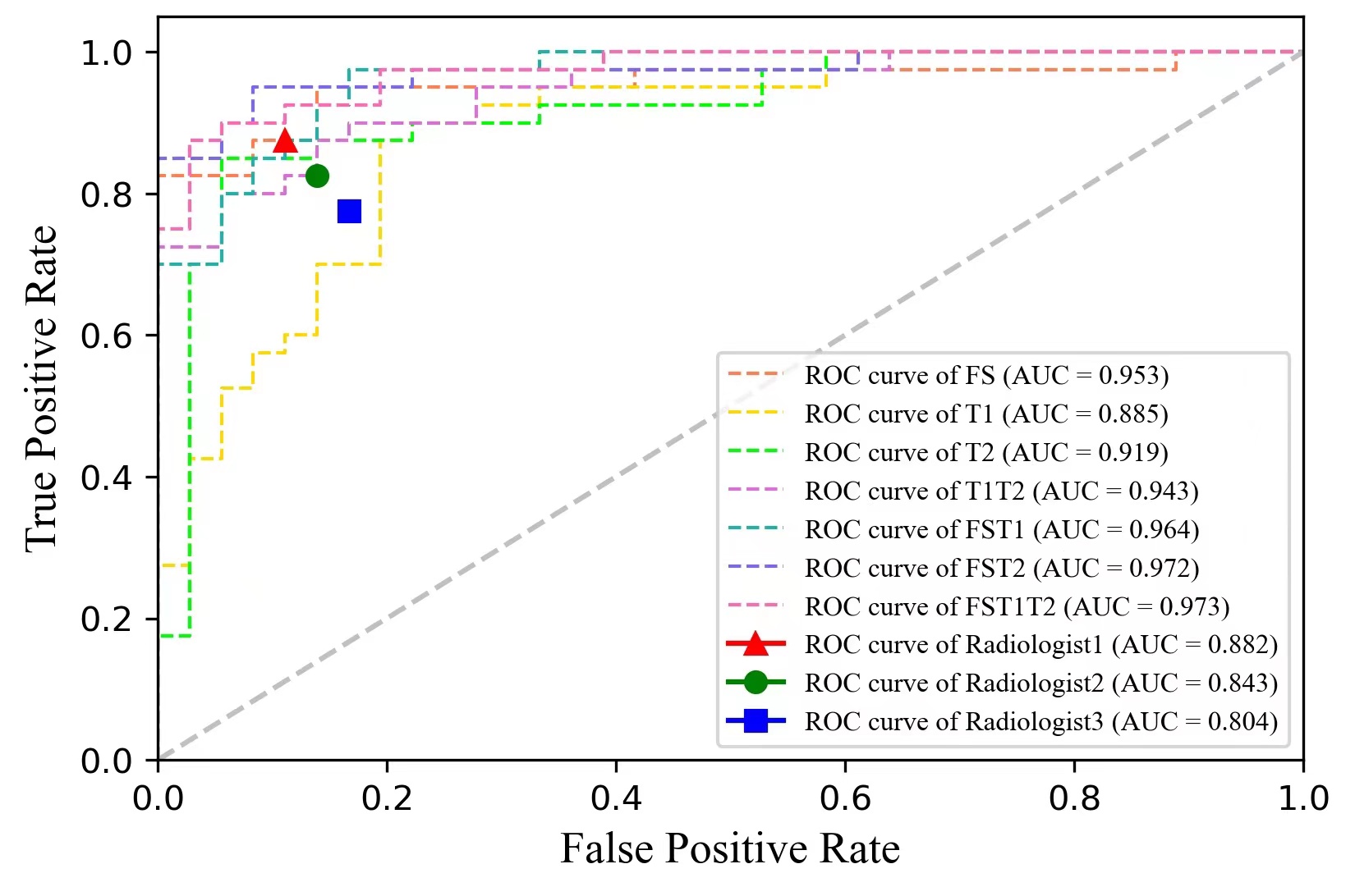
The AUC of T1WI+T2WI+FS T2-model was 0.973, which were higher than the other parameter MRI models including T1WI-model (AUC = 0.885),T2WI-model (AUC = 0.919), FS T2-model (AUC=0.953),T1WI+T2WI-model (AUC=0.943),T1WI+FS T2-model (AUC=0.964) and T2WI+FS T2-model (AUC=0.972).Additionally, the AUC of three musculoskeletal radiologists with different levels of experience were 0.882,0.843,0.804,respectively.Discussion
TS and BS are two prevalent infectious diseases of the spine that are initially caused by bacteremia. In many developing countries, both diseases remain a serious public health problem1,2.Due to similar clinical and imaging presentations,BS is easily misdiagnosed as TS3.Both TS and BS require appropriate treatment with long-term use of appropriate antibiotics. Inadequate treatment due to delayed diagnosis or misdiagnosis may result in neurological deficits or spinal deformities4.Therefore, early and correct diagnosis is essential.MRI is the modality of choice for spine imaging because of its high sensitivity to soft tissue and bone marrow oedema5.Bone marrow oedema is thearliest MR finding, showing low signal in T1WI and high signal T2WI along the endplate6,7.FS T2 is more sensitive and reliable in the detection of water protons in contrast to T1WI and T2WI.Liu et al.applied the FS T2 technique to compare the height and signal intensity of the vertebral bodies and discs in the acute and subacute phases to identify BS and TS8.They concluded that extensive homogeneous high signal within the vertebral body in BS and inhomogeneous high signal near the endplates in TS are characteristic manifestations of FS T2, and these results are consistent with the pathological facts9.Recently, deep learning(DL) has been rapidly evolving. Constant, Caroline et al. outlined the use of DL in medical imaging to improve spine care, noting the preferred DL technique was CNNs, with the most commonly used MRI sequences being T2-weighted alone, T1-weighted alone, and a combination of T1-weighted and T2-weighted10.Yeh et al. applied the ResNet50 algorithm to develop a decision support system for the differential diagnosis of benign and malignant spinal fractures on MRI11.Liu et al. proposed a multi-model weighted fusion framework based on MRI and age information to effectively diagnose benign and malignant spinal tumours12.In this study, we set up a series of CNN models to compare the diagnosis ability among various single MRI sequences (namely T1WI, T2WI and FS T2) and their combinations. The FS T2 sequence realized the highest AUC with 0.953 in distinguishing BS and TS, which aligns with pathological findings. The fusion of FS T2 can largely enhance the performance of TIWI and T2WI, increasing the AUC from 0.885 to 0.964 and 0.919 to 0.972, respectively. Additionally, the fusion of T1WI, T2WI and FS T2 achieved the best AUC of 0.973, suggesting that multiple sequences may provide more information for TS and BS discrimination. Furthermore, the all presented seven deep-learning based models outperformed the diagnosis ability of the involved radiologists.
This study had several limitations. First, although our study had a larger sample size than previous studies, multicentre analyses with larger sample sizes are needed in the future.Second, each ROI was manually drawn on MRI sequences. Therefore, an automated or semi-automated ROI to reduce inter-observer variability is urgently needed.
Conclusion
CNN based on VGG19 can help discriminate between TS and BS using MRI with different parameters, which outperformed the radiologist's assessment and could help guide clinical decision making.Acknowledgements
No acknowledgement found.References
(1) Jiang, H.; O’Callaghan, D.; Ding, J.-B. Brucellosis in China: History, Progress and Challenge. Infect. Dis. Poverty 2020, 9 (1), 55. https://doi.org/10.1186/s40249-020-00673-8.
(2) Shanmuganathan, R.; Ramachandran, K.; Shetty, A. P.; Kanna, R. M. Active Tuberculosis of Spine: Current Updates. North Am. Spine Soc. J. NASSJ 2023, 16, 100267. https://doi.org/10.1016/j.xnsj.2023.100267.
(3) Wang, W.; Fan, Z.; Zhen, J. MRI Radiomics-Based Evaluation of Tuberculous and Brucella Spondylitis. J. Int. Med. Res. 2023, 51 (8), 03000605231195156. https://doi.org/10.1177/03000605231195156.
(4) Gao, M.; Sun, J.; Jiang, Z.; Cui, X.; Liu, X.; Wang, G.; Li, T.; Liang, C. Comparison of Tuberculous and Brucellar Spondylitis on Magnetic Resonance Images. Spine 2017, 42 (2), 113–121. https://doi.org/10.1097/BRS.0000000000001697.
(5) Torres, C.; Zakhari, N. Imaging of Spine Infection. Semin. Roentgenol. 2017, 52 (1), 17–26. https://doi.org/10.1053/j.ro.2016.05.013.
(6) Diehn, F. E. Imaging of Spine Infection. Radiol. Clin. North Am. 2012, 50 (4), 777–798. https://doi.org/10.1016/j.rcl.2012.04.001.
(7)Yang, X.; Zhang, Q.; Guo, X. Value of Magnetic Resonance Imaging in Brucellar Spondylodiscitis. Radiol. Med. (Torino) 2014, 119 (12), 928–933. https://doi.org/10.1007/s11547-014-0416-x.
(8) Liu, X.; Li, H.; Jin, C.; Niu, G.; Guo, B.; Chen, Y.; Yang, J. Differentiation Between Brucellar and Tuberculous Spondylodiscitis in the Acute and Subacute Stages by MRI. Acad. Radiol. 2018, 25 (9), 1183–1189. https://doi.org/10.1016/j.acra.2018.01.028.
(9) Sharif HS, Aideyan OA, Clark DC, et al. Brucellar and tuberculous spondylitis: comparative imaging features. Radiology. 1989;171(2):419-425. doi:10.1148/radiology.171.2.2704806
(10) Constant, C.; Aubin, C.-E.; Kremers, H. M.; Garcia, D. V. V.; Wyles, C. C.; Rouzrokh, P.; Larson, A. N. The Use of Deep Learning in Medical Imaging to Improve Spine Care: A Scoping Review of Current Literature and Clinical Applications. North Am. Spine Soc. J. NASSJ 2023, 15, 100236. https://doi.org/10.1016/j.xnsj.2023.100236.
(11) Yeh, L.-R.; Zhang, Y.; Chen, J.-H.; Liu, Y.-L.; Wang, A.-C.; Yang, J.-Y.; Yeh, W.-C.; Cheng, C.-S.; Chen, L.-K.; Su, M.-Y. A Deep Learning-Based Method for the Diagnosis of Vertebral Fractures on Spine MRI: Retrospective Training and Validation of ResNet. Eur. Spine J. 2022, 31 (8), 2022–2030. https://doi.org/10.1007/s00586-022-07121-1.
(12) Liu, H.; Jiao, M.; Yuan, Y.; Ouyang, H.; Liu, J.; Li, Y.; Wang, C.; Lang, N.; Qian, Y.; Jiang, L.; Yuan, H.; Wang, X. Benign and Malignant Diagnosis of Spinal Tumors Based on Deep Learning and Weighted Fusion Framework on MRI. Insights Imaging 2022, 13 (1), 87. https://doi.org/10.1186/s13244-022-01227-2.
Figures