3785
Machine-learning-based multimodality radiomics analysis for the preoperative prediction for local relapse in osteosarcoma1Department of Radiology, The University of Hong Kong-Shenzhen Hospital, ShenZhen, China, 2Department of Radiology, Zhongshan Hospital of Traditional Chinese Medicine, Zhongshan, China, 3Department of Radiology, State Key Laboratory of Oncology in South China, Collaborative Innovation Center for Cancer Medicine, Sun Yat-sen University Cancer Center, Guangzhou, China, 4GE Healthcare, MR Research, Beijing, China, 5Department of Radiology, The University of Hong Kong - Shenzhen Hospital, Shenzhen, China
Synopsis
Keywords: Diagnosis/Prediction, Bone, Osteosarcoma
Motivation: To compare the efficacy of radiomics models using four machine learning algorithms in predicting for local relapse of osteosarcoma before surgery.
Goal(s): This study established a robust prediction model of local relapse to improve prognosis efficacy of osteosarcoma and aid a personalized treatment planning.
Approach: Comparison of four algorithms in classifying high-risk local-relapse patients from low-risk ones based on only preoperative radiographic, MR, and both radiomic features.
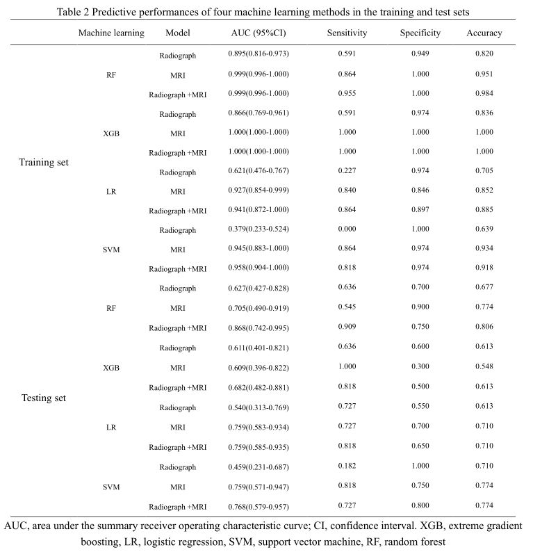
Results: The random-forest based prediction model using both radiograph-MRI radiomic features had the best performance on differentiating patients with local relapse from those with non-local relapse with AUC of 0.868, sensitivity of 0.909, specificity of 0.750.
Impact: This study facilitated early identification of high-risk local-relapse osteosarcoma patients who may benefit from model-guided therapeutic interventions and have better long-term outcomes.
Introduction
Osteosarcoma is the most prevalent primary malignant bone tumor. [1] Despite the advanced treatment strategies such as surgery and chemotherapy, the prognosis for osteosarcoma patients remains challenging due to the high incidence of local relapse and distant metastasis following surgical resection. [2] Accurate presurgical prediction of relapse risk is critical in guiding treatment decisions and improving patient outcomes. The prognostic factors such as tumor size, grade, and histological subtypes have limited prognosis for osteosarcoma. Preoperative radiograph has distinguished capacities in display of bone structure and presence of metastasis while magnetic resonance images (MRI) give insights into tumor characteristics, including cell size, cell distribution, and vascularity. [3] We aimed to dig out one robust prediction model to accurately stratify osteosarcoma relapse risk via using preoperative radiograph only, or MRI only, and radiograph-MRI radiomic features as model training inputs and four different machine-learning approaches including extreme gradient boosting (XGB), logistic regression (LR), support vector machine (SVM) and random forest (RF) for relapse risk classifiers.Materials and methods
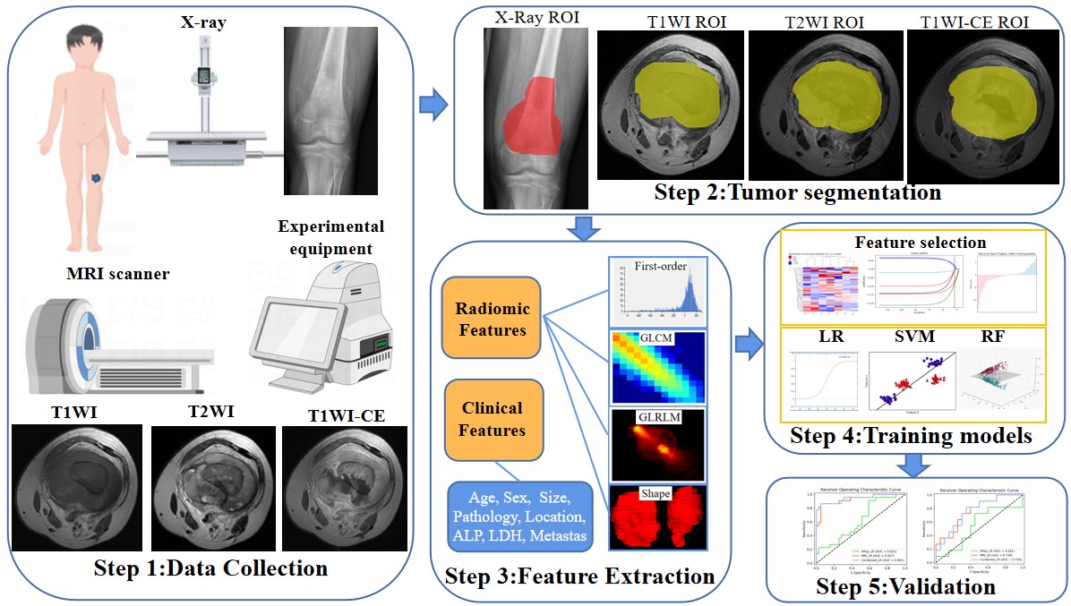
A retrospective study of 92 consecutive patients (a training and a testing set, n=61 and 31) with osteosarcoma were enrolled. The imaging features of each patient were extracted from radiograph and multiparametric MRI (T1WI, T2WI and T1WI-CE). Three-step feature redundancy reduction including minimal-redundancy-maximum-relevance (mRMR), least absolute shrinkage and selection operator (LASSO) regression and the random forest recursive feature elimination (RF-RFE) were used. The reliability, performance and performance difference of the built diagnostic models was respectively accessed by the area under the receiver-operating characteristic curve (AUC), Hosmer–Lemeshow test and DeYong test. Figure 1 depicts a flow diagram that illustrates the process of radiomics analysis.Results
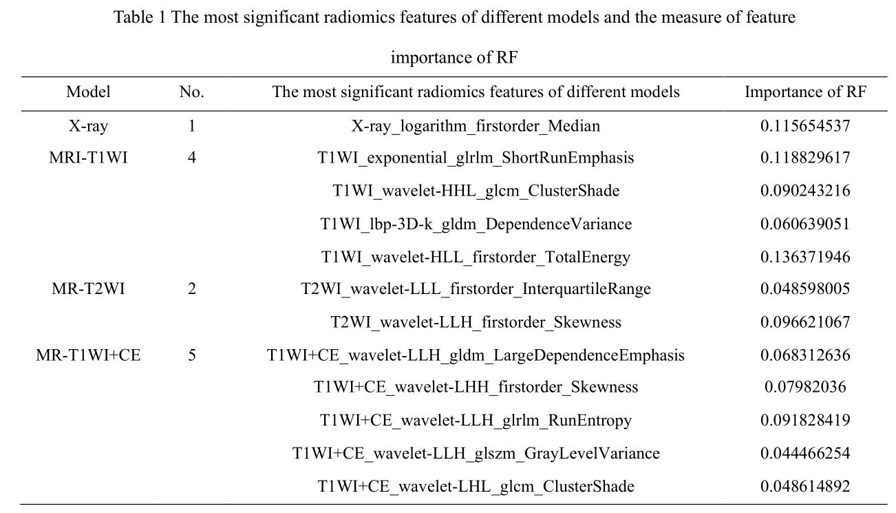
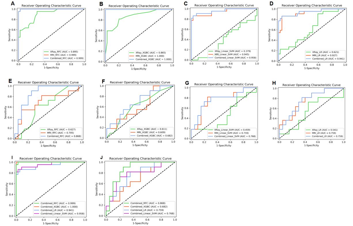
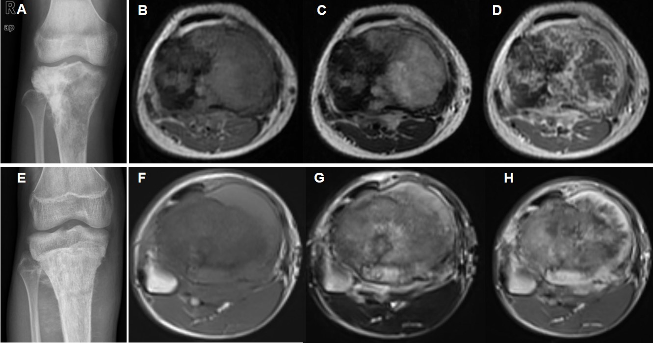
The most significant radiomics features of different models and the measure of feature importance of RF were showed in Table 1. The performance (AUC, sensitivity, specificity, and accuracy) of four classifiers (RF, SVM, LR and XGB) using radiograph-MRI as image inputs were stable (all Hosmer–Lemeshow index > 0.05) with the fair to good prognosis efficacy (Table 2 and Figure 2). The RF classifier using radiograph-MRI features as training inputs exhibited better performance (AUC = 0.806, 0.868) than that using MRI-only (AUC = 0.774, 0.771) and radiograph-only (AUC = 0.613 and 0.627) in the training and testing sets (p < 0.05) while the other three classifiers showed no difference between MRI-only and radiograph-MRI models. Figure 3 shows an example of two correctly classified lesions with different local relapse.Discussion and Conclusion
The AUC obtained from radiomics based on radiograph were not very high. The reason for this may be related to our investigation include 2D segmentation. Previous studies have demonstrated improved results with 3D tumor segmentations,[4, 5] because they provide more radiomic features and points of heterogeneity, but 3D tumor segmentations could not be performed on a single radiograph in this study. The AUC obtained through the utilization of MRI-based radiomics, specifically employing the LR, SVM, and RF models, exhibited a slight superiority over the utilization of radiograph in the prediction of local relapse (LR) in osteosarcoma. The diagnostic performance of the radiograph-MRI model showed better than the other single-modality models because of tumors segmented on 2D-projection radiographs with relatively limited spatial resolution and tissue complexity (e.g., intratumoral heterogeneity). We identified eleven statistically significant high-level features from the MRI images that revealed a substantial association with local relapse as a contrast enhanced T1-weighted radiomics study demonstrated osteosarcoma relapse.[6-8] Nine of selected 11 MR features are associated with wavelet-transformed texture features in display of temporal, spatial, and frequency scale for tissue microstructures (i.e., heterogeneity); the other two features associated with gray-level dependence matrix (GLDM) and gray-level run-length matrix (GLRLM) matrices respectively indicated the number of consistent-signal-intensity center-surrounded voxels and the number of consistent-intensity-and-direction lines. In addition, the only one radiographic feature associated with histogram-retrieved logarithm median reflected the intratumoral distribution of grayscale intensity.The eleven MR radiomic features together with one radiographic feature constituted a more efficient model than other single-modality model. [9, 10] RF classifier among four machine learning algorithms had the best predictive efficiency (AUCs for the training and testing set 0.999 vs. 0.868) in demonstration of non-linear relationships between image and target characteristics despite a limited number of samples.[11] Therefore, RF could particularly handle small-sample radiomics studies with better identification of predominant image features and enhancement of model interpretability in good consistent with previous literature. [12-15]Machine-learning-based multimodality radiomics analyses holds great potential for a preoperative prediction of local relapse in osteosarcoma. Radiograph-MRI radiomics model improved prediction accuracy, helped identify patients at high risk of local relapse and enabled tailored treatment approaches.
Acknowledgements
None to acknowledge.References
1.Lambin P, Leijenaar R, Deist T, Peerlings J, de Jong E, van Timmeren J, et al. Radiomics: the bridge between medical imaging and personalized medicine. Nature reviews Clinical oncology. 2017; 14(12):749-762.
2. van Griethuysen J, Fedorov A, Parmar C, Hosny A, Aucoin N, Narayan V, et al. Computational Radiomics System to Decode the Radiographic Phenotype. Cancer research. 2017; 77(21):e104-e107.
3. Aerts H, Velazquez E, Leijenaar R, Parmar C, Grossmann P, Carvalho S, et al. Decoding tumour phenotype by noninvasive imaging using a quantitative radiomics approach. Nature communications 2014; 5:4006.
4. Wan Q, Zhou J, Xia X, Hu J, Wang P, Peng Y, et al. Diagnostic Performance of 2D and 3D T2WI-Based Radiomics Features With Machine Learning Algorithms to Distinguish Solid Solitary Pulmonary Lesion. Frontiers in oncology. 2021; 11:683587.
5. Gitto S, Cuocolo R, Emili I, Tofanelli L, Chianca V, Albano D, et al. Effects of Interobserver Variability on 2D and 3D CT- and MRI-Based Texture Feature Reproducibility of Cartilaginous Bone Tumors. J Digit Imaging. 2021; 34(4):820-832.
6. Chen H, Liu J, Cheng Z, Lu X, Wang X, Lu M, et al. Development and external validation of an MRI-based radiomics nomogram for pretreatment prediction for early relapse in osteosarcoma: A retrospective multicenter study. European journal of radiology. 2020; 129:109066.
7. Mahrooghy M, Ashraf A, Daye D, McDonald E, Rosen M, Mies C, et al. Pharmacokinetic Tumor Heterogeneity as a Prognostic Biomarker for Classifying Breast Cancer Recurrence Risk. IEEE transactions on bio-medical engineering. 2015; 62(6):1585-1594.
8. Zhang L, Ge Y, Gao Q, Zhao F, Cheng T, Li H, et al. Machine Learning-Based Radiomics Nomogram With Dynamic Contrast-Enhanced MRI of the Osteosarcoma for Evaluation of Efficacy of Neoadjuvant Chemotherapy. Frontiers in oncology. 2021; 11:758921.
9. Zhang Y, He K, Guo Y, Liu X, Yang Q, Zhang C, et al. A Novel Multimodal Radiomics Model for Preoperative Prediction of Lymphovascular Invasion in Rectal Cancer. Frontiers in oncology. 2020; 10:457.
10. Dong F, Li Q, Jiang B, Zhu X, Zeng Q, Huang P, et al. Differentiation of supratentorial single brain metastasis and glioblastoma by using peri-enhancing oedema region-derived radiomic features and multiple classifiers. European radiology. 2020; 30(5):3015-3022.
11. Breiman L. Random forests. Machine Learning. 2001; 45(1):5-32. 12. Schaduangrat N, Malik A, Nantasenamat CJP. ERpred: a web server for the prediction of subtype-specific estrogen receptor antagonists. 2021; 9:e11716.
13. Ye J, Fang P, Peng Z, Huang X, Xie J, Yin X. A radiomics-based interpretable model to predict the pathological grade of pancreatic neuroendocrine tumors. European radiology. 2023.
14. Wu C, Du X, Zhang Y, Zhu L, Chen J, Chen Y, et al. Five machine learning-based radiomics models for preoperative prediction of histological grade in hepatocellular carcinoma. Journal of cancer research. 2023.
15. Wang W, Fan Z, Zhen J. MRI radiomics-based evaluation of tuberculous and brucella spondylitis.Journal of International Medical Research. 2023; 51(8):3000605231195156.
Figures