3782
Assessing the Efficacy of Radiotherapy for Brain Metastases in Advanced Non-Small Cell Lung Cancer through Raidomics Prediction1Department of Radiology, The Affiliated Hospital of Guzhou Medical Unversity, Guiyang, China, 2Philips Healthcare, Xi’an, China, 3Philips Healthcare, Shanghai, China
Synopsis
Keywords: Diagnosis/Prediction, Radiomics
Motivation: Magnetic resonance imaging (MRI) raidomics has shown unique advantages and potential in non-invasive evaluation of therapeutic efficacy in cancer patients.
Goal(s): To construct a model that can predict the prognosis of patients with advanced non-small cell lung cancer (NSCLC) brain metastases after radiotherapy.
Approach: For patients with advanced NSCLC brain metastasis who underwent pre-treatment MRI examination, stable and reproducible raidomics features were quantitatively extracted and screened. Additionally, artificial intelligence methods were utilized to construct raidomics labels.
Results: The potential of the MRI raidomics-based method in predicting the efficacy of radiotherapy for brain metastases from advanced NSCLC was preliminarily confirmed.
Impact: The method based on MRI raidomics (T1, T2, DWI, DCE) can not only enhance the precision of radiation therapy efficacy assessment for patients with NSCLC brain metastasis, but also offer clinicians a more scientific basis for treatment decision-making.
Introduction
Brain metastasis is one of the common complications of advanced NSCLC, which has a significant impact on the survival and quality of life of patients[1]. At present, radiotherapy is one of the main treatment approaches for brain metastases. However, traditional methods for assessing radiotherapy efficacy often suffer from issues like subjectivity and a lack of individualization[2]. Radiomics is a rapidly evolving field of research focused on extracting quantitative metrics, commonly referred to as radiomic features, from medical images. It has the potential power to facilitate better clinical decision making, especially in the treatment of patients in cancer[3-5]. Therefore, this study aims to use the method of MRI based radiomics to analyze multiple feature parameters, predicting the radiotherapy efficacy of advanced N brain metastases, and providing scientific treatment decision-making support for clinical practice.Methods
This study involved the collection of sixty patients with advanced NSCLC brain metastasis. Pre-radiotherapy MRI sequences were acquired from all patients. The parameters of scan protocols are as follows: T1-weighted image with TR (repetition time) = 600 ms, TE (echo time) = 10 ms, TI (inversion time): approximately 200 ms; T2-weighted image sequence: TR = 3000 ms, TE = 80 ms; diffusion-weighted imaging (DWI) sequence: TR = 4000 ms, TE = 60 ms, b-value (diffusion sensitivity): 1000 s/mm²; dynamic contrast enhancement (DCE) sequence: TR = 2 ms, TE = 1ms.Primarily, two experienced radiologists independently and blindly delineate the region of interest (ROI) from primary tumor tissue, following which MRI images and the tumor ROI area are exported for standardization. Subsequently, a high-throughput extraction of radiomics features is conducted[6]. Then, a prediction model is established using machine learning algorithms, and the optimal prediction model is determined through the learning of the training group and the validation of the validation group. Finally, evaluate the model using a test set and calculate indicators such as accuracy and sensitivity of the model.Results
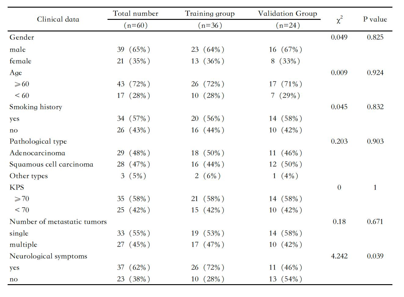
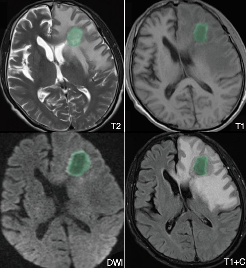
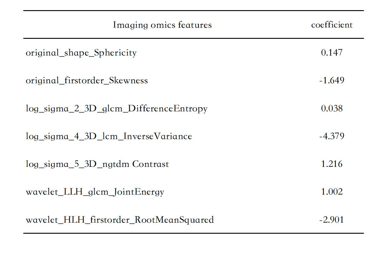
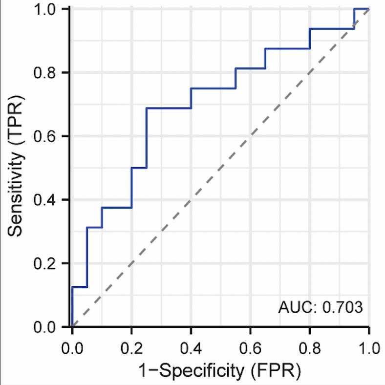
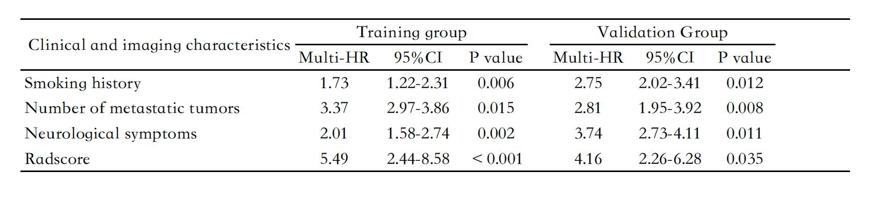
Initially, this study partitioned sixty advanced NSCLC brain metastasis patients into a training set and a validation set (Table 1). Figure 1 showed a representative example of patient with advanced NSCLC brain metastasis and the corresponding ROI placing. Utilizing the method of MRI radiomics, it introduced radiomic features from multiple sequences. In contrast to traditional MRI radiomic studies which only incorporated T1 and T2 sequences[7], this research further integrated DWI and DCE sequences. These provided a wealth of information, enabling a more comprehensive capture of the tumor tissue's morphology, metabolism, and hemodynamic characteristics. Subsequently, through MRI radiomics feature extraction, dimension reduction and selection, we ultimately distilled seven radiomics features that exhibited the most robust correlation with patient PFS(progression-free survival). These characteristics, with their stability and repeatability, were capable of reflecting both the biological attributes of the tumor and the efficacy of radiotherapy (Table 2). Ultimately, by constructing radiomics labels, a quantitative metric was established to evaluate the therapeutic efficacy of radiotherapy in patients by calculating each patient's Radiomics Score (Radscore). In training cohorts, the Radscore demonstrated commendable prognostic predictive power, with sensitivity, specificity, and C-index being 49.8%, 80.1%, and 0.703, respectively. And in validation dataset, the sensitivity, specificity, and C-index were 60.6%, 74.3%, and 0.687, respectively (Figure 2). Univariate and multivariate Cox survival analysis further confirmed that Radscore is a potential prognostic indicator for patients with advanced NSCLC and brain metastases (Table 3).Discussion
In this study, we have preliminarily confirmed the potential of MRI based radiomics methods in predicting the efficacy of radiotherapy for advanced non-small cell lung cancer with brain metastases. This method not only improves the accuracy and personalization level of radiotherapy efficacy evaluation, but also provides more scientific treatment decision-making basis for clinical doctors. However, due to the small sample size and limitations in data sources, this study still has certain limitations. Subsequent research will further expand the sample size and combine it with other imaging and molecular biology indicators to improve the accuracy and stability of the prediction model.Conclusion
The method based on MRI radiomics can predict the radiotherapy efficacy of advanced non-small cell lung cancer patients with brain metastases. This study provides a new approach and tool for clinical decision-making, and supports the development of personalized treatment plans.Summary of Main Findings
This study demonstrated good accuracy and stability in predicting the radiotherapy efficacy of advanced NSCLC patients with brain metastases through a method based on MRI radiomics. This method provides a new approach for personalized treatment decision-making and is expected to play an important role in clinical applications.Acknowledgements
We are grateful to all the participants for their cooperation and patience.References
[1]Waqar, Saiama N., Samson, Pamela P., Robinson, Cliff G., Bradley, Jeffrey., Devarakonda, Siddhartha.. Non-small-cell Lung Cancer With Brain Metastasis at Presentation. Clinical lung cancer, 2018, 19(4):e373-e379.
[2]Giaj-Levra, Niccolò., Borghetti, Paolo., Bruni, Alessio., Ciammella, Patrizia., Cuccia, Francesco.. Current radiotherapy techniques in NSCLC: challenges and potential solutions. Expert review of anticancer therapy, 2020, 20(5):387-402.
[3]Bi, Wenya Linda., Hosny, Ahmed., Schabath, Matthew B., Giger, Maryellen L., Birkbak, Nicolai J.. Artificial intelligence in cancer imaging: Clinical challenges and applications. CA: a cancer journal for clinicians, 2019, 69(2):127-157.
[4]Chetan, Madhurima R., Chetan, Madhurima R., Gleeson, Fergus V., Gleeson, Fergus V.. Radiomics in predicting treatment response in non-small-cell lung cancer: current status, challenges and future perspectives. European radiology, 2020, 31(2):1049-1058.
[5]Visonà, Giovanni., Spiller, Lisa M., Hahn, Sophia., Hattingen, Elke., Vogl, Thomas J.. Machine-Learning-Aided Prediction of Brain Metastases Development in Non-Small-Cell Lung Cancers. Clinical lung cancer, 2023, .
[6]Zhang, Lifei., Fried, David V., Fave, Xenia J., Hunter, Luke A., Yang, Jinzhong.. IBEX: an open infrastructure software platform to facilitate collaborative work in radiomics. Medical physics, 2015, 42(3):1341-53.
[7]Baeßler, Bettina., Weiss, Kilian., Pinto Dos Santos, Daniel.. Robustness and Reproducibility of Radiomics in Magnetic Resonance Imaging: A Phantom Study. Investigative radiology, 2019, 54(4):221-228.
Figures