3780
Radiomics Features on Magnetic Resonance Images Can Predict C5aR1 Expression Levels and Prognosis in High-Grade Glioma.1Department of Radiology, Renmin Hospital of Wuhan University, Wuhan, China
Synopsis
Keywords: Diagnosis/Prediction, Radiomics, C5aR1; high-grade glioma; MRI; prognosis; biomarker
Motivation: High-grade glioma is a complex disease characterized by genome instability caused by the accumulation of genetic alterations. Identifying and evaluating the oncogenes involved is crucial for determining treatment strategies and evaluating prognosis.
Goal(s): We sought to explore whether radiomics models based on MRI features can noninvasively predict C5aR1 expression and the prognosis of patients with high-grade glioma.
Approach: This study uses machine learning approaches based on paired MRI and RNA sequencing data.
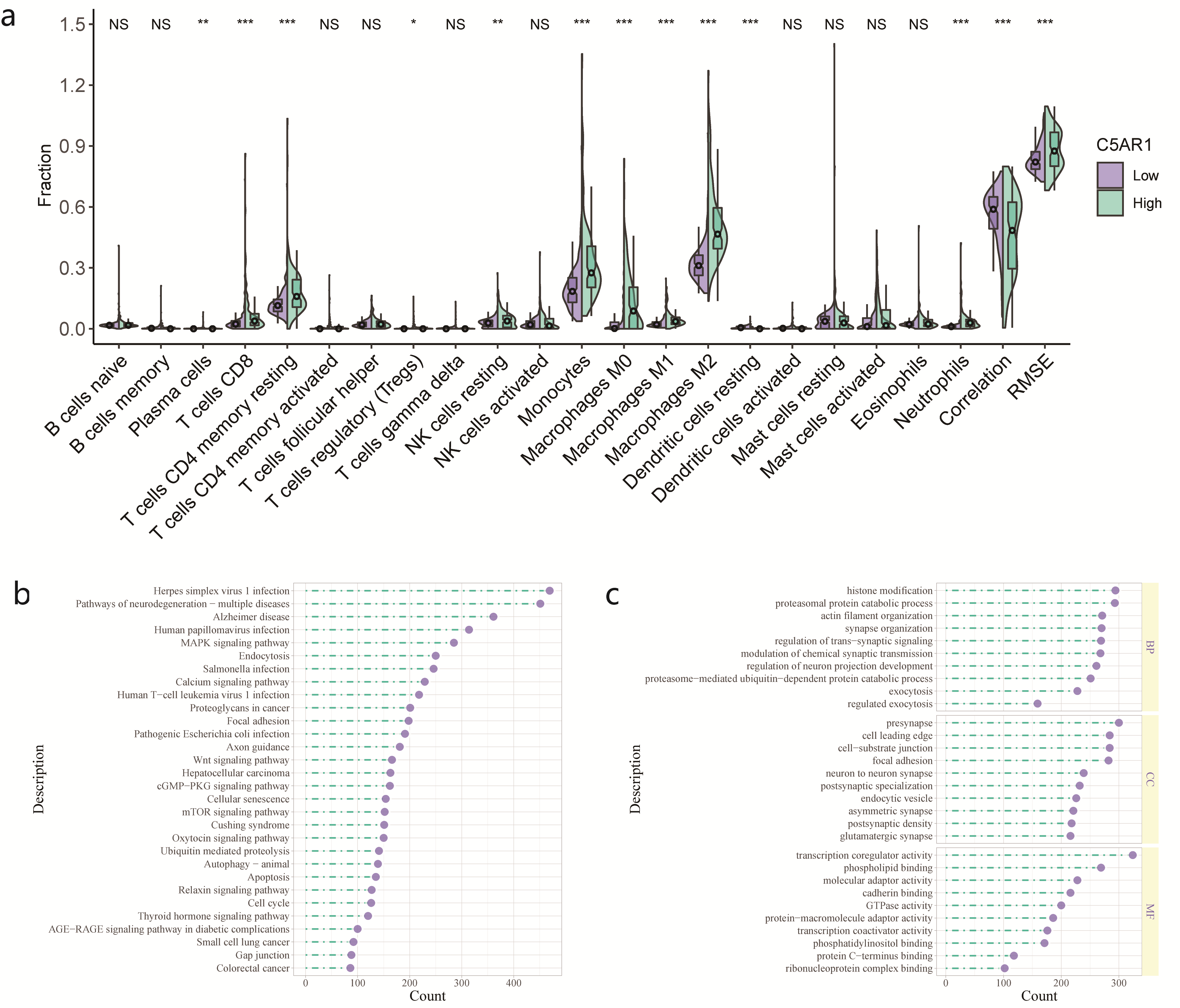
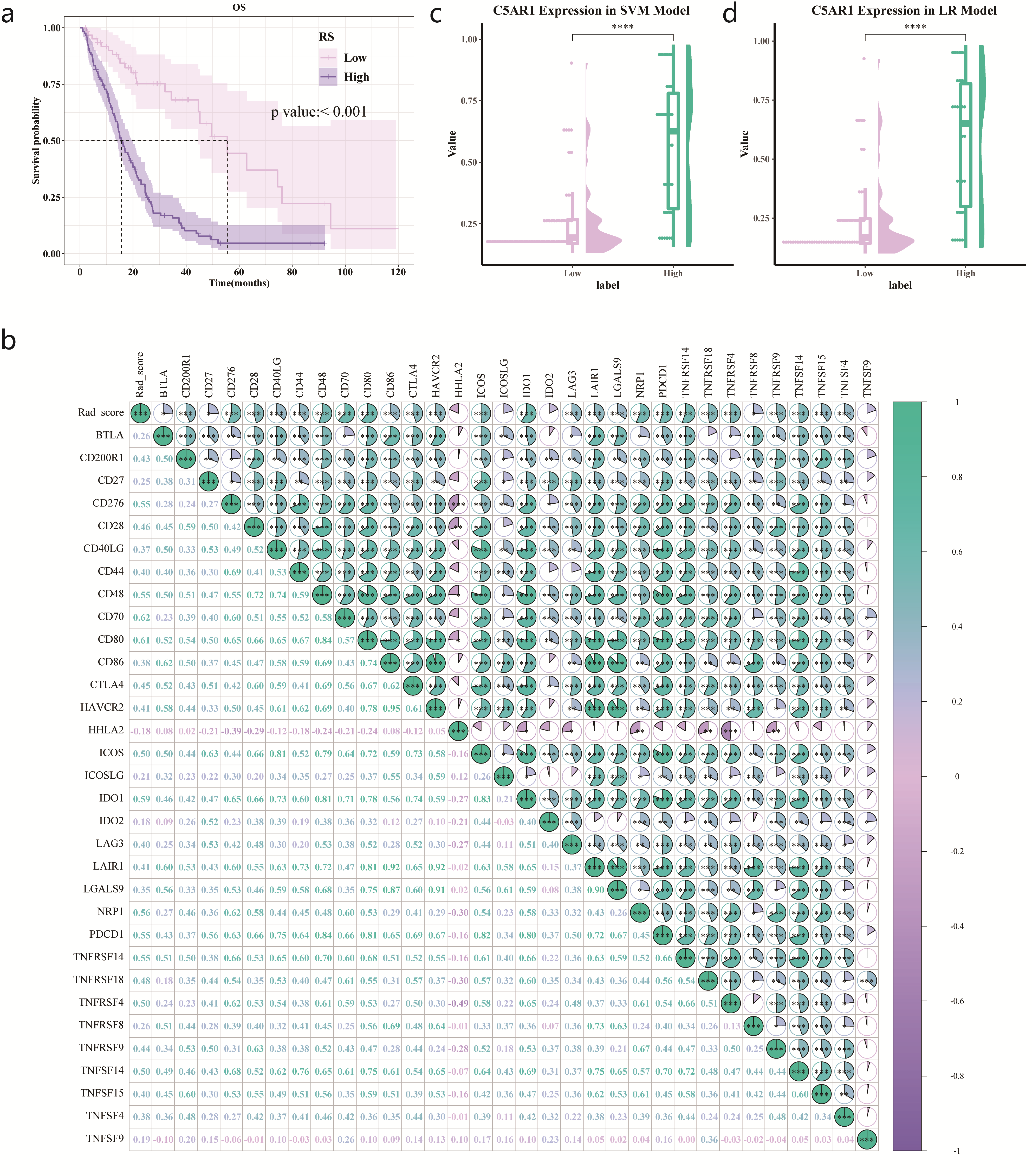
Results: The radiomics models yield satisfactory performances in predicting C5aR1 expression. Our findings also reveal associations between MRI radiomics and immune-related features.
Impact: As an effective and reproducible tool, our radiomics model may support clinical decision making and individualized treatment.
Introduction
The complement component C5a receptor 1 (C5aR1) regulates cancer immunity. This retrospective study aimed to assess its prognostic value in high-grade glioma (HGG) and predict C5aR1 expression using a radiomics approach.Methods
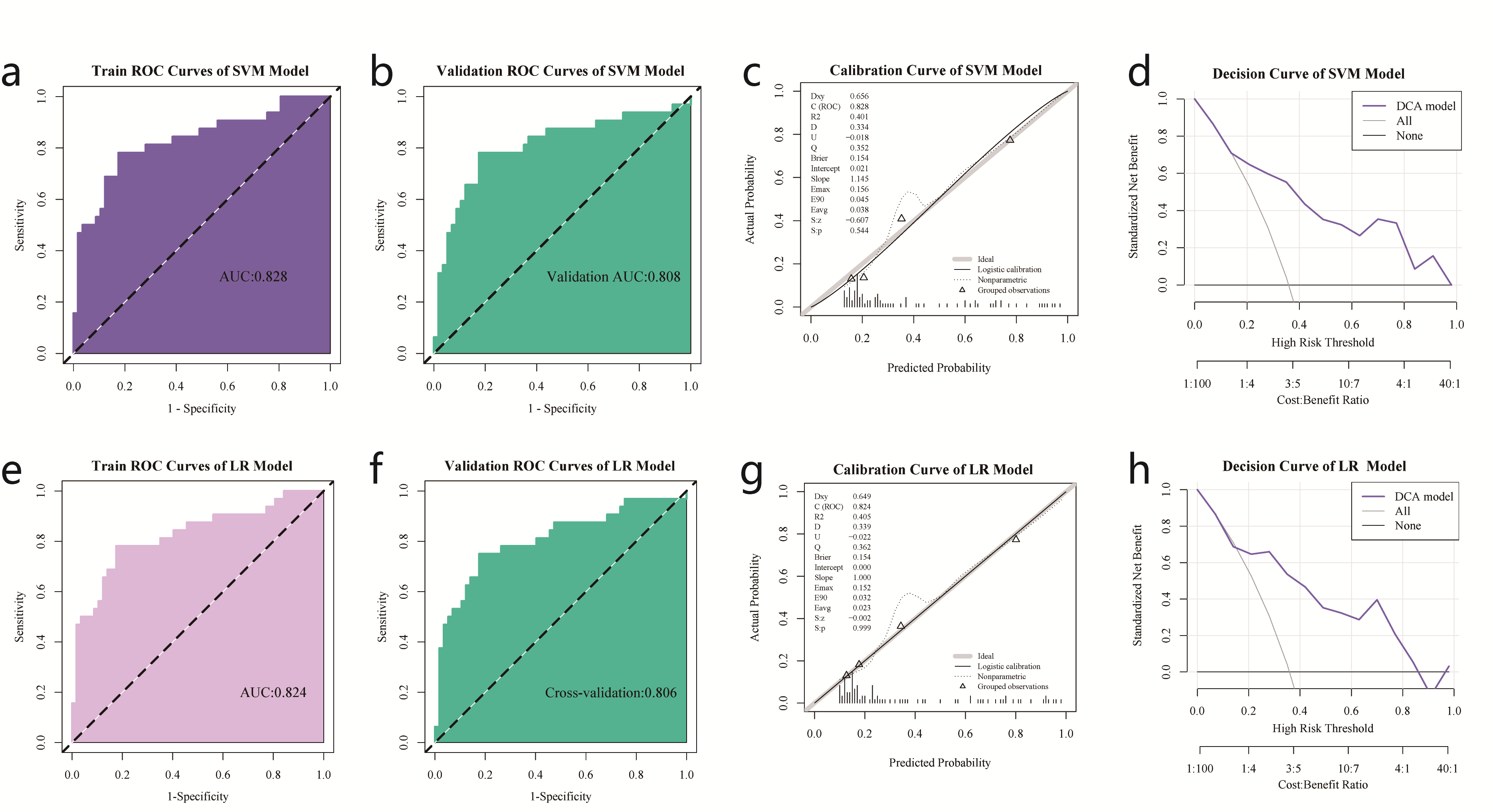
Among 298 patients with HGG, 182 with MRI data were randomly divided into training and test groups for radiomics analysis. We examined the association between C5aR1 expression and prognosis through Kaplan–Meier and Cox regression analyses. We used maximum relevance–minimum redundancy and recursive feature elimination algorithms for radiomics feature selection. We then built a support vector machine (SVM) and a logistic regression model, investigating their performances using receiver operating characteristic, calibration curves, and decision curves.Results
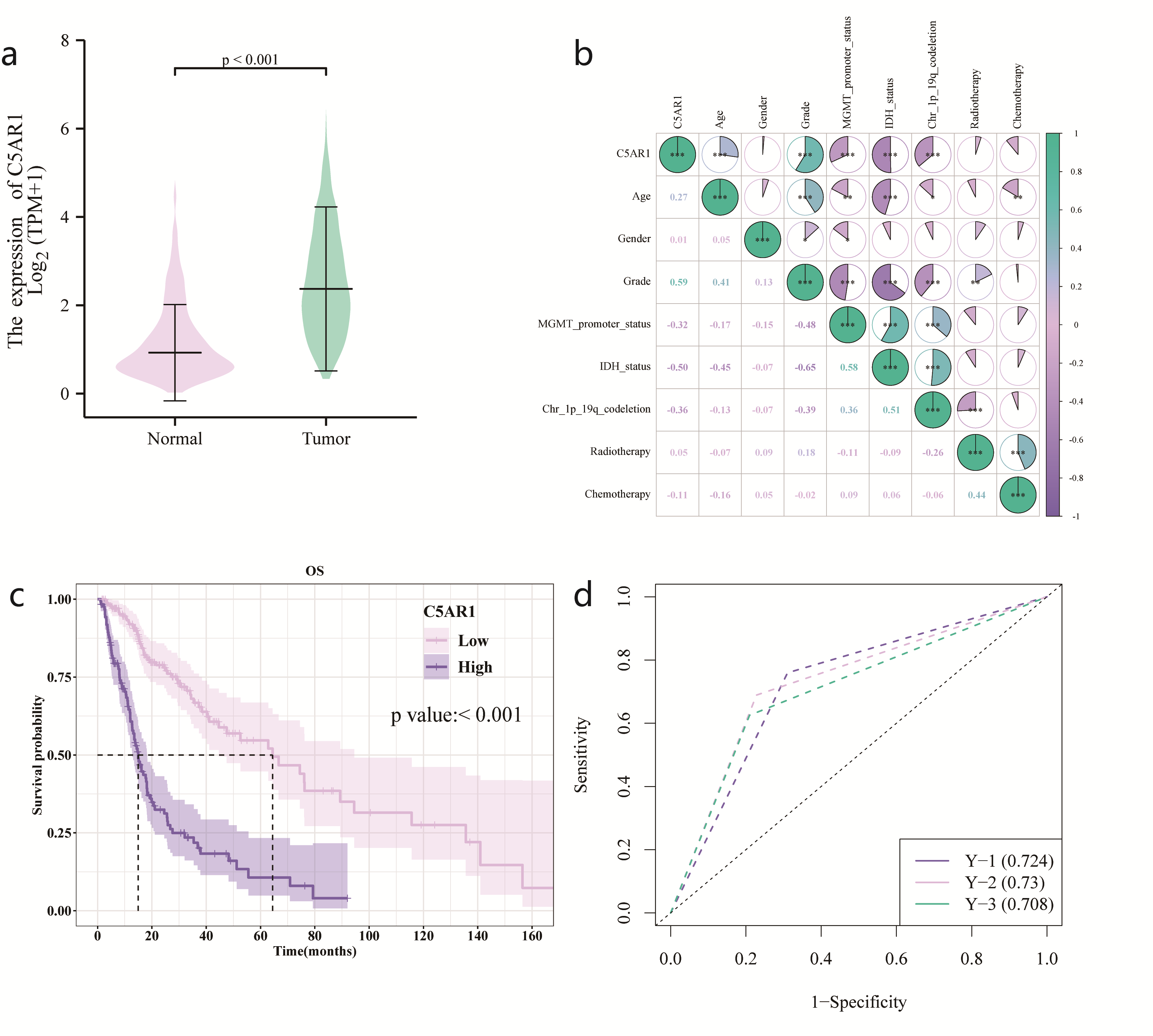
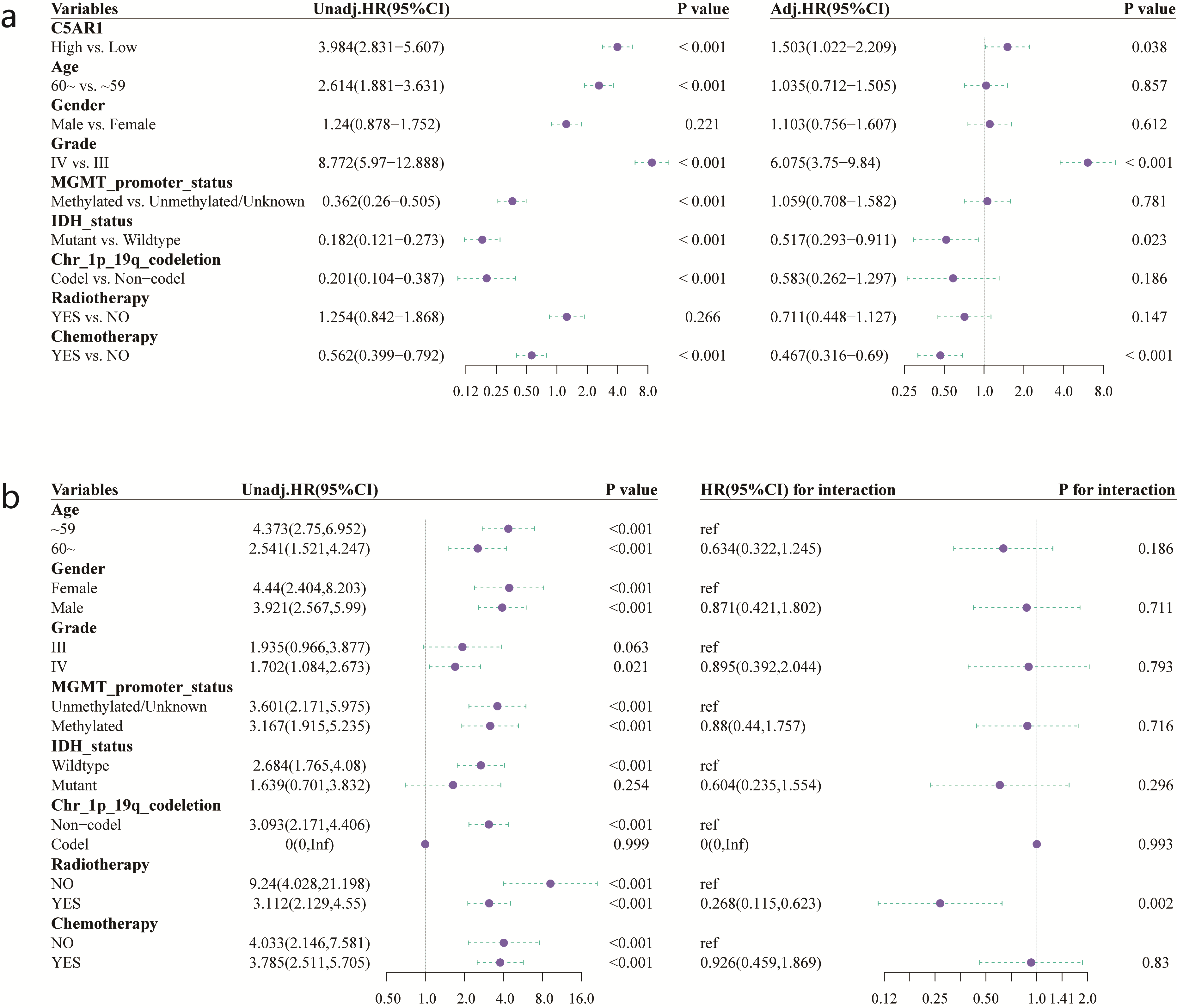
C5aR1 expression was elevated in HGG and was an independent prognostic factor (hazard ratio = 3.984, 95% CI: 2.834–5.607). Both models presented with >0.8 area under the curve values in the training and test datasets, indicating efficient discriminatory ability, with SVM performing marginally better. The radiomics score calculated using the SVM model correlated significantly with overall survival (p < 0.01).Conclusion
Our results highlight C5aR1’s role in HGG development and prognosis, supporting its potential as a prognostic biomarker. Our radiomics model can noninvasively and effectively predict C5aR1 expression and patient prognosis in HGG.Acknowledgements
Funding: This research was funded by the Fundamental Research Funds for Central Universities (grant number: 2042023kf0036) from Wuhan University.References
1. Tan, A.C.; Ashley, D.M.; López, G.Y.; Malinzak, M.; Friedman, H.S.; Khasraw, M. Management of glioblastoma: State of the art and future directions. CA Cancer J. Clin. 2020, 70, 299–312. https://doi.org/10.3322/caac.21613.
2. Wen, P.Y.; Weller, M.; Lee, E.Q.; Alexander, B.M.; Barnholtz-Sloan, J.S.; Barthel, F.P.; Batchelor, T.T.; Bindra, R.S.; Chang, S.M.; Chiocca, E.A.; et al. Glioblastoma in adults: A Society for Neuro-Oncology (SNO) and European Society of Neuro-Oncology (EANO) consensus review on current management and future directions. Neuro-Oncol. 2020, 22, 1073–1113.
3. Ding, P.; Li, L.; Lv, X.; Zhou, D.; Wang, Q.; Chen, J.; Yang, C.; Xu, E.; Dai, W.; Zhang, X.; et al. C5aR1 is a master regulator in Colorectal Tumorigenesis via Immune modulation. Theranostics 2020, 10, 8619–8632.
4. Li, G.; Li, L.; Li, Y.; Qian, Z.; Wu, F.; He, Y.; Jiang, H.; Li, R.; Wang, D.; Zhai, Y.; et al. An MRI radiomics approach to predict survival and tumour-infiltrating macrophages in gliomas. Brain J. Neurol. 2022, 145, 1151–1161.
5. Sun, Q.; Chen, Y.; Liang, C.; Zhao, Y.; Lv, X.; Zou, Y.; Yan, K.; Zheng, H.; Liang, D.; Li, Z.C. Biologic Pathways Underlying Prognostic Radiomics Phenotypes from Paired MRI and RNA Sequencing in Glioblastoma. Radiology 2021, 301, 654–663.
Figures