3778
Quantitative Physiologic MRI Parameters Combined with Innovative Machine Learning to Distinguish Glioblastoma from Solitary Brain Metastases1Department of Neurology & neurosurgery, Mcgill University, Montréal, QC, Canada, 2Translational Neuroimaging Laboratory, The McGill University Research Centre for Studies in Aging, Douglas Hospital, McGill University, Montréal, QC, Canada, 3Department of Radiology, Perelman School of Medicine at the University of Pennsylvania, Philadelphia, PA, United States
Synopsis
Keywords: Diagnosis/Prediction, Tumor, Glioblastomas, Solitary_Brain_Metastases, Quantitative_Physiologic, MRI, Innovative_Hyper-tuned_Machine_Learning
Motivation: There is an unmet need to develop advanced MRI based prediction models to distinguish glioblastomas (GBMs) from solitary brain metastases (BMs) with high-accuracy, as conventional MRI-techniques often yield ambiguous results.
Goal(s): The objective is to discriminate GBMs from solitary BMs using physiologic MRI parameters and machine-learning based novel-methods.
Approach: Employing diffusion-tensor-imaging (DTI) and dynamic-susceptibility contrast-perfusion weighted imaging (DSC-PWI), the study uses a novel machine-learning approach that integrates multiple features with hyper-tuned models to enhance pattern-recognition and prediction.
Results: The innovative-method combining interacted and non-interacted features via hyper-tuned machine-learning models significantly outperformed traditional-methods, thus achieving high accuracy and reliability in differentiating GBMs from BMs.
Impact: The integration of quantitative and physiologically-sensitive MRI-parameters with novel machine-learning based algorithms may be promising in distinguishing glioblastomas from solitary brain metastases. This approach may be useful in making prognostication and guiding optimal, personalized patient-treatments in the era of personalized-medicine.
Introduction
Accurate and early distinction of glioblastomas (GBMs) and solitary brain metastases (BMs) provide a window of opportunity for reframing treatment strategies enabling optimal and timely therapeutic interventions in these patients (1). However, conventional MRI findings are often ambiguous and unreliable in distinguishing these common malignancies (2). The purpose of this study was to leverage the physiologically sensitive, and quantitative derived parameters from diffusion tensor imaging (DTI), dynamic susceptibility contrast-perfusion weighted imaging (DSC-PWI), along with machine-learning based complex feature interactions and classification algorithms in distinguishing GBMs from solitary brain metastases (3).Methods
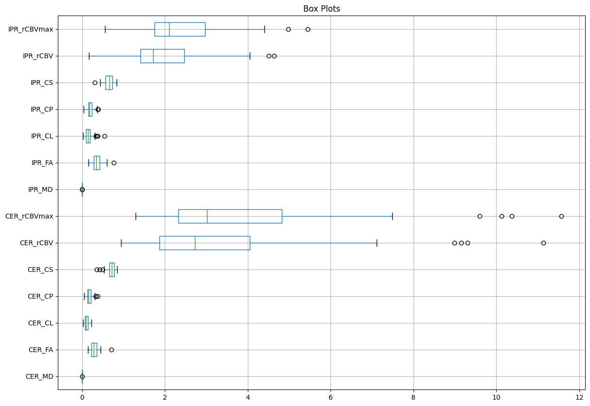
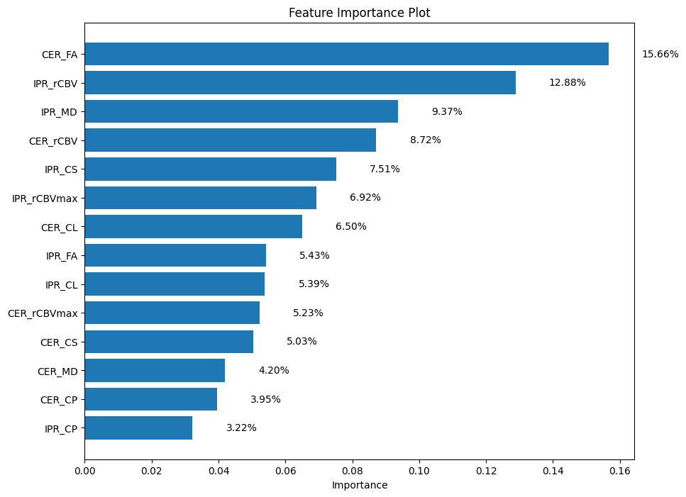
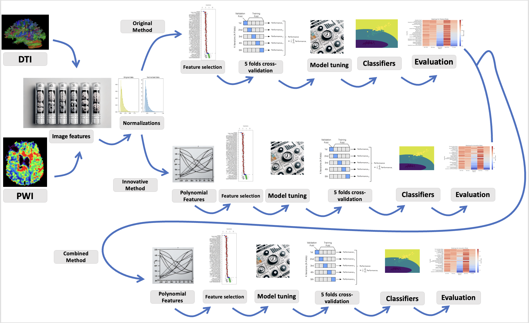
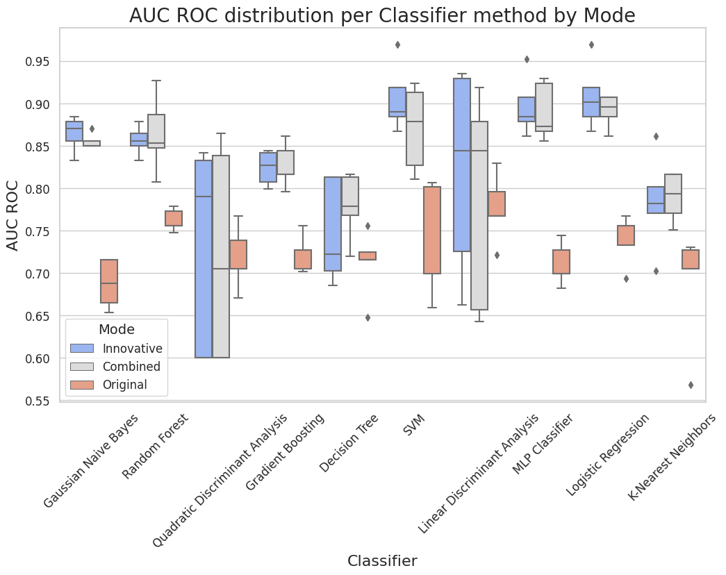
Histopathology confirmed patients with GBM (n=62) and BMs (n=26) exhibiting solitary enhancing lesions underwent anatomical imaging, DTI and DSC-PWI on a 3T magnet prior to treatment. Pixel-wise mean diffusivity (MD), fractional anisotropy (FA), coefficient of linear anisotropy (CL), planar anisotropy (CP), and spherical anisotropy (CS) maps from DTI and leakage-corrected cerebral volume (CBV) maps were generated from DSC-PWI using methods described previously (4). The DTI derived maps, CBV maps, and T2-FLAIR images were resliced and co-registered to postcontrast T1-weighted images. A semiautomatic approach was used to segment the contrast-enhancing regions (CER) and 4-mm thick immediate peritumoral regions (IPR) beyond the contrast enhancement in each case by using a signal intensity-based thresholding method. The median values of DTI metrics (MD, FA, CL, CP, and CS) from the CER and IPR were computed. The CBV values from these regions were normalized by corresponding values from contralateral normal white matter regions to obtain relative CBV (rCBV). The top 90th percentile rCBV values were also measured from the enhancing regions and were reported as rCBVmax. It is essential to place dataset features into a scale equivalent when they are available on various scales (5). Data scaling was performed by using the MinMaxScaler normalization technique for scaling DTI and DSC-PWI feature in the range of 0-1 (Figure 1). In the next step, feature-selection was performed using techniques such, as Mutual Information, ANOVA F value, c2 and model-based selections [Logistic regression (LR) and Random Forest (RF)] to identify the most relevant features (Figure 2). Subsequently, Scikit learn library was used to establish various higher-order feature interactions among features by automatically varying the number of interactions from zero (no-interactions) to 10. This approach has been shown to greatly enhance the classifiers’ ability to recognize specific patterns in the data and then using those patterns to make binary predictions (6). Finally, 10 various machine-learning classifiers including Gaussian Naive Bayes (GNB), RF, Quadratic Discriminant Analysis (QDA), Gradient Boosting (GB), Decision Tree (DT), Support Vector Machine (SVM), Linear Discriminant Analysis (LDA), Multi-layer Perceptron (MLP), LR, K-Nearest Neighbors (KNN) were employed involving non-interacted (Original method), interacted (Innovative method) and combination of interacted and non-interacted features (Combined method) in distinguishing GBMs and metastases as shown in figure 3. Each classifier was carefully adjusted through Grid Search to find the combination of settings ensuring that each model performed at its optimal potential. To establish reliability and generalizability, 5-fold cross validation tests were performed.Results
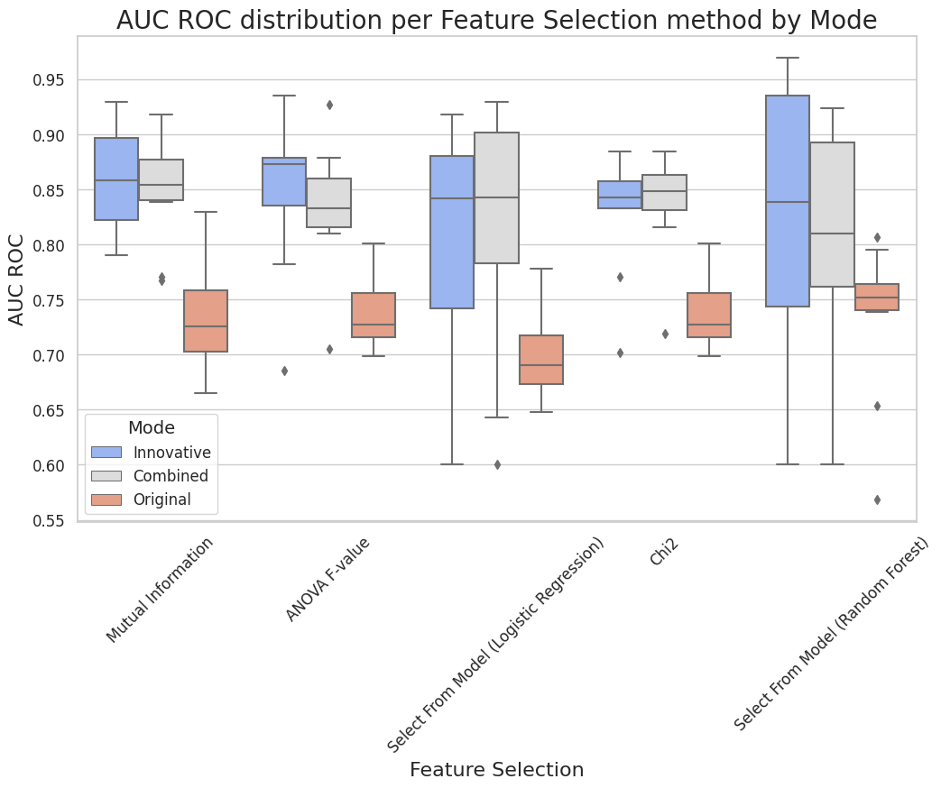
The combination of interacted and non-interacted features (combined method) provided the best discriminatory performance as evident by various metrics such as AUC, F1 Score, accuracy, sensitivity, and specificity in distinguishing GBMs from BMs (Figure 4 & 5). The summary of top performances across each of three methods (Original, Innovative, and Combined) is described. (a) Original Method: Classifier: Random Forest, Feature-Selection: Select from Model (LR), F1 Score: 0.7, Accuracy: 77.84%, AUC-ROC: 77.77%, Sensitivity: 63.64%, Specificity: 87.5%.(b) Innovative Method: Classifier: Gaussian Naive Bayes, Feature-Selection: Mutual Information, F1 Score: 0.82, Accuracy: 84.07%, AUC-ROC: 85.56%, Sensitivity: 81.81%, Specificity: 78.75%.(c) Combined Method: Classifier: Random Forest, Feature-Selection: ANOVA F-value, F1 Score: 0.8, Accuracy: 87.78%, AUC-ROC: 92.67%, Sensitivity: 73.64%, Specificity: 97.5%.Discussion
With the aim of evaluating influence of multiple interactions among MRI parameters and hyper-tuning machine learning models in the diagnostic performance of classification models, our results indicate that combined approach of exploiting interacted and non-interacted DTI and DSC-PWI derived features has the potential for improving classification accuracy in distinguishing GBMs from BMs. Instead of using high-dimensional radiomic features including shape and high-order texture based features, our study also emphasized the importance of using quantitative DTI and DSC-PWI derived parameters, which are directly related to tumor biology and provide meaningful physiologically sensitive information commonly used for clinical interpretation.Conclusion
Machine learning based approach involving combined use of interacted and non-interacted physiologic MRI parameters may be promising in distinguishing GBMs from BMs.Acknowledgements
No acknowledgement found.References
1. Hosseini SA, Hosseini E, Hajianfar G, Shiri I, Servaes S, Rosa-Neto P, et al. MRI-Based Radiomics Combined with Deep Learning for Distinguishing IDH-Mutant WHO Grade 4 Astrocytomas from IDH-Wild-Type Glioblastomas. Cancers. 2023;15(3):951.2. Hosseini SA, Shiri I, Hajianfar G, Bagley S, Nasrallah M, O’Rourke DM, et al., editors. MRI based Radiomics for Distinguishing IDH-mutant from IDH wild-type Grade-4 Astrocytomas. Proceedings of the 31st Annual Meeting of ISMRM, London, UK; 2022.3. Chiu F-Y, Yen Y. Imaging biomarkers for clinical applications in neuro-oncology: current status and future perspectives. Biomarker Research. 2023;11(1):1-23.4. Chawla S, Wang S, Mohan S, Nasrallah M, Verma G, Brem S, et al. Differentiation of brain infection from necrotic glioblastoma using combined analysis of diffusion and perfusion MRI. Journal of Magnetic Resonance Imaging. 2019;49(1):184-94.5. Naeem A, Anees T, Ahmed KT, Naqvi RA, Ahmad S, Whangbo T. Deep learned vectors’ formation using auto-correlation, scaling, and derivations with CNN for complex and huge image retrieval. Complex & Intelligent Systems. 2023;9(2):1729-51.6. Li Y, Chu X, Tian D, Feng J, Mu W. Customer segmentation using K-means clustering and the adaptive particle swarm optimization algorithm. Applied Soft Computing. 2021;113:107924.Figures