3777
A fusion model based on preoperative MRI radiomics features can predict potential bone invasion of meningiomas1Department of Radiology, West China Hospital of Sichuan University, Chengdu, China, 2Institute of Research and Clinical Innovations, Neusoft Medical Systems, Shanghai, China
Synopsis
Keywords: Diagnosis/Prediction, Radiomics, bone invasion,meningiomas
Motivation: Bone invasion is a common problem in meningioma surgery and is associated with patient prognosis. However, 10-26% of patients with potential bone invasion are difficult to identify by preoperative imaging.
Goal(s): To develop an artificial intelligence based preoperative diagnostic model.
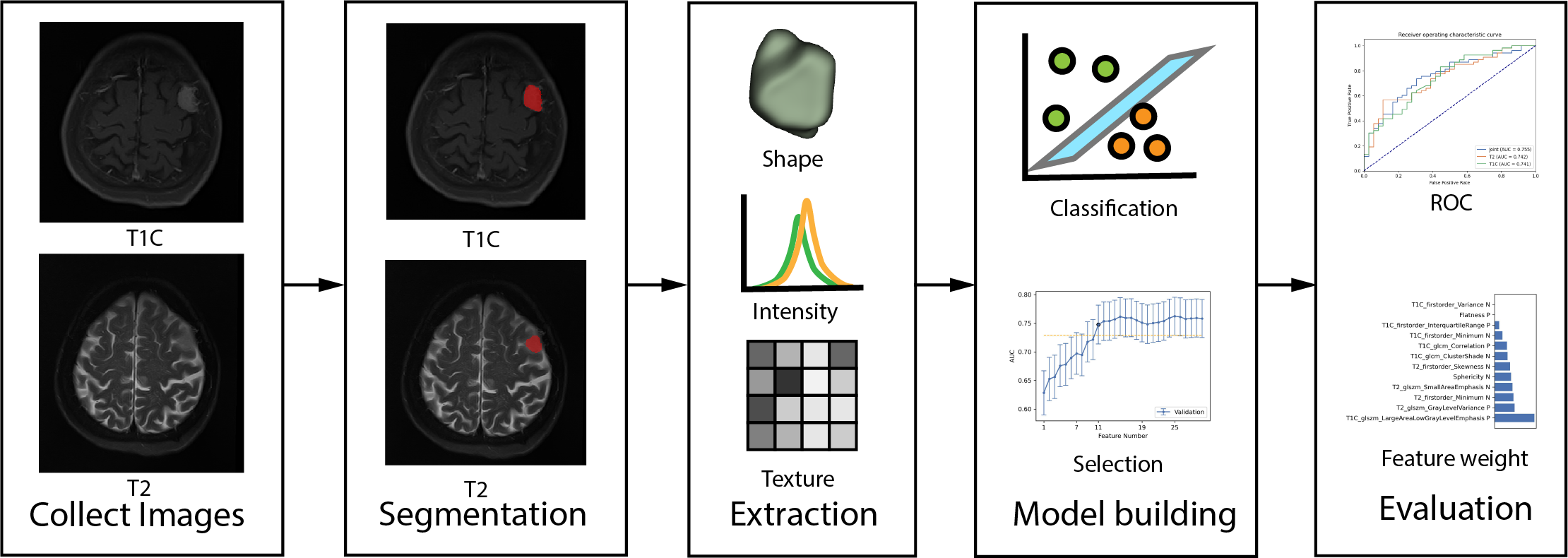
Approach: Radiomics features were extracted from preoperative, contrast-enhanced T1-weighted (T1C) and T2-weighted (T2) MR images of 296 patients. Candidate radiomics were selected by applying feature reduction and 5-fold cross validation.
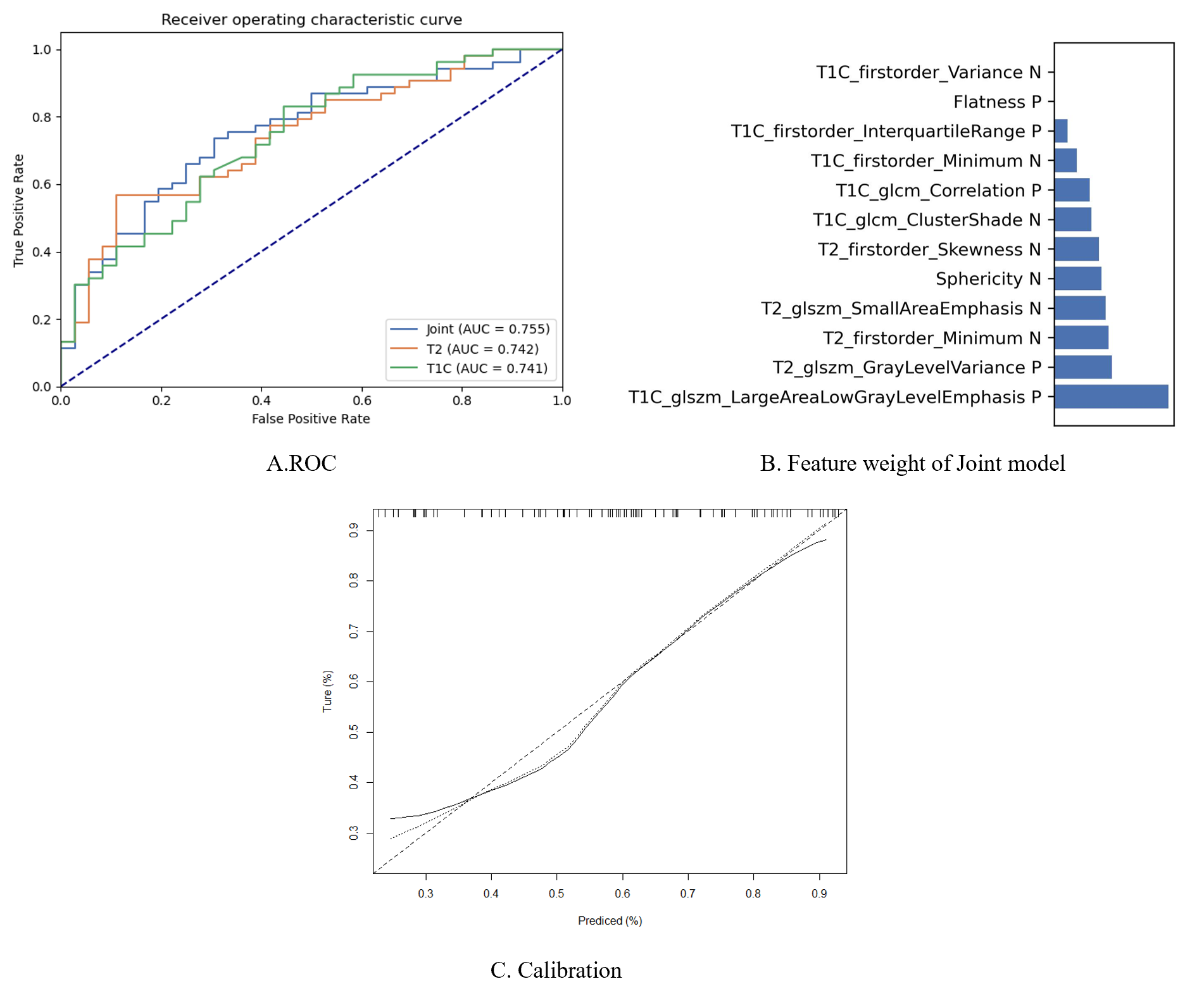
Results: A more accurate and robust fusion radiomics model was built based on T1C and T2 MR images with AUC of 0.755.
Impact: Our results have demonstrated that radiomics features extracted from T1C and T2 MR images may be employed as effective preoperative biomarkers for predicting potential bone invasion in meningiomas.
Introduction
Meningiomas are the most frequent primary intracranial tumors in adults, and account for 36% of all intracranial tumors1. In the 2021 World Health Organization (WHO) classification scheme2, meningiomas are divided into three grades, benign (WHO grade I), atypical (WHO grade II), and anaplastic (WHO grade III), accounting for about 65–80 %, 20–25 %, 1–3 % of meningiomas, respectively. Bone invasion is a common problem in meningioma surgery, up to 20-69%3-5, and is not contained in the WHO classification criterion for meningiomas. However, in long-term follow-up, tumor infiltration of the bone flap may lead to a high recurrence rate even in completely resected WHO grade I or II meningiomas6. Accordingly, bone invasion has been considered as an independent predictor of recurrence and is related with reduced progression free survival and overall survival4. This seems to be preventable, as invasive bone - if identified - can be safely removed in most cases, especially in convex meningiomas. Preoperative imaging findings of bone hyperplasia may indicate bone invasion. However, 10-26% of patients without hyperplasia present with bone involvement3,5, making imaging inadequate in predicting bone invasion. Therefore, quantitative analysis is significant for improving the predictive efficiency of bone invasion. Radiomics analysis is becoming a comprehensive quantitative method for assessing brain tumors7, extracting parameters associated with potential anatomical microstructures and small-scale biophysical processes such as gene expression, tumor cell proliferation, and neovascularization dynamics [8]. In addition, radiomics analysis has been proven to be able to provide predictive markers for the diagnosis, prognosis, and treatment planning of brain tumors7-11. So far, only one application of radiomics analysis in prediction of bone invasion has been reported and it has shown the value of radiomics in meningiomas12; however, there is still room to improve the accuracy of that model. Therefore, we aim to develop a more robust radiomics model for bone invasion prediction in meningiomas.Methods
Data acquisition: We collected 296 patients with meningioma, 178 cases of whom were diagnosed with bone invasion, and split to training and test cohort randomly by 7:3. MR examinations were performed using Siemens Skyra or Trio clinical scanners (Siemens Healthcare, Erlangen, Germany). The main MRI sequences were T1WI and T2WI. The contrast agent gadolinium-diethylenetriaminepentaacetic acid (0.1 mmol/kg body weight, via forearm vein injection) was used in enhanced MR scanning. A radiologist with 12 years of experience delineated region of interest (ROI) of lesions manually on T1C and T2 MR images.Feature selection: The radiomics features were extracted by PyRadiomics. A total of 107 features were extracted for analysis, including shape, intensity and texture features. The Spearman’s correlation was conducted between each radiomics features to select features which are not correlated. In addition, a feature selection algorithm Relief with 5-fold cross-validation was employed for feature dimension reduction in the training cohort.
Machine learning model construction: Supported vector machine (SVM) and logistic regression (LR) were both used as classifiers, of which the best performance model was ultimately selected as the final model. The above steps were integrated in the open source radiomics analysis software FAE13. The performance of the model was evaluated using receiver operating characteristic (ROC) curve analysis. The area under the ROC curve (AUC) and calibration curve were calculated for quantification, and the Delong test was used for comparison of ROC.
Results
The proposed fusion model that combined radiomics feature from T1C and T2 MR images achieved the best performance in the test cohort with an AUC of 0.756(0.653-0.856). Texture features from T1C and T2 MR had higher weight for classification. Importantly, prediction reliability was confirmed in the calibration curve as well. Table 2 shows 6 radiomics features that demonstrated significant difference between bone invasion and no-invasion group, including texture and shape features.Discussion
The fusion model achieved the best performance with AUC of 0.755, in which those selected radiomics features were mostly textural features from T1C and T2 MR images, suggesting that they are related to microscopic environment of the meningiomas. Our results also showed that 3 texture and 3 intensity features of T1C MR images, 2 texture and 2 intensity features of T2 images and 2 shape features were significantly associated with bone invasion, indicating that these simple radiomic features might be used as a novel biomarker to predict potential bone invasion of meningioma prior to surgery12.Conclusion
Preoperative detection of potential bone invasion is crucial for meningioma patients to improve clinical decision making and prognosis prediction. Our results have demonstrated satisfactory results in predicting bone invasion by using the proposed fusion radiomics model, which provides a valuable tool for choosing optimal treatment options in meningioma patients.Acknowledgements
We would like to acknowledge the equal contributions of Hongjing and Jing Zhang to this work. Both authors contributed equally to the experimental design, data analysis, and manuscript preparation.References
1. Park YW, Oh J, You SC, et al. Radiomics and machine learning may accurately predict the grade and histological subtype in meningiomas using conventional and diffusion tensor imaging. Eur Radiol. 2019 Aug;29(8):4068-4076.
2. Louis DN, Perry A, Wesseling Pet et al.The 2021 WHO Classification of Tumors of the Central Nervous System: a summary. Neuro Oncol. 2021 Aug 2;23(8):1231-1251.
3. Goyal N, Kakkar A, Sarkar C, et al. Does bony hyperostosis in intracranial meningioma signify tumor invasion? A radio-pathologic study. Neurol India. 2012 Jan-Feb;60(1):50-4.
4. Lemée JM, Corniola MV, Da Broi M,et al. Extent of Resection in Meningioma: Predictive Factors and Clinical Implications. Sci Rep. 2019 Apr 11;9(1):5944.
5. Pieper DR, Al-Mefty O, Hanada Y,et al. (1999) Hyperostosis associated with meningioma ofthe cranial base: secondary changes or tumor invasion. Neurosurgery 44(4):742–747
6. Lam Shin Cheung V, Kim A, Sahgal A,et al. Meningioma recurrence rates following treatment: a systematic analysis. J Neurooncol. 2018 Jan;136(2):351-361. doi: 10.1007/s11060-017-2659-6. Epub 2017 Nov 15. PMID: 29143273.
7. Zhou M, Scott J, Chaudhury B, et al. Radiomics in Brain Tumor: Image Assessment, Quantitative Feature Descriptors, and Machine-Learning Approaches. AJNR Am J Neuroradiol. 2018 Feb;39(2):208-216.
8. Gatenby RA, Grove O, Gillies RJ. Quantitative imaging in cancer evolution and ecology. Radiology. 2013 Oct;269(1):8-15.
9. Zhang J, Zhang G, Cao Y, et al. A Magnetic Resonance Imaging-Based Radiomic Model for the Noninvasive Preoperative Differentiation Between Transitional and Atypical Meningiomas. Front Oncol. 2022 Jan 21;12:811767.
10. Hsieh HP, Wu DY, Hung KC,et al. Machine Learning for Prediction of Recurrence in Parasagittal and Parafalcine Meningiomas: Combined Clinical and MRI Texture Features. J Pers Med. 2022 Mar 24;12(4):522.
11. Park CJ, Choi SH, Eom J,et al. An interpretable radiomics model to select patients for radiotherapy after surgery for WHO grade 2 meningiomas. Radiat Oncol. 2022 Aug 22;17(1):147.
12. Zhang J, Sun J, Han T, et al. Radiomic features of magnetic resonance images as novel preoperative predictive factors of bone invasion in meningiomas. Eur J Radiol. 2020 Nov;132:109287.
13. Song Y, Zhang J, Zhang Yd, et al. (2020) FeAture Explorer (FAE): A tool for developing and comparing radiomics models. PLOS ONE 15(8): e0237587. https://doi.org/10.1371/journal.pone.0237587
Figures