3770
Detection of Early-stage Primary Central Nervous System Lymphoma Manifesting Atypical Radiological Phenotype Using Multi-Task Neural Network1Sichuan University West China Hospital, chengdu, China, 2GE Healthcare, MR Reasearch, Beijing, China, 3Infervision Medical Technology, Beijing, China
Synopsis
Keywords: Diagnosis/Prediction, Machine Learning/Artificial Intelligence
Motivation: In clinical practice, distinguishing atypical radiological features of primary central nervous system lymphoma (PCNSL) from certain glioma or demyelinating disease patients is challenging and often lead to delayed or incorrect treatment.
Goal(s): To develop deep learning model to identify PCNSL with atypical radiological features.
Approach: Developing a multi-task, multi-modal deep learning model capable of end-to-end identification of early-stage atypical PCNSL.
Results: The multi-task, multi-modal deep learning model accurately discriminates early-stage atypical PCNSL from other radiologically similar diseases, significantly enhancing the diagnostic accuracy of radiologists in clinical practice.
Impact: The practical clinical application of the model demonstrates its diagnostic value in identifying challenging cases suspected of early-stage atypical PCNSL. This research shifts academic attention towards distinguishing specific subtypes prone to misdiagnosis, rather than solely focusing on disease-level differentiations.
Introduction
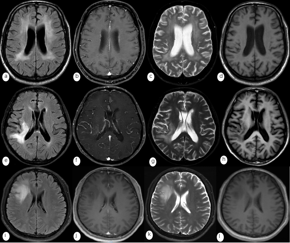
Typical primary central nervous system lymphoma (PCNSL) can be easily identified in clinical practice due to its distinct radiological manifestations[1,2]. However, a subset of early-stage PCNSL may exhibit patchy signal abnormalities and lack diagnostic specificity. These atypical early-stage PCNSL cases often pose challenges as they can be confused with gliomas or demyelinating diseases that share similar radiological features. This confusion frequently leads to incorrect diagnosis or treatment[3-5]. The objective of this study is to develop a fully automated multi-task neural network model capable of accurately identifying early-stage atypical PCNSL based on MRI images of conventional sequences.Method
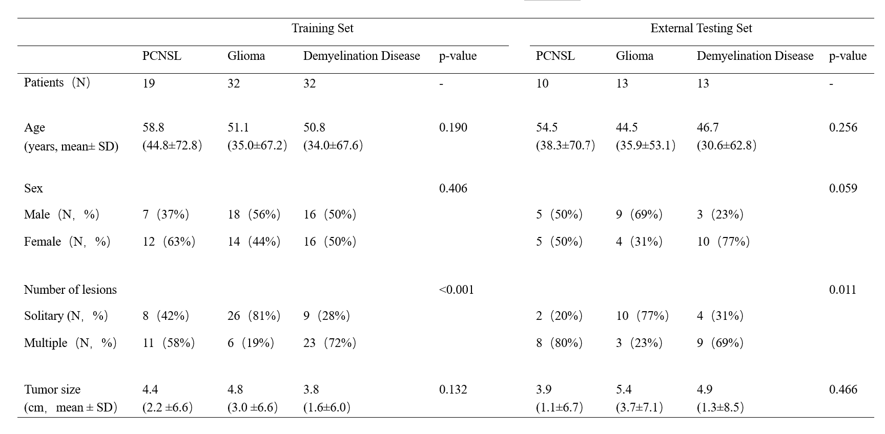
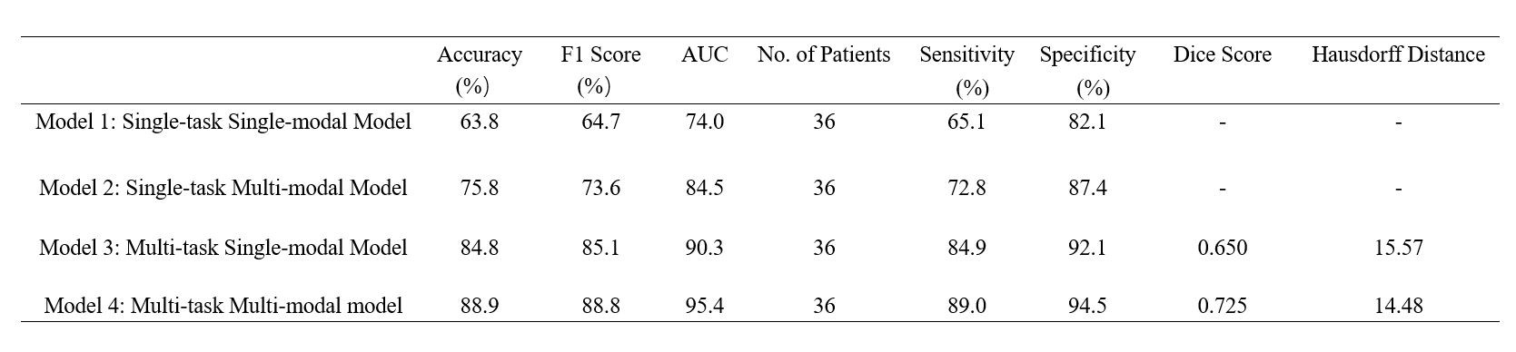
Two cohorts comprising of atypical early-stage PCNSL patients, as well as patients with gliomas and demyelinating diseases exhibiting similar radiological manifestations, were retrospectively recruited from West China Hospital of Sichuan University (WCHSU; n=83) and Chengdu Shangjin Nanfu Hospital (CSNH; n=36) to serve as the training and external testing sets, respectively. We developed a multi-task multi-modal neural network model that simultaneously achieved lesion segmentation and disease diagnosis. The performance of the model was evaluated using ablation analysis and a series of classical performance metrics. Additionally, we have assessed the practical value of the model by implementing it in real-world clinical practice.Result
The multi-task multi-modal neural network model achieved an Area Under the Receiver Operating Characteristic (AUROC) of 0.95 and an accuracy of 85.4% on the independent external testing set. The results of the ablation analysis provided compelling evidence of the substantial enhancement in diagnostic capabilities resulting from the incorporation of the multi-task neural network architecture and the integration of multi-modal image fusion techniques. Furthermore, the application of the model in real-world clinical practice has significantly enhanced the diagnostic proficiency of radiologists in identifying atypical early-stage PCNSL casesDiscussion
The exceptional performance of the multi-task multi-modal neural network model on the external testing set demonstrates the potential of deep learning techniques in accurately identifying early-stage atypical PCNSL through meticulous analysis of MRI images. The model's successful application in real-world clinical practice highlights its remarkable ability to bridge the diagnostic proficiency gaps among radiologists with diverse expertise, thereby offering significant clinical utility as an auxiliary toolConclusion
The developed neural network model can accurately identify early-stage atypical PCNSL based on conventional MRI sequence images, and it can provide effective assistance to radiologists in real-world clinical practiceAcknowledgements
No acknowledgement found.References
1.Coulon A , Lafitte F , Hoang-Xuan K ,et al. Radiographic findings in 37 cases of primary CNS lymphoma in immunocompetent patients[J]. Eur Radiol, 2002,12(2):329-340.
2.Erdag N , Bhorade RM , Alberico RA ,et al. Primary lymphoma of the central nervous system: typical and atypical CT and MR imaging appearances[J]. AJR Am J Roentgenol, 2001,176(5):1319-1326.
3.Ferreri AJ, Reni M. Primary central nervous system lymphoma[J]. Crit Rev Oncol Hematol, 2007,63:257–268
4. Prados MD, Levin V. Biology and treatment of malignant glioma[J]. Seminars in Oncology, 2000, 27(3 Suppl 6):1-10.
5.Latov N. Diagnosis and treatment of chronic acquired demyelinating polyneuropathies[J]. Nature Reviews Neurology, 2014, 10(8):435-46.
Figures