3764
Radiological Image Quality Assessment of Synthetic 3T MRI Image from 64mT MRI1Monash Biomedical Imaging, Monash University, Clayton, Victoria, Australia, 2Australian National Imaging Facility, Queensland, Australia, 3Neuroscience, Monash University, Clayton, Victoria, Australia, 4Radiology, Alfred Hospital, Victoria, Australia, 5Herston Imaging Research Facility, University of Queensland, Queensland, Australia, 6School of Electrical Eng. and Computer Science, University of Queensland, Queensland, Australia, 7School of Clinical Science, Queensland University of Technology, Queensland, Australia, 8David Hartley Chair of Radiology, Royal Perth Hospital, Western Australia, Australia, 9Medical School, University of Western Australia, Western Australia, Australia, 10South Australian Health and Medical Research Institute, South Australia, Australia, 11Data Science and AI, Monash University, Clayton, Victoria, Australia
Synopsis
Keywords: Synthetic MR, Low-Field MRI
Motivation: The necessity to enhance the quality of portable low-field MRI images, which are crucial for wider accessibility but lack high-resolution, drives this research.
Goal(s): This study aims to determine whether Synthetic 3T technology can elevate low-field image quality to that of high-field 3T standards, making it diagnostically adequate.
Approach: We employed a generative network to transform low-field images to higher quality, maintaining pixel-level accuracy and structural integrity. The study involved a paired dataset from 37-healthy subjects and an evaluation on 20-images by two neuroradiologists.
Results: Synthetic 3T demonstrated improved clarity, structure, and contrast, aligning closer to the 3T-quality than the original low-field images.
Impact: This investigation highlights the potential of Synthetic 3T to bridge the gap in portable MRI imaging, enabling broader clinical utility. Further research could pivot on its application in pathological cases, with an aim to enhance diagnostic capabilities in resource-limited settings.
Introduction
Generative Adversarial Networks (GAN) have shown promise in generating synthetic medical imaging data1. This study examines the capability of synthetic 3T images to transform low-field MRI (64mT) images to resemble 3T MR image quality, with a focus on the clinical significance and diagnostic accuracy of the synthetic T1-weighted 3T MR images.Methodology
Model, Data, and Image Generation:Synthetic 3T, a specialized 3T image generator framework for MRI modalities, employs a conditional GAN approach for image-to-image translation2. The architecture comprises a generator that translates input 64mT MRI data to 3T MRI domain, guided by a noise vector, producing an output that closely reflects the 3T domain. In parallel, the discriminator differentiates between authentic and synthetic images, providing feedback essential for refining the generator's output. A distinguishing feature of Synthetic 3T is its emphasis on conditional translations, ensuring generated outputs retain the structural intricacies of the input. Moreover, it incorporates L1 loss for pixel fidelity and structural similarity index measure (SSIM) loss for structural congruence, resulting in a complete approach to quality and structural precision in MRI image translation. Synthetic 3T was trained on paired datasets of 37 healthy subjects, institutional review board approved, using Hyperfine Swoop (64mT) and Siemens Biograph mMR (3T) systems. T1 synthetic MRI images were generated from the test dataset, and 20 images were selected. This study employed a randomized, double-blind methodology, where image datasets were randomly allocated, and the evaluating neuroradiologists were blinded to the origin of the images, ensuring an unbiased assessment of image quality.
Image Assessment:
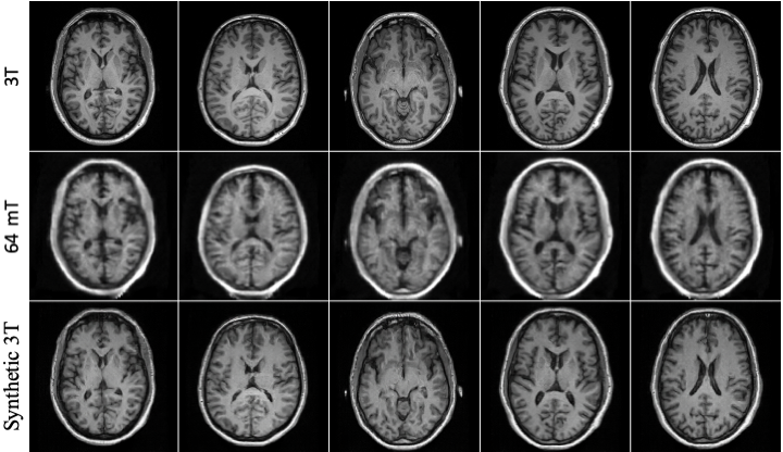
Two board-certified neuroradiologists independently evaluated the synthetic MRI images. Refer to Figure 1 for a visual comparison of T1-weighted images obtained from 3T, 64mT, and images synthesized by Synthetic 3T.
Scoring System:
A five-point visual grading scale was utilized for a thorough analysis of image quality, evaluating aspects like clarity, structural definition, contrast and brightness, and artifacts3,4,5. Each neuroradiologist reviewed ten data sets for 64mT, 3T, and synthetic 3T images.
Results
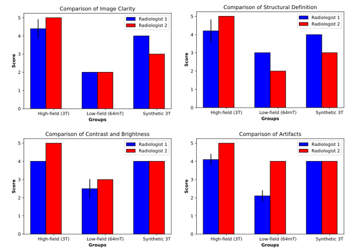
Evaluation Scores:Evaluation Scores: Refer to Figure 2 for visual comparison and evaluation of image quality across high-field, low-field, and synthetic images produced by Synthetic 3T. High-field images received consistently high ratings across all criteria. In contrast, low-field images showed variability in scores due to their inferior quality. Images generated by Synthetic 3T stand between the high-field standard of care and low-field scans, underlining the model's effectiveness in generating synthetic images.
Comparative Analysis:
Comparative Analysis: In evaluating the three imaging modalities – 3T, 64mT, and Synthetic 3T – across multiple criteria, statistical assessments were conducted to discern the distributional equivalences among these groups (Figure 2). Notably, most of the data did not conform to the assumptions of normality, necessitating the application of non-parametric tests. The Kruskal-Wallis test, a non-parametric analogue to the one-way ANOVA, was employed to discern any significant distribution disparities across the three imaging modalities. The results evidenced statistically significant differences in all criteria: Image Clarity (H = 52.7, p < 0.0001), Structural Definition (H = 42.9, p < 0.0001), Contrast and Brightness (H = 50.8, p < 0.0001), and Artifacts (H = 31.0, p < 0.0001). These findings indicate that at least two modalities exhibit divergent performance in each criterion.
Due to the significant differences in the Kruskal-Wallis tests, Dunn's post-hoc analyses were executed to determine pairwise contrasts among the imaging modalities. For Image Clarity, the results demonstrated statistically significant differences between 3T and 64mT (p < 0.00001), and 3T and Synthetic 3T (p = 0.0014), with 64mT and Synthetic 3T also differing significantly (p < 0.00001). In evaluating structural definition, both 3T and 64mT (p < 0.00001), as well as 3T and Synthetic 3T (p = 0.00165) showcased marked differences. Interestingly, 64mT and Synthetic 3T did not differ significantly in this metric. For contrast and brightness, substantial disparities were observed between 3T and 64mT (p < 0.00001), 3T and Synthetic 3T (p = 0.048), and 64mT and Synthetic 3T (p < 0.00001). Finally, in the context of artifacts, significant contrasts were evident between 3T and 64mT (p < 0.00001), 3T and Synthetic 3T (p = 0.0065), and 64mT and Synthetic 3T (p = 0.0088).
Conclusion
Portable low-field MRI represents an emerging avenue for accessible high-acuity imaging, with Synthetic 3T T1 images substantially elevating its quality in normal participants. While these images are not yet on par with high-field MRI, they show promising diagnostic potential, particularly if future adaptations for pathologic cohorts are successful. Ongoing refinement of the model is anticipated to further close the gap with high-field MRI, broadening the horizon for accessible diagnostic imaging.Acknowledgements
This research receives funding from the National Imaging Facility (NIF) and Hyperfine Inc. The authors acknowledge the facilities and scientific and technical assistance of the NIF, a National Collaborative Research Infrastructure Strategy (NCRIS) capability, at the Monash Biomedical Imaging, Monash University.References
1. Goodfellow, I., Pouget-Abadie, J., Mirza, M., Xu, B., Warde-Farley, D., Ozair, S., ... & Bengio, Y. (2020). Generative adversarial networks. Communications of the ACM, 63(11), 139-144.
2. Isola, P., Zhu, J. Y., Zhou, T., & Efros, A. A. (2017). Image-to-image translation with conditional adversarial networks. In Proceedings of the IEEE conference on computer vision and pattern recognition (pp. 1125-1134).
3. Pawar, K., Chen, Z., Seah, J., Law, M., Close, T., & Egan, G. (2020). Clinical utility of deep learning motion correction for T1 weighted MPRAGE MR images. European Journal of Radiology, 133, 109384.
4. Burmeister, H. P., Baltzer, P. A. T., Möslein, C., Bitter, T., Gudziol, H., Dietzel, M., ... & Kaiser, W. A. (2011). Visual grading characteristics (VGC) analysis of diagnostic image quality for high resolution 3 Tesla MRI volumetry of the olfactory bulb. Academic Radiology, 18(5), 634-639.
5. Ludewig, E., Richter, A., & Frame, M. (2010). Diagnostic imaging–evaluating image quality using visual grading characteristic (VGC) analysis. Veterinary research communications, 34, 473-479.
Figures