3763
The value of synthetic MRI in differentiating metastatic and non-metastatic lymph nodes in squamous cell carcinoma of head and neck1Department of Diagnostic Radiology, National Cancer Center/National Clinical Research Center for Cancer/Cancer Hospital, Chinese Academy of Medical Sciences and Peking Union Medical College, Beijing, China, 2MR Research China, GE Healthcare, Beijing, Beijing, China
Synopsis
Keywords: Synthetic MR, Head & Neck/ENT, Differential diagnosis
Motivation: The presence of metastatic lymph nodes in HNSCC indicates a worse prognosis, biopsy is invasive and has a high incidence of false-negative results. The diagnosis value of synthetic MRI needs to be studied further.
Goal(s): To explore the value of the features derived from synthetic MRI in distinguishing between benign and malignant lymph nodes.
Approach: This study included lymph nodes of HNSCC which were confirmed by pathology, and utilized multiple methods to select meaningful features.
Results: Features derived from synthetic MRI have satisfactory discrimination efficiency.
Impact: Synthetic MRI may be able to be used as a method to help assemnet the lymph nodes in clinical practice.
Introduction
Head and neck cancer is the sixth most common malignancy all over the world , among which squamous cell carcinoma (SCC) accounts for more than 90%1.The presence of metastatic lymph nodes (LNs) in HNSCC significantly affect the stage , therapeutic schedule and follow up of patients after therapy and the prognosis is worse when lymph node metastasis occurs2. Biopsy is the most accuracy method to recognize the metastatic LNs , but it’s invasive and might have a significant sampling error. Several studies focus on non-invasive imaging techniques to identify metastatic cervical LNs. Synthetic MRI (syMRI), a relatively novel quantitative MRI technique , based on multi-echo and multi-delay acquisition method , can simultaneously generate a comprehensive set of relaxometry mapping of longitudinal relaxation time (T1), transverse relaxation time (T2), and proton density (PD) in a single scan. In HNSCC, a recent study has also suggested that histogram parameters derived from syMRI may serve as a potential biomarker for evaluating relevant histopathological features which might influence the prognosis3. To our knowledge, there is no previous literature to explore the function of syMRI in distinguishing metastatic LNs in patients with HNSCC. Therefore , we did the preliminary research to explore the potential value of syMRI.Methods
This study included 61 LNs , 31 nodes were proven to be histologically malignant , 22 were by surgery and 9 were biopsy, 30 nodes were proven to be benign by surgery . Two radiologists manually delineated the volume of malignant and benign nodes independently , excluding any visible necrosis and cyst areas . Sixteen first-order texture features (05th Percentile, 10th Percentile, 25th Percentile, 50th Percentile, 75th Percentile, 90th Percentile, 95th Percentile, Kurtosis, Skewness, Entropy, InterquartileRange, Median, Maximum, Mean, Minimum, Variance) were obtained from T1, T2, and PD maps through Pyradiomics.Interobserver agreement was tested by using inter-class correlation coefficients (ICC). The Mann-Whitney U test or Student's t-test was used after assessing the normality distribution of data by using the Kolmogorov-Smirnov test. The receiver operating characteristic (ROC) curves were performed for all significant variables and Pearson correlation coefficients (PCC) were calculated , features that have a high correlation (PCC > 0.80) and a relatively lower area under the curve (AUC) were removed. The univariate analysis and multivariate logistic regression analysis with a forward stepwise selection procedure were used, odds ratios and 95% confidence intervals (CIs) were calculated. Furthermore, these models were enrolled in the pairwise ROC curve comparison to identify the optimal model.
Results
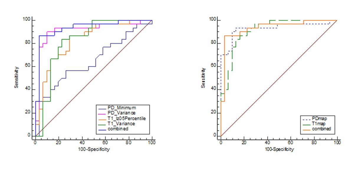
All parameters showed excellent inter-rater consistency (all ICC ≥ 0.831). Several parameters from T1_map and PD_map were significantly correlated with the status of lymph nodes(Fig.1). After multivariate logistic regression analysis T1_05th Percentile, T1_Variance, PD _Variance and PD_Minimum were identified as the significant independent predictors. The AUC of T1_map model , PD_map model and Combined model was 0.915, 0.930, 0.934 respectively, and there was no significantly different among three models ( P≥0.465 ) (Fig.2 and Fig.3).Discussion
The presence of metastatic LNs in HNSCC indicates the worse prognosis, and differentiation between metastatic and benign LNs is challenging. Non-invasive and accurate techniques are urgent needed. This preliminary study discovered that several first-order texture features may be helpful, the Combined model yielded the highest AUC of 0.934. Of course, we need further research to improve our viewpoint and we are indeed in process.Conlusion
The information derived from syMRI may have a high diagnostic efficiency in assement of cervical lymph nodes in patients with HNSCC before surgery.Acknowledgements
No acknowledgement found.References
1. Siegel RL, Miller KD, Jemal A. Cancer Statistics, 2017. CA Cancer J Clin. 2017;67(1):7-30. doi:10.3322/caac.21387
2. Dünne AA, Müller HH, Eisele DW, Kessel K, Moll R, Werner JA. Meta-analysis of the prognostic significance of perinodal spread in head and neck squamous cell carcinomas (HNSCC) patients. Eur J Cancer. 2006;42(12):1863-1868. doi:10.1016/j.ejca.2006.01.062
3. Yang F, Li Y, Lei H, et al. Histogram analysis of synthetic magnetic resonance imaging: Correlations with histopathological factors in head and neck squamous cell carcinoma. Eur J Radiol. 2023;160:110715. doi:10.1016/j.ejrad.2023.110715
Figures
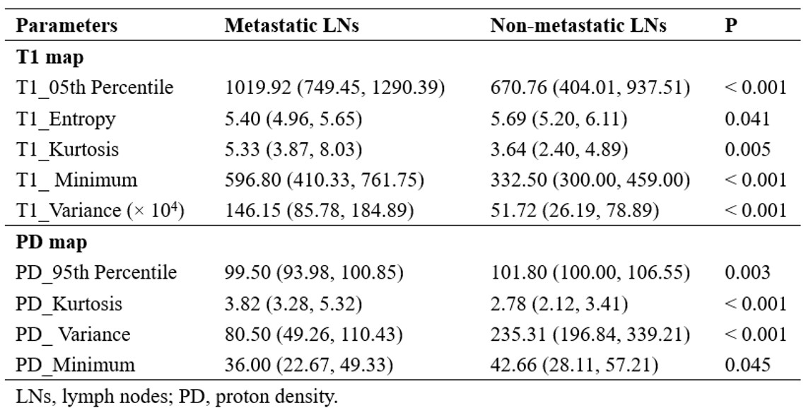
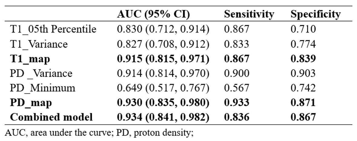
Fig.2 Diagnostic performance of models based on single and combined functional maps in differentiating metastatic LNs from non-metastatic LNs.