3760
Deep learning imaging-based reconstruction improved the image quality of synthetic high b-value DWI for prostate lesion detecting1Second Affiliated Hospital of Naval Medical University, Shanghai, China, 2MR Research, GE Healthcare, Beijing, China
Synopsis
Keywords: Synthetic MR, Prostate, Deep learning reconstruction, diffusion weighted imaging, cancer
Motivation: Synthetic-DWI (SyDWI) at high-b-value, derived from low-b-value DWI, might be beneficial for prostate cancer evaluation due to better conspicuity of lesions. Relative to conventional reconstruction (ConR), a vendor-provided deep learning reconstruction (DLR) has been reported for improving imaging quality in aspects of higher SNR and imaging sharpness.
Goal(s): Investigate the impact of DLR on the image quality of SyDWI for prostate lesion detection.
Approach: Low-b-value DWI was reconstructed with DLR and ConR, separately. SyDWIs were generated from DWI_DLR and DWI_ConR. The image quality and lesion conspicuity were compared.
Results: SyDWI generated from DWI_DLR showed improved image quality and enhanced prostate lesion detection.
Impact: The enhancement of prostate lesion detection would be beneficial for clinical examination.
Introduction
Diffusion weighted imaging (DWI) is an essential component of PI-RADS in prostate cancer evaluation [1]. The PI-RADS recommends the use of high b-value (>1400 s/mm2), attributing to the improved conspicuity of malignant lesions in comparison with low b-value [1-2]. However, the acquisition of DWI at high b-value is usually challenging because of low signal-to-noise (SNR) and increased image distortions [2].Synthetic DWI (SyDWI), also accepted by PI-RADS [1], generates high b-value diffusion images based on DWI acquired at multiple low b-values in a standard mono-exponential fit [2]. Previous study of prostate cancer has reported that SyDWI was less distorted than the actual DWI acquired at the identical high b-value and provided better conspicuity of lesions compared to low b-value DWI [2]. With these advantages, it is however, noteworthy that the image quality of SyDWI highly depends on that of the acquired low b-value DWI.
It has been reported that the vendor-provided deep learning reconstruction (DLR) technique (AIRTM ReconDL; GE Healthcare) can notably improve the image quality of DWI for prostate and liver, including significantly reduced image noise level, increased imaging sharpness as well as eliminated imaging artifact, in comparison with conventional reconstruction (ConR) [3-4]. However, the impact of DLR on the SyDWI for prostate tumor detection remains unknown. In this study, we aimed to investigate if DLR DWI could improve the image quality of SyDWI and impact the detection of prostate tumor. The 5-points Likert scale, SNR, and contrast-to-noise ratio (CNR) were used to assess the image quality and were compared for SyDWIs generated with DLR and ConR DWI.
Materials and Methods
Patients72 patients (44 PI-RADS<4; 28 PI-RADS ≥4) were recruited in this study.
MRI Acquisition
All MRI examinations were scanned at a 3.0T scanner (SIGNA Premier; GE Healthcare, CA, USA) using 21-channel AIRTM flexible coil (GE Healthcare). DWI was imaged using FOCUS technique with TR/TE=4500ms/48.6ms, FOV=180mm×90mm, matrix size=100×50, slice thickness=3mm, number of slices=22, b values=0/50/800 s/mm2, NEX=1/1/4. Each DWI was reconstructed with DLR (DWI_DLR) and ConR (DWI_ConR), separately.
Data Analysis
SyDWIs at b-value of 1500 s/mm2 (i.e., sB1500_DLR and sB1500_ConR) were computed from DWI_DLR and DWI_ConR, separately, using the vendor-provided workstation (AW4.7; GE Healthcare). The 5-points Likert scale of overall image quality and lesion conspicuity, SNR and CNR of lesions were used to assess the image quality of sB1500_ConR, sB1500_DLR. The acquired DWIs with b-value of 800 s/mm2 (aB800_DLR and aB800_ConR) were also evaluated.
Statistical analysis
The Friedman test was separately used to assess the differences of 5-points Likert scale, SNR, and CNR value among aB800_DLR, aB800_ConR, sB1500_DLR and sB1500_ConR. Dunn-Bonferroni post-hoc test was used to adjust all paired comparisons with significantly difference. P <0.05 was considered statistically significant.
Results
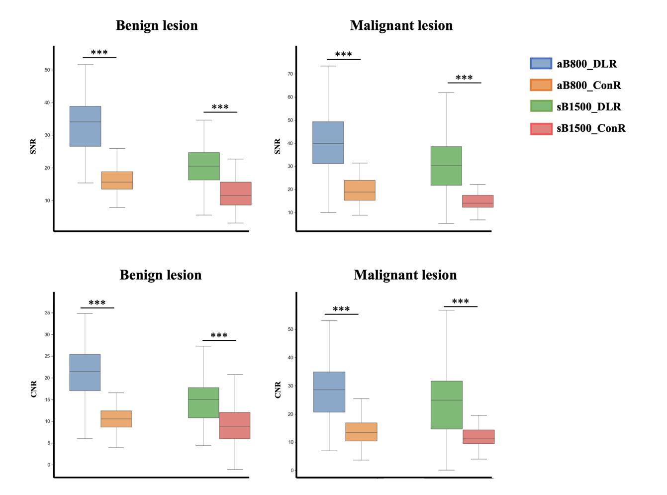
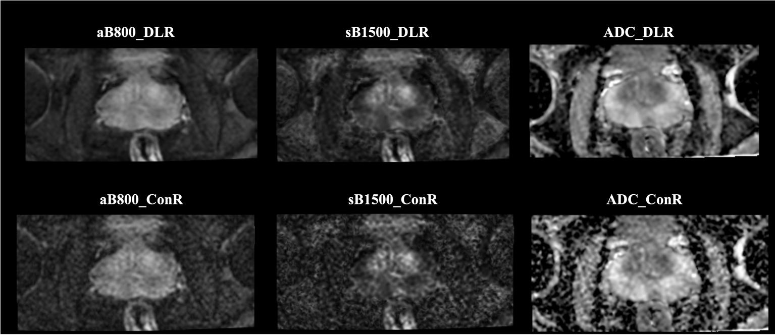
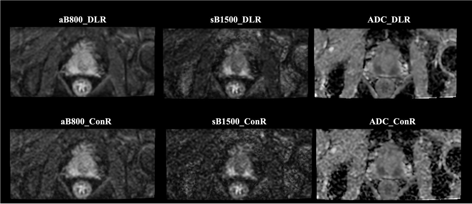
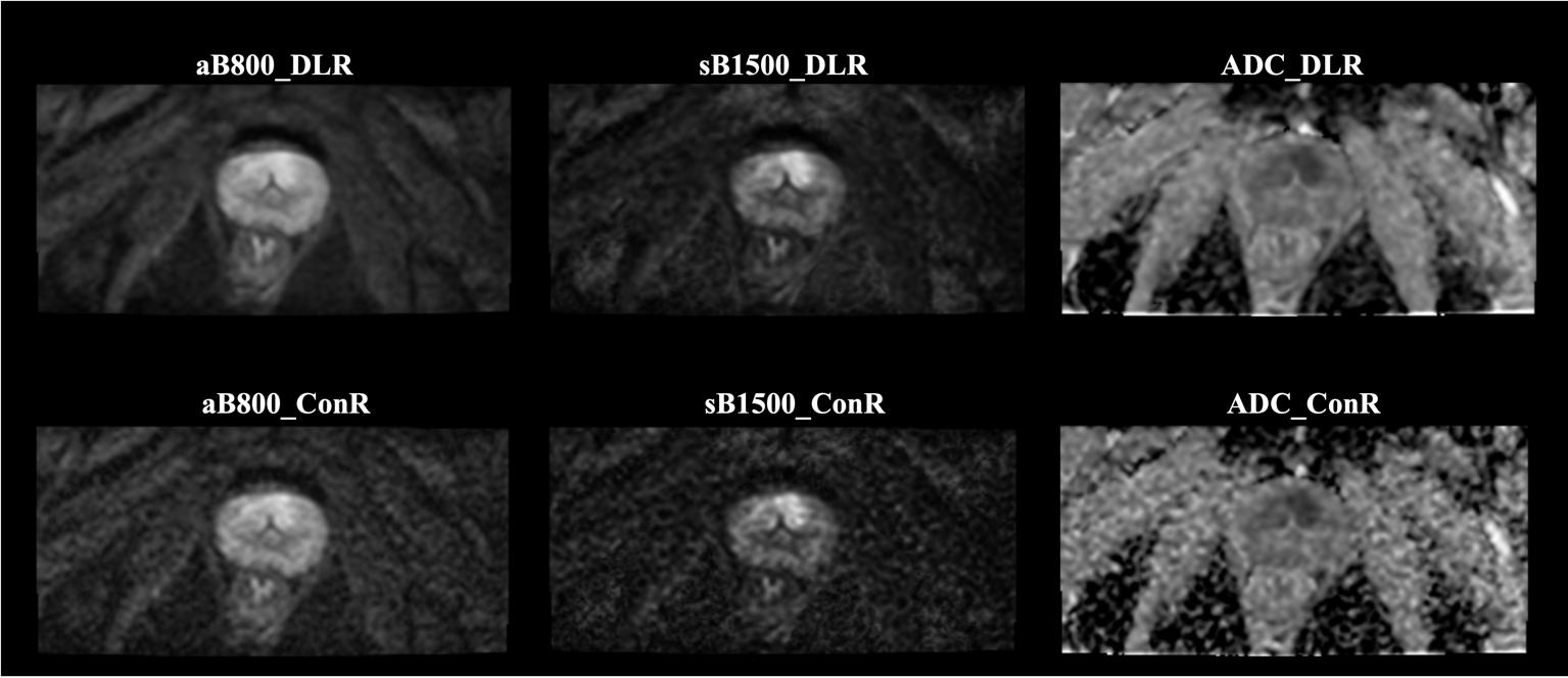
As shown in Table 1, DLR, relative to ConR, significantly improved the overall image quality, benign lesion conspicuity, and malignant lesion conspicuity of the acquired low b-value DWI as well as the synthetic high b-value DWI (all P<0.001). Notably, sB1500_DLR had the highest image quality in detecting malignant lesions. The SNR and CNR of benign and malignant lesions were significantly higher in the aB800_DLR than in the aB800_ConR (Figure 1). The same trend was also observed in the SyDWI. Figure 3-5 showed synthetic high b-value DWI, particularly in which the one computed from DWI_DLR, can enhance the detection of prostate lesions.Discussion and Conclusion
This study compared the image quality of synthetic high b-value DWIs computed from DLR and ConR reconstructed low b-value DWI. The results showed DLR can significantly improve the image quality of acquired low b-value DWI as well as the derived SyDWI, in terms of SNR, CNR and image quality evaluation. SyDWI is mathematically derived from acquired DWI images by projecting signal intensity at non-acquired b-values using a standard mono-exponential fit [2]. The improved image quality of acquired DWI can inherently transmit to the derived SyDWI. As a result, SyDWI computed from DLR reconstructed DWI showed improved image quality and enhanced prostate tumor detection.In conclusion, SyDWI derived from DWI_DLR was superior to that computed from DWI_ConR and may be beneficial for prostate lesion detection.
Acknowledgements
References
[1] Turkbey B, Rosenkrantz AB, Haider MA, et al. Prostate imaging reporting and data system version 2.1: 2019 update of prostate imaging reporting and data system version 2. Eur Urol 2019;(76):340–51.
[2] Andrew B Rosenkrantz 1, Hersh Chandarana, Nicole Hindman, et al. Computed diffusion-weighted imaging of the prostate at 3 T: impact on image quality and tumor detection. Eur Radiol 2013;23(11):3170-7.
[3] Kang-Lung Lee, Dimitri A Kessler, Simon Dezonie, et al. Assessment of deep learning-based reconstruction on T2-weighted and diffusion-weighted prostate MRI image quality. Eur J Radiol. 2023; 166:111017.
[4] Marta Zerunian, Francesco Paucciarelli, Damiano Caruso, et al. Artificial intelligence-based image quality enhancement in liver MRI: a quantitative and qualitative evaluation. Radiol Med. 2022; 127(10):1098-1105.
Figures