3757
Pushing MP2RAGE boundaries: ultimate time-efficient parameterization combined with exhaustive T1 synthetic contrasts.1CEA, NeuroSpin, Gif-sur-Yvette, France, 2Neuroradiology, Henri Mondor University Hospital, Creteil, France, 3Siemens Healthcare SAS, Saint Denis, France, 4Paris Brain Institute, ICM, Sorbonne Université, Paris, France, 5Medical Imaging, Henri Mondor University Hospital, Créteil, France
Synopsis
Keywords: Synthetic MR, Brain, Acquisition Methods, Challenges, High-Field MRI MR, Fingerprinting/Synthetic MR, MR Value, Multiple Sclerosis, Neuro, Parallel Transmit & Multiband, Relaxometry
Motivation: Redefining MP2RAGE sequence for clinical efficiency.
Goal(s): To provide a time-efficient MP2RAGE parameterization with on-demand synthetic T1-weighted contrasts.
Approach: Sequence parameters are chosen to minimize idle time while maximizing CNRWM/GM. Synthetic contrasts are derived from T1 map. Experimental validation is carried for 7T brain imaging using plug-and-play parallel transmission.
Results: Time-efficient MP2RAGE reduced acquisition time by up to 40% compared to reference, while maintaining contrast quality. Multiple sclerosis patients benefited from enhanced lesion visualization with a 10min, (0.67mm)3 time-efficient protocol. This optimization enabled shorter acquisition times or higher resolution within a given time budget, while providing an exhaustive set of brain T1-w contrasts.
Impact: A time-efficient MP2RAGE sequence parameterization is proposed, resulting in faster, higher-resolution 3D T1-weighted brain imaging compared to conventional settings, combined with a complete set of synthetic T1-w contrasts generated online, with meaningful potential in clinical and research practice.
Introduction
MP2RAGE sequence is widely employed for T1-weighted brain structural imaging at 7T1 and 3T2. Standard parameter optimization achieves uniform T1-w contrast (UNI) with high contrast-to-noise ratio (CNR)1 and derives a T1 map3. FLAWS parameterization4 differs from MP2RAGE notably by inversion time (TI) choices and yields additional contrasts such as: WM-nulled (FGATIR), GM-highlighted (FLAWSmin) or high-contrast T1-w (FLAWShco5). While potentially useful in pathological contexts (e.g., epilepsy6,7,8, multiple sclerosis (MS)9,10) acquiring all MP2RAGE/FLAWS contrasts in a single acquisition is challenging12. In this study, the central idea shifts focus away from the radiological relevance of acquired or combined contrasts and instead uses T1 mapping as an intermediate step to generate desired synthetic T1-based contrasts13. Consequently, this approach allowed investigating a new “time-efficient” parameterization for MP2RAGE with TI and TR minimization. Experimental validation was conducted at 7T with parallel transmission (pTx) on healthy subjects and MS patients at various spatial resolutions.Methods
With MP2RAGE, a direct relationship between UNI signal intensity and T1 values can be numerically calculated for any voxel1. This function depends on a handful of key MP2RAGE parameters11. The core idea of T1-based synthetic imaging consists in creating additional mathematical relationships between T1 values and desired synthetic UNI signals. It relies on the simplified forward solution of the UNI signal equation1 based on the acquired T1 map and by retrospectively changing the MP2RAGE parameters. The non-linear dynamic of signal with respect to T1 can be fully controlled to tune the tissue contrast and generate synthetic T1-w images according to any specific clinical needs. Time-efficient MPR2AGE parameterization was designed as follows: 1/ spatial resolution sets the number of partitions, 2/ echo spacing is adjusted through bandwidth to reach desired SNR and 3/ TI and TR are set as short as possible. FA values are optimized with EPG simulations12 to maximize CNRWM/GM while ensuring a bijective T1-to-UNI function for the T1 range of interest. Synthetic imaging process was implemented on the MR system’s online reconstructor, generating sUNI, sFGATIR, sEDGE, sFLAWSMIN and sFLAWSHCO contrasts (Figure1). Methodological validation on four healthy volunteers was performed on a whole-body investigative 7T system (Magnetom, Siemens Healthineers) with pTx at (1mm)3, (0.80mm)3, (0.65mm)3 and (0.45mm)3. Clinical evaluation was performed on six MS patients comparing a time-efficient MP2RAGE versus ‘conventional’ MP2RAGE (Figure2). Universal RF pulses (UP)14 allowed calibration-free imaging. CNR between brain tissues and lesions were measured. Image quality and cortical lesion depiction were assessed by an experimented neuroradiologist.Results
UNI-to-T1 relationships were monotonous in physiological T1 ranges for both conventional and time-efficient protocols at all resolutions (Figure3A). T1-to-synthetic UNI relationships are presented on Figure 3B. On healthy volunteers, time-efficient approach reduced acquisition time by 40%, 30% and 19% for (1mm)3, (0.80mm)3 and (0.65mm)3 resolutions respectively, compared to conventional settings. sUNI images had comparable CNR values, and image quality remained similar. Flexibility in parameter selection enabled (0.65mm)3 imaging without partial Fourier and (0.45mm)3 whole-brain imaging in 19min56s (Figure4). For MS patients, a (0.67mm)3 time-efficient acquisition enhanced cortical lesion visualization compared to a conventional (0.80mm)3 protocol, while decreasing scan time by 15% (Figure5). Visual analysis showed high lesion-to-tissue contrasts similarity, supported by CNR differences <15%. The increased resolution improved delineation of small MS lesions near the cortex.Discussion
The T1 synthetic imaging framework allowed to fundamentally redefine the MP2RAGE parameterization. This has enabled a "single-acquisition/multi-contrast” approach, overcoming usual dichotomous representation between MP2RAGE&FLAWS, while saving time and improving spatial resolution. Decoupling final contrasts from acquisition parameters paves the way for unified MP2RAGE imaging that overcomes many challenges encountered in previous optimizations (such as fixed TI values limiting FLAWS spatial resolution or limited contrasts with MP2RAGE). Efficiency gain was demonstrated at various resolutions, including achieving for the first time a whole-brain imaging at (0.45mm)3. Preliminary results on MS patients suggest accessible high-resolution 3D T1-w imaging to provide increase comfort to the radiologist for lesion load assessment. PTx with UP was employed to homogenize signal throughout the entire brain. This method was helpful but not mandatory, as time-efficient parameterization could be optimized as well for single-channel use. Since image quality was not compromised, time-efficient approach could likely be combined with advanced reconstruction techniques like Compressed-sensing15. This time-efficient approach will also be applied at 3T, expanding diagnosis possibilities. The measured CNRWM/GM were comparable between with sUNI and conventional UNI, suggesting robust T1 estimation with time-efficient parameterizations. However, T1 mapping accuracy and reproducibility were not evaluated, making this approach not intended at this stage for highly-reproducible quantitative imaging.Conclusion
Time-efficient MP2RAGE combined with synthetic T1-based imaging enables fast high-resolution multi-contrast 3D T1-w imaging for clinical and research purposes.Acknowledgements
Blanche Bapst is a recipient of the 2022 Alain Rahmouni SFR-CERF research grant provided by the French Society of Radiology and the French Academic College of Radiology. This research received funding from the RETP program (Research Equipment and Technology Platforms) of the Leducq Foundation. French FLUMASEPT and ENERGYSEP cohorts are supported by grants from Merck-Serono-GMSI, ARSEP and FISM.The authors would like to thank Dr. Dokumacı for fruitful discussions and for providing MP2RAGE EPG Matlab open-access code.References
1. Marques JP, Kober T, Krueger G, van der Zwaag W, Van de Moortele PF, Gruetter R. MP2RAGE, a self bias-field corrected sequence for improved segmentation and T1-mapping at high field. NeuroImage. 2010;49(2):1271-1281.
2. Trotier AJ, Dilharreguy B, Anandra S, et al. The Compressed Sensing MP2RAGE as a Surrogate to the MPRAGE for Neuroimaging at 3 T. Invest Radiol. 2022;57(6):366-378.
3. Marques JP, Gruetter R. New Developments and Applications of the MP2RAGE Sequence - Focusing the Contrast and High Spatial Resolution R1 Mapping. Yacoub E, ed. PLoS ONE. 2013;8(7):e69294.
4. Tanner M, Gambarota G, Kober T, et al. Fluid and white matter suppression with the MP2RAGE sequence. J Magn Reson Imaging. 2012;35(5):1063-1070.
5. Beaumont J, Gambarota G, Saint‐Jalmes H, et al. High‐resolution multi‐T 1 –weighted contrast and T 1 mapping with low sensitivity using the fluid and white matter suppression (FLAWS) sequence at 7T. Magn Reson Med. 2021;85(3):1364-1378.
6. Demerath T, Kaller CP, Heers M, et al. Fully automated detection of focal cortical dysplasia: Comparison of MPRAGE and MP2RAGE sequences. Epilepsia. 2022;63(1):75-85.
7. Feldman RE, Delman BN, Pawha PS, et al. 7T MRI in epilepsy patients with previously normal clinical MRI exams compared against healthy controls. Bernhardt BC, ed. PLOS ONE. 2019;14(3):e0213642.
8. Chen X, Qian T, Kober T, et al. Gray-matter-specific MR imaging improves the detection of epileptogenic zones in focal cortical dysplasia: A new sequence called fluid and white matter suppression (FLAWS). NeuroImage Clin. 2018;20:388-397.
9. Beck ES, Sati P, Sethi V, et al. Improved Visualization of Cortical Lesions in Multiple Sclerosis Using 7T MP2RAGE. Am J Neuroradiol. 2018;39(3):459-466.
10. Kolber P, Droby A, Roebroeck A, et al. A “kissing lesion”: In-vivo 7T evidence of meningeal inflammation in early multiple sclerosis. Mult Scler J. 2017;23(8):1167-1169.
11. Tao S, Zhou X, Lin C, Patel V, Westerhold EM, Middlebrooks EH. Optimization of MP2RAGE T1 mapping with radial view-ordering for deep brain stimulation targeting at 7 T MRI. Magn Reson Imaging. 2023;100:55-63.
12. Dokumacı AS, Aitken FR, Sedlacik J, et al. Simultaneous Optimization of MP2RAGE T 1 ‐weighted ( UNI ) and FLuid And White matter Suppression ( FLAWS ) brain images at 7T using Extended Phase Graph ( EPG ) Simulations. Magn Reson Med. 2023;89(3):937-950.
13. Massire A, Seiler C, Troalen T, et al. T1-Based Synthetic Magnetic Resonance Contrasts Improve Multiple Sclerosis and Focal Epilepsy Imaging at 7 T. Invest Radiol. 2021;56(2):127-133.
14. Gras V, Vignaud A, Amadon A, Bihan D, Boulant N. Universal pulses: A new concept for calibration‐free parallel transmission. Magn Reson Med. 2017;77(2):635-643.
15. Mussard E, Hilbert T, Forman C, Meuli R, Thiran J, Kober T. Accelerated MP2RAGE imaging using Cartesian phyllotaxis readout and compressed sensing reconstruction. Magn Reson Med. 2020;84(4):1881-1894.
Figures
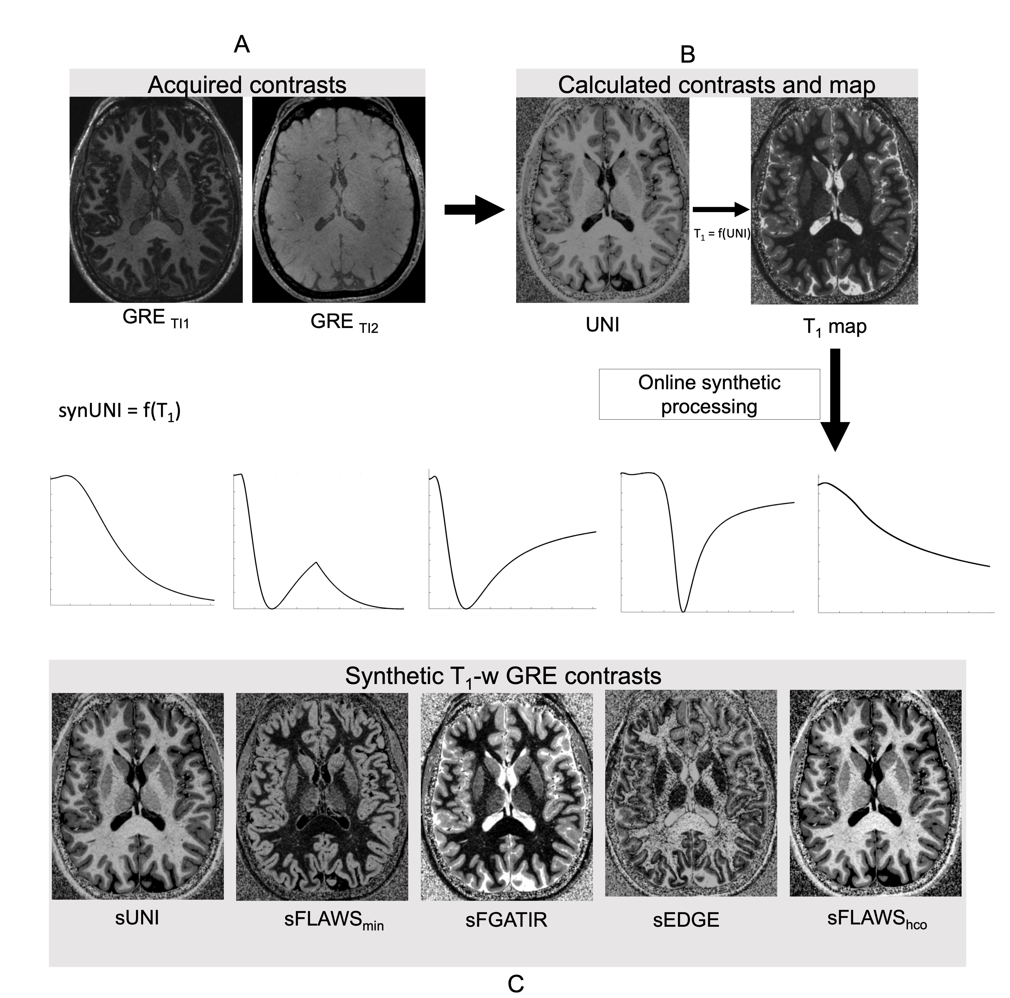
Figure1: Synthetic T1-weighted imaging using time-efficient MP2RAGE parameterization workflow.
A. MP2RAGE sequence parameterization is chosen to maximize CNRWM/GM while keeping the acquisition at minimum for a given spatial resolution. GRETI1 and GRETI2 are not used for radiological analysis.
B. UNI image generated with MP2RAGE signal equation is used only to generate T1 map (tissue contrast might be suboptimal for radiological analysis).
C. Signal equation is re-integrated using the T1 map to generate synthetic contrasts to be used for radiological analysis.
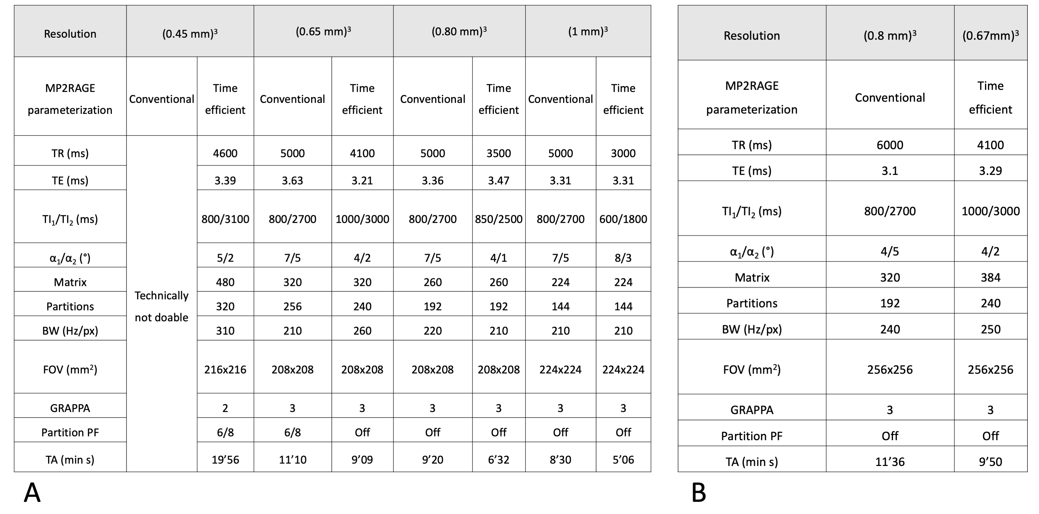
Figure 2: Sequence parameters used on healthy volunteers (A) and MS patients (B)
Note that in this MR sequence implementation all gradients were in ‘Whisper’ mode to avoid any peripheral nerve stimulation.
TR: Repetition time; TE: Echo time; TI: Inversion time; BW: Receiver bandwidth; FOV: Field of view; GRAPPA: Generalized autocalibrating partially parallel acquisitions; TA: acquisition time.
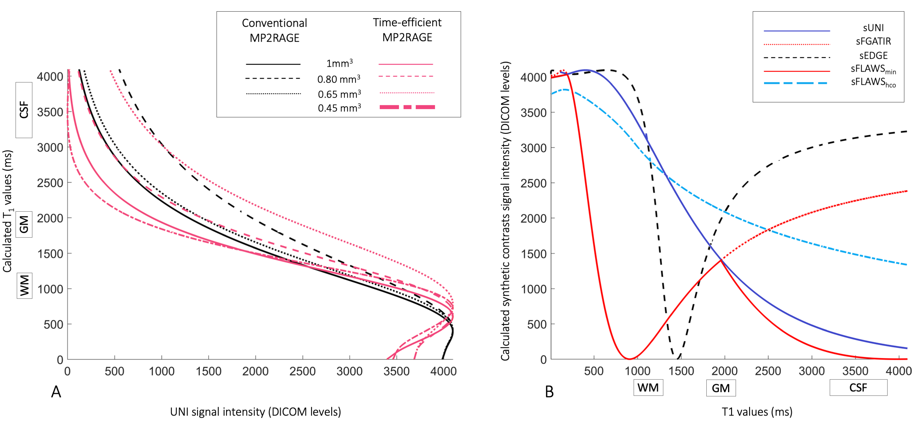
Figure 3. Simulated relationship between UNI signal and T1 values.
A. Evaluated T1 values (output) obtained based on UNI signal (input) for conventional (black) and time-efficient (pink) MP2RAGE parameterizations at different spatial resolutions. Note how all curves monotonously sample the brain T1range.
B. Generated synthetic UNI signal intensities (output) versus input T1 values for synthetic MP2RAGE protocols. The synthetic MP2RAGE parameters were chosen empirically or based on the literature.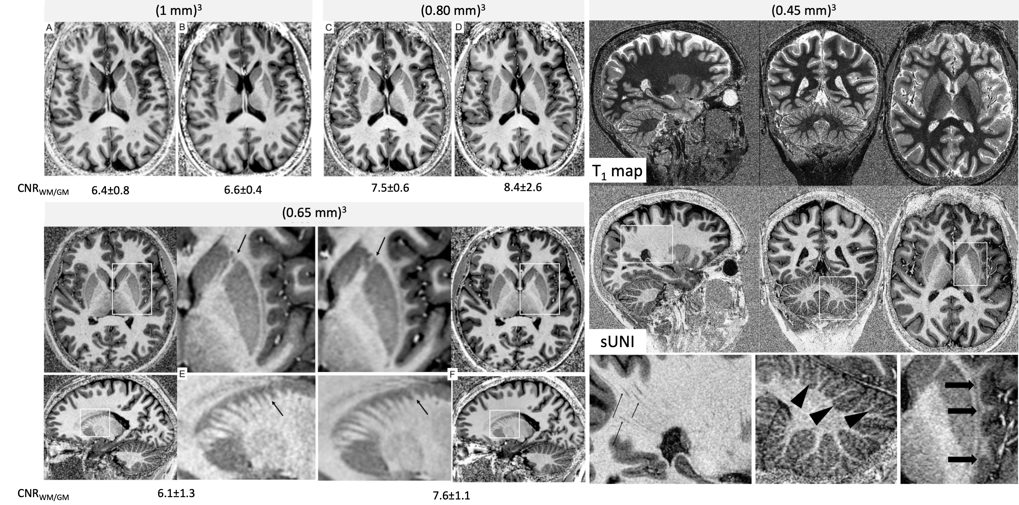
Figure 4: Comparison of synthetic and conventional UNI contrasts at (1mm)3, (0.80mm)3 and (0.65mm)3 resolutions (A-F), and illustration of a whole-brain (0.45mm)3 acquisition using the time-efficient MP2RAGE parameterization.
(A,C,E) time-efficient MP2RAGE acquisitions with sUNI contrast
(B,D,F) conventional MP2RAGE acquisitions with UNI contrast
The difference in true spatial resolution due to Partial Fourier removal is visible at (0.65mm)3 (arrows). At (0.45mm)3, high spatial resolution allows visualization of fine anatomical details (arrows).
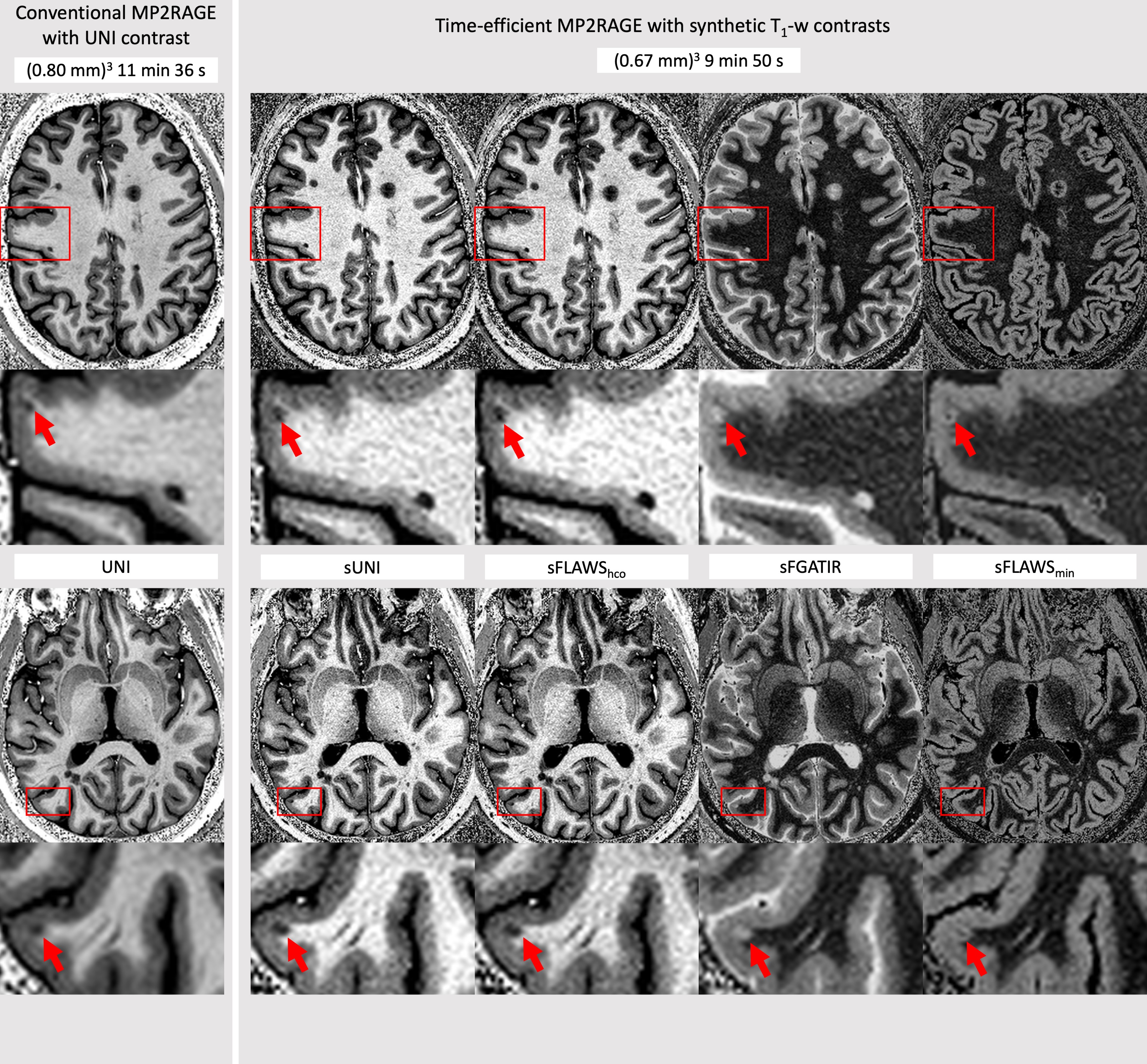
Figure 5: Comparison of conventional and time-efficient MP2RAGE with on-demand synthetic T1-w contrasts on a MS subject.
On the left: Conventional MP2RAGE protocol with UNI contrast.
On the right: Time-efficient protocol with multiple synthetic contrasts.
Arrows on zoomed-images indicate the better delineation of two juxtacortical lesions with time-efficient parameterization ( notably with sUNI and sFLAWShco) and their appearance on various synthetic contrasts.