3754
Synthesis of 4D Flow MRI Data using Particle-based Bloch Simulations in CMRsim1University and ETH Zurich, Zurich, Switzerland
Synopsis
Keywords: Synthetic MR, Simulations
Motivation: Signal model-based synthesis of 4D flow MRI data from CFD is promising to study image acquisition, reconstruction methods and augment data for inference. However, the approach assumes idealized encoding, and particle-based Bloch simulations are needed to include complex effects.
Goal(s): Implement a particle-based Bloch simulation in CMRsim for generating synthetic multi-venc 4D flow data.
Approach: Using pulsatile, stenotic U-bend CFD data as input, simulations of moving magnetization in CMRsim generate synthetic 4D flow MRI data.
Results: 4D flow simulation is feasible in CMRsim, demonstrating tractable simulation times and good agreement with ground truth and magnetization history-based effects, namely acceleration and intra-voxel dephasing.
Impact: Numeric Bloch simulations in CMRsim permit a detailed study of imaging protocol-dependent flow-related artifacts in 4D flow MRI of complex flows and can thereby assist in improving acquisition and reconstruction approaches.
Introduction
Simulation of synthetic MR images is becoming increasingly important for the evaluation and optimization of acquisition, reconstruction, and post-processing algorithms, facilitating access to ground truth information and synthetic data for model evaluation and training. Specifically, phase-contrast (PC) based methods may benefit significantly from data driven methods. However, these methods generally require large datasets for training. As such, synthetic PC-MRI datasets may help to address the demand. Additionally, velocity ground-truth data for these methods is often unavailable. Recent works on super-resolution1-4 have demonstrated the possibility of using computational fluid dynamics (CFD) to generate ground-truth data for inference purposes. Typically, synthetic PC-MRI data are obtained from CFD simulations using a signal model5, which assumes ideal encoding and does not include effects such as ghosting, magnetization history effects, and motion during encoding. Recent work has improved on this by coupling CFD and MR simulation6, however these methods inherently require re-calculation of CFD results for each simulated MR image.We recently presented CMRsim7 (gitlab.ethz.ch/ibt-cmr/mri_simulation/cmrsim), a particle-based Bloch simulation framework, focused on complex motion and flow. In this work we demonstrate initial results of multi-venc 4D flow MRI simulations using CMRsim and evaluate the quality of the simulated results. Bayesian velocity combination8 is performed and compared to ground truth.
Methods
Ground truth flow data were generated for a stenotic U-bend geometry with a main diameter of 3cm, a stenotic area of 0.75cm2 and a peak velocity of 3.5ms-1 (representing a moderate to severe stenosis9) using pulsatile large eddy simulation5 (LES).Resulting time-resolved flow fields were subsequently used to generate synthetic PC-MRI images using CMRsim. Figure 1A shows an overview of the pipeline.
A spoiled gradient-echo, 7-point 4DFlow sequence was generated using CMRseq10 (gitlab.ethz.ch/ibt-cmr/mri_simulation/cmrseq) for an MR system with 80mT/m and 400T/m/s gradients. Magnetization was carried between TRs to allow for possible magnetization history effects. Sequence parameters are shown in Figure 1B. Spoiling was achieved by both RF spoiling (quadratic phase increment) and gradient spoiling in the frequency-encode and slice-select directions. The velocity-encoding strengths (Venc) were set to 3.5m/s and 1m/s for all directions, with a separate simulation run performed for each direction and Venc. A pseudo spiral Cartesian trajectory11 was used, with 20 cardiac phases and an undersampling factor of 3. The frequency-encode direction (M) was oriented parallel to the inlet and stenotic jet. Relaxation parameters were T1=1300ms, T2=250ms, T2*=50ms. The simulations were performed with 3 million particles (9.5/mm3) and a temporal resolution of 10us on GPUs (Titan RTX 24Gb), taking ~24 hours per simulation.
Data was retrospectively binned and reconstruction was performed using BART12 inverse FFT. Bayesian combination was performed to combine low and high Venc data, and three regions of interest were used to evaluate velocity, shown in Figure 1C.
Results and Discussion
Figure 2 shows high and low Venc magnitude images resulting from velocity encoding along M, as well as velocity images for all directions (M,P,S). Magnitude images show magnetization history effects, including inflow contrast and signal loss due to intravoxel dephasing. As expected, low Venc data result in phase wrapping in regions of high velocity, specifically in the stenotic jet.Extracted velocity curves from the ROIs are shown in Figure 3, comparing high and low Venc and Bayesian combination to ground truth.

Figure 4 shows flow in all directions for Bayesian combination and ground truth. Figure 5 shows velocity error images compared to ground truth for Bayesian, high Venc and low Venc.
Overall, there is a good agreement in regions of high flow when the encoding direction matches the dominant flow direction, as seen in Figure 3(A,E), however in the jet region there are errors in directions orthogonal to the jet (D,G). As magnetization history effects are present, these errors may be due a combination of factors, including incomplete spoiling, acceleration, or spatial encoding errors.
As expected, difference images show large errors for high Venc, with reduced errors and wrapping for low Venc. Bayesian combination provides the lowest error overall.
One interesting result is that, while no noise was added, images show significant noise-like artifacts. Previous testing suggests that this is due to a combination of RF/gradient spoiling, magnetization history and insufficient particle densities. Another possible contributing factor is undersampling, as sampling patterns are incoherent between cardiac phases, with undersampling increasing for higher spatial frequencies.
Conclusion
This initial work demonstrates that numeric Bloch simulation of 4D flow MRI is feasible using CMRsim, producing promising results, specifically in relation to magnetization history related effects. Further work is necessary to refine the simulation settings to remove numeric instabilities and better understand sources of velocity/encoding errors.Acknowledgements
References
1. Ferdian E, Suinesiaputra A, Dubowitz DJ, et al. 4DFlowNet: Super-Resolution 4D Flow MRI Using Deep Learning and Computational Fluid Dynamics. Front. Phys. 2020;8:1–14 doi: 10.3389/fphy.2020.00138.
2. Ferdian E, Marlevi D, Schollenberger J, et al. Cerebrovascular super-resolution 4D Flow MRI – Sequential combination of resolution enhancement by deep learning and physics-informed image processing to non-invasively quantify intracranial velocity, flow, and relative pressure. Med. Image Anal. 2023;88:102831 doi: 10.1016/j.media.2023.102831.
3. Shit S, Zimmermann J, Ezhov I, et al. SRflow: Deep learning based super-resolution of 4D-flow MRI data. Front. Artif. Intell. 2022;5 doi: 10.3389/frai.2022.928181.
4. Nath R, Kazemi A, Callahan S, Stoddard MF, Amini AA. 4Dflow-VP-Net: A deep convolutional neural network for noninvasive estimation of relative pressures in stenotic flows from 4D flow MRI. Magn. Reson. Med. 2023;90:2175–2189 doi: 10.1002/mrm.29791.
5. Dirix P, Buoso S, Peper ES, Kozerke S. Synthesis of patient-specific multipoint 4D flow MRI data of turbulent aortic flow downstream of stenotic valves. Sci. Rep. 2022;12:16004 doi: 10.1038/s41598-022-20121-x.
6. Puiseux T, Sewonu A, Moreno R, Mendez S, Nicoud F. Numerical simulation of time-resolved 3D phase-contrast magnetic resonance imaging.; 2021. doi: 10.1371/journal.pone.0248816.
7. Weine J, McGrath C, Kozerke S. CMRsim - A Python package for MRI simulations incorporating complex organ motion and flow. 2023 ISMRM&ISMRT Annual Meeting & Exhibition, 2393
8. Binter C, Knobloch V, Manka R, Sigfridsson A, Kozerke S. Bayesian multipoint velocity encoding for concurrent flow and turbulence mapping. Magn. Reson. Med. 2013;69:1337–1345 doi: 10.1002/mrm.24370.
9. Baumgartner H, Hung J, Bermejo J, et al. Recommendations on the echocardiographic assessment of aortic valve stenosis: A focused update from the European Association of Cardiovascular Imaging and the American Society of Echocardiography. Eur. Heart J. Cardiovasc. Imaging 2017;18:254–275 doi: 10.1093/ehjci/jew335.
10. Weine J, McGrath C, Kozerke S. CMRSeq - A Python package for intuitive sequence design. 2023 ISMRM&ISMRT Annual Meeting & Exhibition, 2398
11. Gottwald LM, Peper ES, Zhang Q, et al. Pseudo-spiral sampling and compressed sensing reconstruction provides flexibility of temporal resolution in accelerated aortic 4D flow MRI: A comparison with k-t principal component analysis. NMR Biomed. 2020;33:1–13 doi: 10.1002/nbm.4255.
12. Uecker M, Ong F, Tamir JI, et al. Berkeley advanced reconstruction toolbox. Proc Intl Soc Mag Reson Med. 2015;23:2486.
Figures
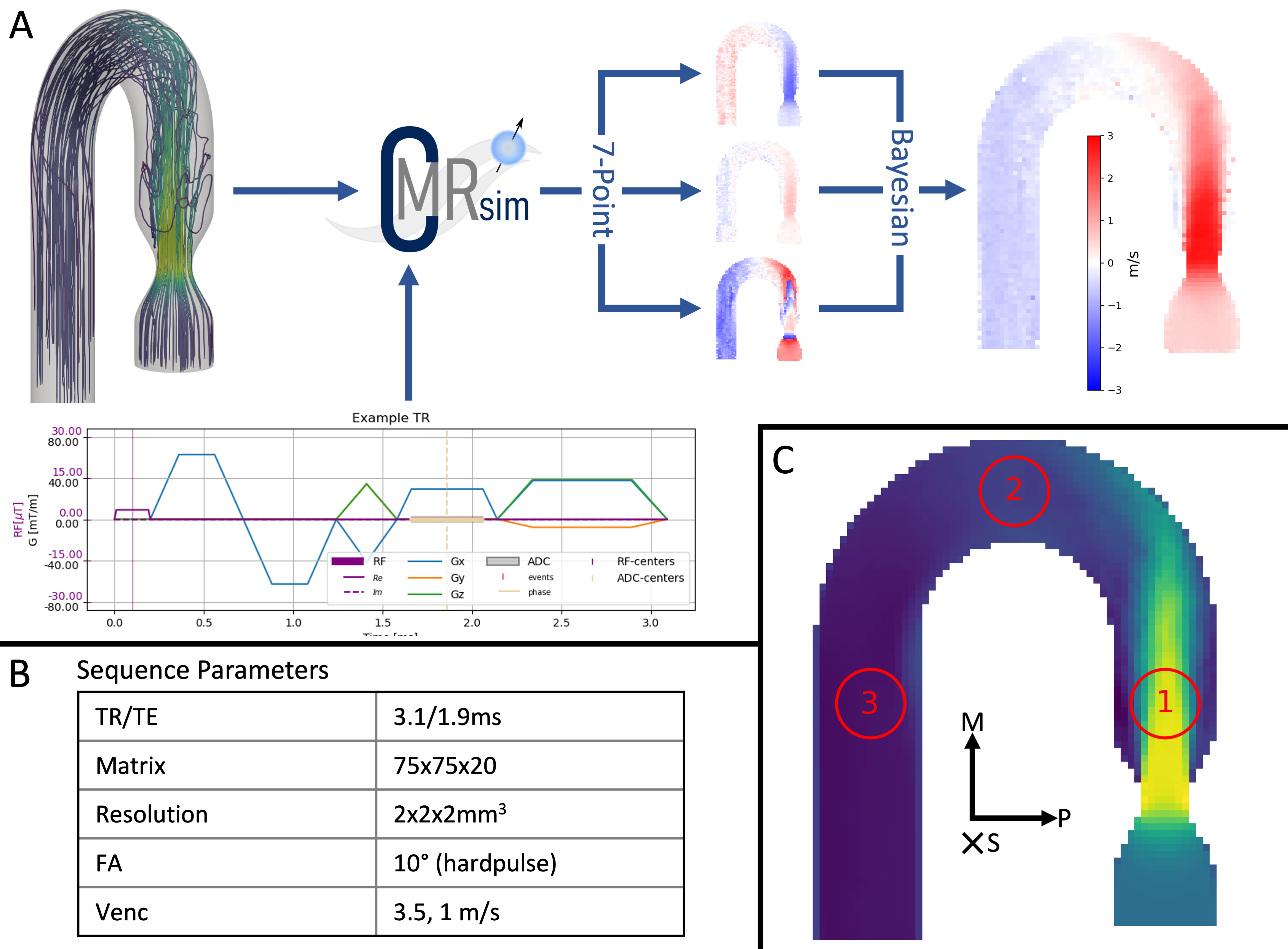

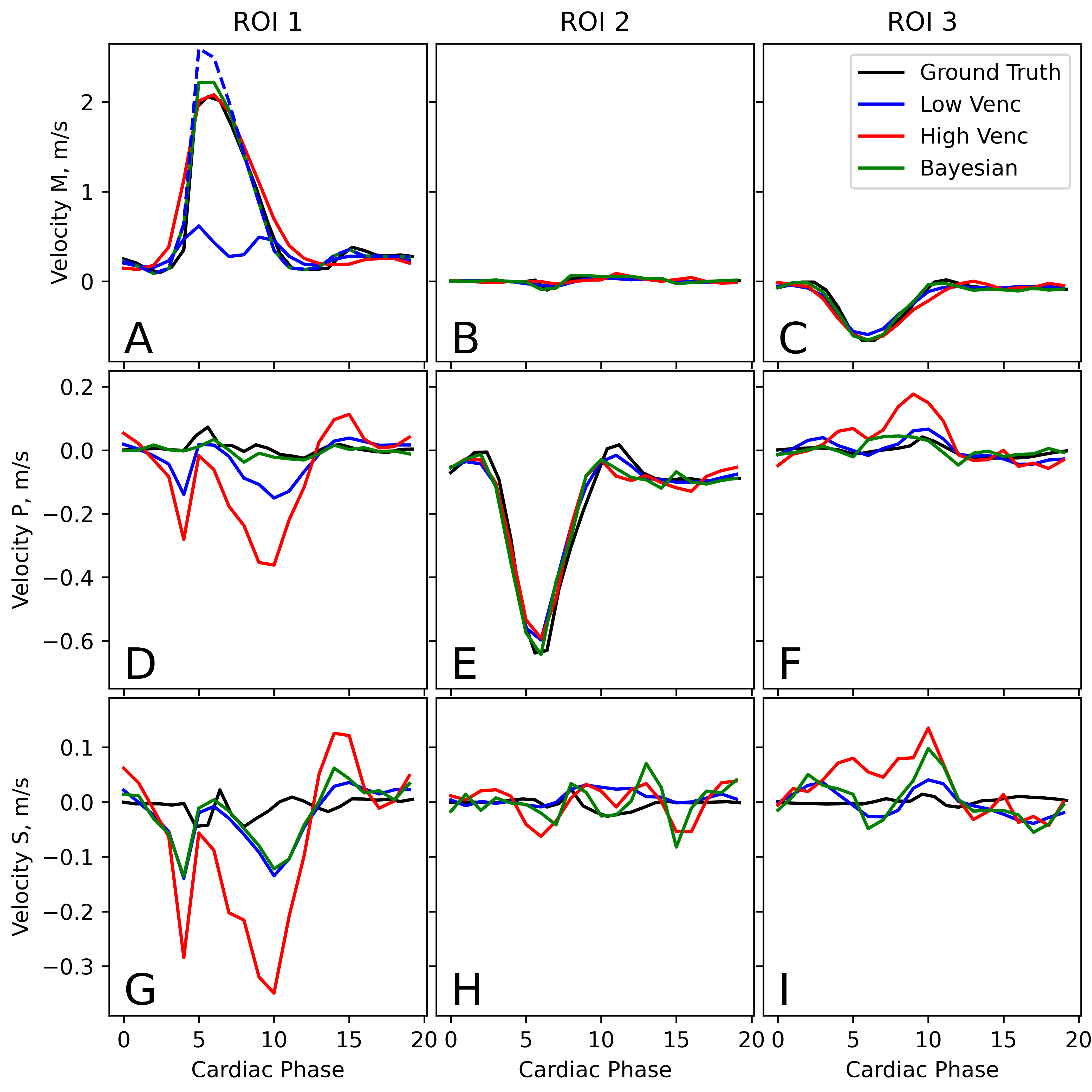

Figure 4: (Click for GIF) Comparison of Bayesian velocity combination (A-C) and ground truth (D-F) for MPS directions. Good agreement is seen, however some small details in regions of low flow are lost (pink arrow). Additionally, Bayesian combination shows some single voxel artifacts at peak flow.