3752
Numerical Evaluation of the Specific Absorption Rate Change of Transmit Arrays at 9.4T due to Presence of B0 Field Probes1High-field MR Center, Max Planck Institute for Biological Cybernetics, Tuebingen, Germany, 2Department of Diagnostic and Interventional Neuroradiology, University Hospital Tuebingen, Tuebingen, Germany, 3Department of Biomedical Magnetic Resonance, University of Tübingen, Tuebingen, Germany
Synopsis
Keywords: Safety, Safety, Electroencephalography (EEG)
Motivation: Imperfections of B0-field can reduce the MR image quality. To track the B0 change during imaging, NMR field-probes inside the RF coil can be used. Insertion of the field-probes may lead to alteration of the B1+ and pSAR.
Goal(s): To evaluate alterations of B1+ field and pSAR of the 16-channel Tx-array in the presence of NMR field-probes at 9.4T.
Approach: To reach the goal, we simulated the Tx-part of the 16Tx32Rx array loaded by a phantom and human voxel model in the presence of 11 field-probes.
Results: Insertion of field-probes led to a small drop of B1+ and a slight change of pSAR.
Impact: We showed that inserting field-probes doesn’t significantly alter the B1+ field and pSAR of the 16Tx32Rx array coil at 9.4T. Therefore, field-probes can be safely used in-vivo to evaluate B0.
Introduction
Ultra-high field (UHF, B0 higher than 7T) MRI is a powerful research instrument providing a significant improvement in signal- and contrast-to-noise ratios[1]. However, the benefit of higher resolution could be disrupted by volunteer movements or B0-field imperfections of another source. To track the B0 change during imaging, local NMR probes placed near the sample can be used[2]. Since UHF MRI requires local transmit (Tx) coils (arrays), field-probes need to be placed inside the Tx-array. To avoid coupling to the Tx-elements, coaxial cables connected to the field probes must be carefully routed. Still, presence of the probes and cables may increase local sample heating commonly evaluated by numerically calculating the peak of the Specific Absorption Rate (pSAR). Therefore, before using field-probes, the in-vivo safety of the setup must be carefully evaluated. According to our safety procedure[3], each home-built coil must be simulated using realistic human voxel models. After numerical evaluation, pSAR will be used to limit the RF power in in-vivo experiments. As demonstrated previously[4], presence of the B0-probes led to a small decrease of B1+. This work, however, did not evaluate the SAR change and did not consider cable routings, which may also alter pSAR.In this work, we performed a numerical evaluation of B1+ and SAR of the double-row 16-loop Tx-array[5] both on phantom and with voxel models to estimate the effect of the field-probes and cables inserted inside the RF coil.
Methods
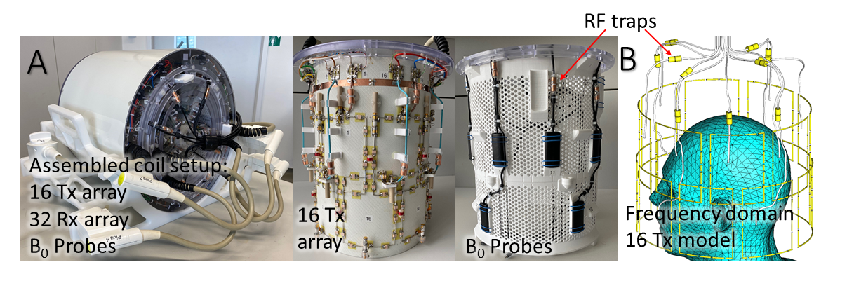
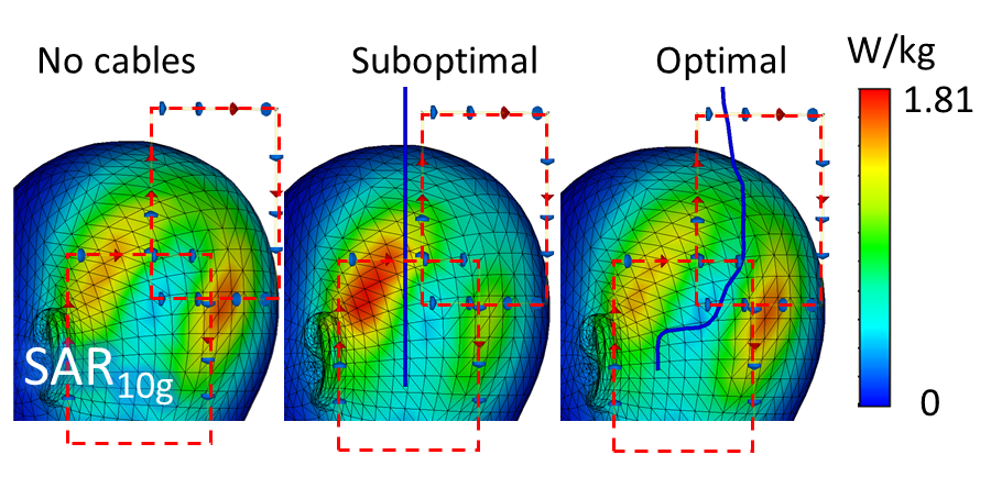
Eleven field-probes were inserted inside the 400-MHz double-row 16-loop Tx-array. A photo of the experimental setup is shown in Fig.1A. Each field-probe consisted of a small solenoid inside a plastic body and a coaxial cable connected to the console. To minimize interaction with the RF field of Tx-loops, probes were positioned in the loop centers, and field-probe cables were routed close to the loop centers with a minimum of E1-field. Therefore, we limited our model to probe cables (simulated as solid 2.5-mm silver tubes), cable traps, and the Tx-array itself. Fig.1B shows the entire numerical model of Tx-array loaded by the head and shoulders (HS) phantom. Floating ground cable traps were placed on probe cables to reduce the cable effect. Accurate positions of the cables in the final design were determined by CT scanning with 1.2x1.2x0.5 mm³ resolution. For comparison, we also simulated the array without cable traps and without cables. In addition, we simulated a two-loop array with optimal and suboptimal (through the E1-maximum) cable routing.The finite elements method in the frequency domain relied on CST Studio 2022 was used for numerical simulations of the array with the phantom. Final simulations of the array loaded by the Duke voxel model were performed in CST using finite integration in the time domain. In all cases, SAR10g was calculated using the CST Legacy averaging method. B1+ and SAR were evaluated for the CP-mode excitation (-45◦ between adjacent elements and -22.5◦ between rows).
Results and Discussion
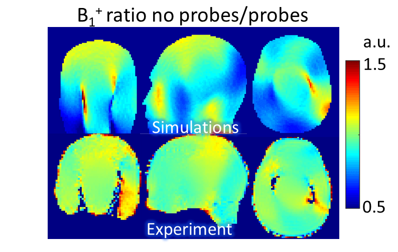
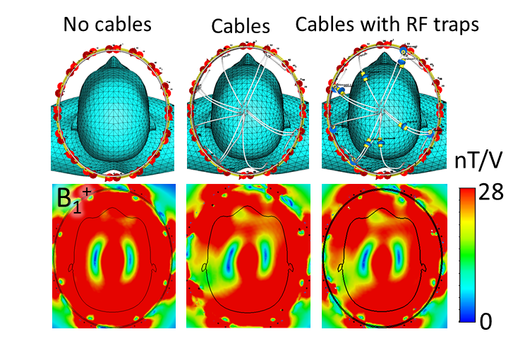
Fig.2 presents transversal B1+ maps obtained using the HS phantom. As seen in the figure, presence of cables alters the RF field, and cable traps do not significantly change the field distribution. Mean B1+ averaged over the 215-mm transversal slab for the case with probes and cable traps vs array w/o probes measured 31 and 37 nT/V, respectively. Alteration of the SAR10g distribution due to suboptimal cable routing is shown in Fig.3. Suboptimal cable routing led to 9% higher pSAR.Fig.4 shows simulated and experimentally measured ratios of B1+ maps obtained using the HS phantom without and with B0-probes. Experimentally measured drop in the average B1+ was ~ 5%. To start with, we see a greater (17% vs 5%) B1+ decrease in simulations than in the experiment.
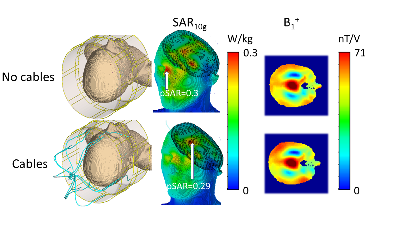
At the same time, simulations using the Duke voxel model showed insignificant changes of the B1+ and pSAR. Fig.5 presents results of SAR and B1+ simulations with and without field-probes for the array loaded to the Duke voxel model. This could be explained by insufficient meshing of the probe cables in the time-domain simulations.
Conclusion
We demonstrated that presence of the field-probes can influence both the B1+ field and SAR10g distributions. At the same time, we see certain discrepancies between simulated and measured phantom data as well as between simulations using frequency and time domains.Acknowledgements
No acknowledgement found.References
[1] Ladd, Mark E., et al. "Pros and cons of ultra-high-field MRI/MRS for human application." Progress in nuclear magnetic resonance spectroscopy 109 (2018): 1-50.
[2] De Zanche N, Barmet C, Nordmeyer-Massner JA, Pruessmann KP. NMR probes for measuring magnetic fields and field dynamics in MR systems. Magnetic Resonance in Medicine. 2008;60(1):176-186.
[3] Hoffmann J, Henning A, Giapitzakis IA, Scheffler K, Shajan G, Pohmann R, Avdievich NI. Safety testing and operational procedures for self-developed radiofrequency coils. NMR Biomed 2016;29(9):1131-1144.
[4] Son Ch., Gras V., Boulant N., and Shajan G. ISMRM & ISMRT Annual Meeting & Exhibition • 03-08 June 2023 • Toronto, ON, Canada, p. 4589.
[5] Nikulin AV, Scheffler K, Avdievich NI. 30th Annual Meeting and Exhibition of ISMRM 2022, London, UK, p.3315.
Figures
Numerically simulated SAR10g for two loop setups: no field probes, field probes with non-optimal and optimal routing