3741
Transmit uniformity and SAR optimization by a deep-learning method in UHF imaging1Shenzhen Institute of Advanced Technology, Chinese Academy of Sciences, Shenzhen, China, 2Key Laboratory for Magnetic Resonance and Multimodality Imaging of Guangdong Province, Shenzhen, China, 3Magnetic Resonance Imaging Research center, University of Nottingham Ningbo China, Ningbo, China, 4United Imaging Research Institute of Innovative Medical Equipment, Shenzhen, China, 5Lauterbur Research Center for Biomedical Imaging, Shenzhen Institute of Advanced Technology, Chinese Academy of Sciences, Shenzhen, China, 6University of Nottingham, Nottingham, United Kingdom, 7Department of Biomedical Engineering, State University of New York at Buffalo, Buffalo, NY, United States
Synopsis
Keywords: Safety, Safety
Motivation: UHF imaging is limited by both transmit uniformity and local SAR. Information on RF electric field is unavailable in conventional MR scan procedure.
Goal(s): This work aims to provide real-time RF electric field for joint optimization of imaging uniformity and peak local SAR.
Approach: A deep-learning method is proposed to predict the real-time EM field distribution using B1+ data obtained in routine prescan of the imaging procedure. The output field data is used in combined optimization of transmit uniformity and local SAR.
Results: In the torso imaging case, this method achieved both improvement of transmit field uniformity and reduction of peak local SAR.
Impact: This work studied the feasibility of machine-learning methods for RF field estimation and simultaneous optimization of transmit homogeneity and peak local SAR, aiming to reduce the estimation error of local SAR and increase the available maximum B1.
Introduction
With the development of ultra-high-field(UHF) magnetic resonance hardware and system software, applications have been evolving not only for research purposes but also in clinical scenarios including high-resolution anatomical imaging and metabolic spectroscopy for neurological diseases1. However, the RF transmit system has suffered from two aspects. First, image uniformity is seriously degraded in UHF system due to the high frequency transmit field distribution in human tissue. Second, patient safety constraints due to RF power deposition have been limiting the pulse-sequence design in the total B1RMS system limit. Multi-channel RF transmit system is introduced to help solve the above problems by providing localized transmit field distribution and independent channel weighting for optimized combination. However, only RF transmit field (B1+) is measurable in MR scanner for real-time B1+ shimming. Electric field information necessary for SAR estimation can only be obtained by EM simulation with pre-defined patient models.In this research, a deep-learning method is used to predict the real-time, channel-independent full electric RF field information for the estimation of local SAR. Along with the B1+ map obtained in routine prescan, the complete RF electric and magnetic field can be used in the optimization algorithm for simultaneous transmit uniformity improvement and peak local SAR reduction.
Methods
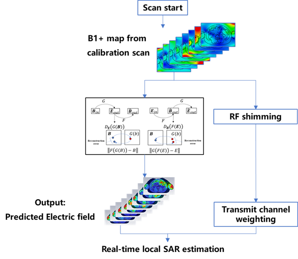
The complete RF shimming procedure is shown in Figure 1. In the prescan stage, B1+ map is obtained for each individual transmit channel with both amplitude and relative phase information. A deep-learning model based on cycleGAN is trained with a set of simulated RF electric and magnetic field datasets for the 8-channel volume transmit coil developed on the 5.0T MR scanner by United Imaging Healthcare. The model exports the full complex vector electric field that can be used for local SAR estimation. In order to reduce the calculation burden for local SAR distribution, the Q-matrix2 for the specific case is calculated to evaluate the local SAR distribution for any given transmit channel weighting factors. The obtained peak local SAR value is added as an additional penalty term for the optimization of transmit field uniformity.The deep-learning model is based on our previous study, but several modifications were made to improve the field estimation accuracy. First, the electric field is much larger in air than in human tissue. Voxels at the human model surface will have abnormally large field values in the simulation result that affect the model training process. Second, the combination of transmit channels uses a set of complex weighting factors so the phase information of the local electric field is also important in evaluating the local SAR distribution. Consequently, abnormal surface voxels are excluded in data preprocessing, and the input electric field is separated into a pair of real and imaginary datasets to include phase information.
Joint optimization of transmit uniformity and local SAR is evaluated with the simulation dataset and deep-learning model. The B1+ field data of human models in the test dataset is used as input information. Local SAR evaluated by the electric field information is added as an extra penalty term in the optimization of the transmit field uniformity.
Results
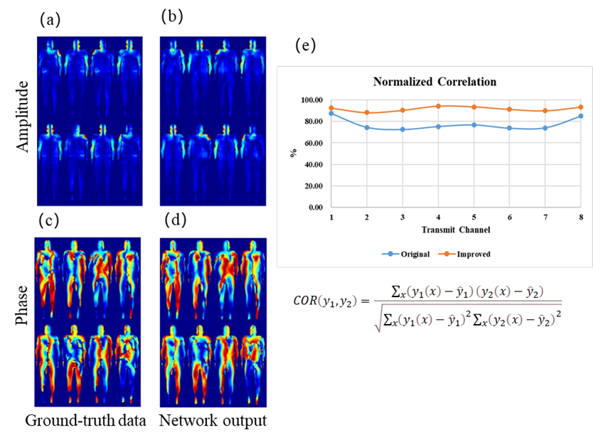
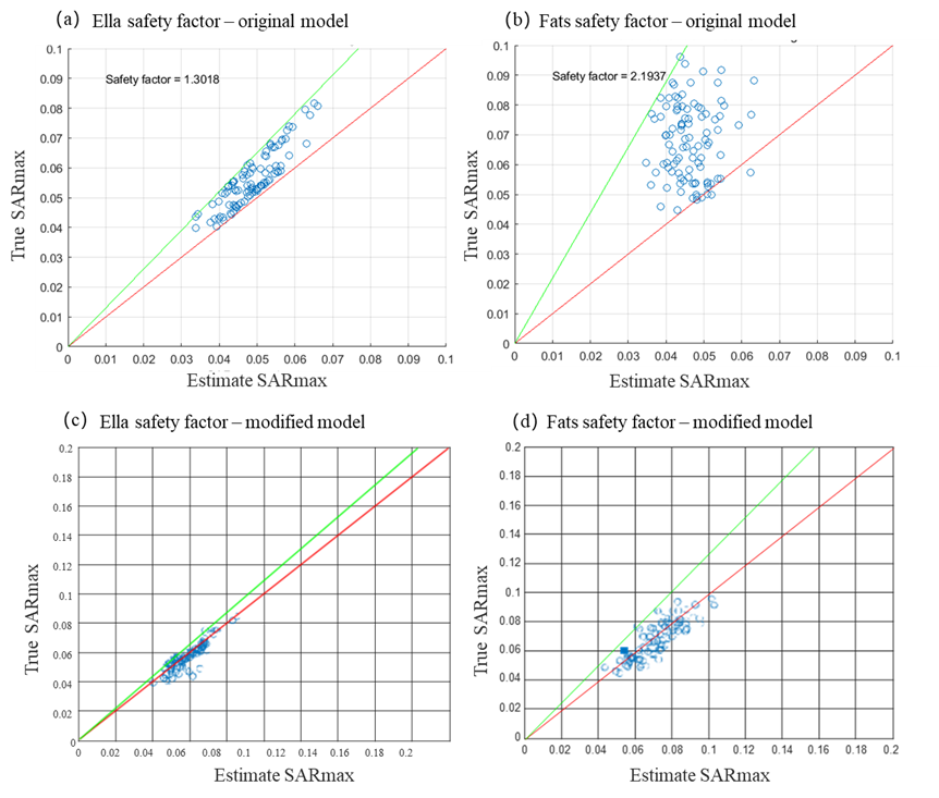
Improvement of field estimation accuracy is achieved with the above modifications. In the simulation model Fats with the largest body size, the estimated electric field agrees both in amplitude and phase patterns for all individual transmit channels as shown in Figure 2.Estimation accuracy of peak local SAR for random transmit weights is also improved. Figure 3 shows two typical human models, one female and one male. The safety factor estimated in both models decreased with the result of the modified deep-learning model.
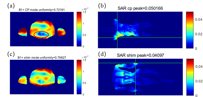
The results of joint optimization are shown in Figure 4. In the torso imaging scenario for model Fats, the dark spot in CP mode indicated by the red ellipse gains substantial improvement in the shim mode as well as the uniformity index value. The peak SAR was shifted from the underarm to the wrist with its value decreased to approximately 82% of the original one.
Discussions
With the modification in the training dataset preprocessing, the estimation accuracy of the model in the electric field distribution and peak local SAR value are both improved. Further research is needed to cover more human model cases. Realistic B1+ input data should be tested for the effectiveness of the current model.Conclusions
In this study, the feasibility and potential gain of the deep-learning model in UHF RF transmit calibration is evaluated. The method could provide an alternative to the current SAR prediction method based on the pre-defined simulation model.Acknowledgements
This work was supported in part by the National Key Research and Development Program of China, 2021YFF0501502, Project on Global Common Challenges of Chinese Academy of Sciences(No. 321GJHZ2022081GC), the Key Laboratory for Magnetic Resonance and Multimodality Imaging of Guangdong Province (2023B1212060052), the Funding Program of Shenzhen, China (RCYX20200714114735123), the Chinese Academy of Sciences Youth Innovation Promotion Association funded project (Y2021098), National Natural Science Foundation of China, U22A20344References
1, Barisano G, Sepehrband F, Ma S, et al. Clinical 7 t MRi: Are we there yet? A review about magnetic resonance imaging at ultra-high field. Br J Radiol. 2019;92(1094). doi:10.1259/bjr.20180492
2, Graesslin I, Homann H, Biederer S, et al. A specific absorption rate prediction concept for parallel transmission MR. Magn Reson Med. 2012;68(5):1664-1674. doi:10.1002/mrm.24138
Figures