3729
Heating Observed in EEG Amplifiers with Different EPI Slew Rates at 7T
R. Allen Waggoner1, Makoto Uji1, Kenichi Ueno1, Chisato Suzuki1, Fumiaki Sato1, Xuemei Li1, Hiroyuki Kamiguchi1, and Masako Tamaki1,2
1RIKEN Center for Brain Science, Wako-shi, Japan, 2RIKEN Cluster for Pioneering Research, Wako-shi, Japan
1RIKEN Center for Brain Science, Wako-shi, Japan, 2RIKEN Cluster for Pioneering Research, Wako-shi, Japan
Synopsis
Keywords: Multimodal, Multimodal, EEG, fMRI
Motivation: Evaluate the impact of EPI read-out gradient slew-rate on EEG amp heating, for EEG-fMRI experiments at 7T.
Goal(s): Identify slew-rates that will minimize heating of EEG amps, for EEG-fMRI experiments at 7T.
Approach: Test a range of EPI slew-rates while measuring the temperature of the EEG amp, during phantom tests at 7T.
Results: EPI read-out slew-rates of 120, 88, 61 and 46 T/m/s were tested and the slew-rates of 61 and 46 T/m/s were found to induce minimal heating of the EEG amp during EEG-fMRI experiments at 7T.
Impact: These results should help neuroscientists doing EEG-fMRI studies at 7T, to conduct their experiments without damaging the EEG amps.
Introduction
Whenever non-standard devices are introduced into the MRI environment, testing to ensure that the MRI environment will remain safe for both subjects and equipment is important. There are numerous studies combining EEG and fMRI at 3T and lower fields. These experiments demonstrate that with appropriate caution, EEG-fMRI studies can be performed safely at these field strengths. Moving to higher magnetic fields requires further safety testing. Jorge et al. reported that heating of the electrodes in the EEG cap were minimal, but significant heating of the RF amp was observed.[1] In this study we explore the impact of the slew rate of the EPI read-out gradient pulses, on heating induced in the EEG amps at 7T.Methods
A phantom EEG-MRI experiment was conducted on a 7 Tesla MRI system (Signa 7T, GE HealthCare). The Brian Products EEG cap was placed on a spherical water phantom, which was place in the 32-channel head coil (Nova Medical). The EEG amplifiers (BrainAmp MR Plus and BrainAmp ExG MR; Brain ProductsGmbH) were placed behind the RF coil with the EEG cap attached. A fiber optic temperature probe (Rugged Monitoring model L201), was placed between the EEG and ExG amps, as illustrated in Figure 1. Previous measurements had shown this to be the location of maximum heating. EPI images were acquired for at least 5 minutes, with read-out gradient slew rates of 120, 88, 61, and 46 T/m/s, while the temperature between the EEG amps was continually monitored with the fiber optic temperature probe.In addition, to evaluate the impact of changing the slew rate on image quality, EPI images were acquired with a human subject (30 yr. old male). Data sets were acquired with slew rates of 120 and 61 T/m/s. The data sets were acquired with an isotropic resolution of 1.5mm3, TE=28ms, multi-band factor=6, and TR = 800, 900ms respectively.
Results
Figure 2 shows the measured temperature changes, for the EPI scans with each slew rate. Both the slew rates of 120 and 80 T/m/s showed substantial heating of the EEG amps over the 5 min. test periods. For the slew rate of 61 T/m/s, only very modest heating was observed, with a maximum of 0.25 °C. For the slew rate of 46 T/m/s, the observed temperature change, never rose above baseline.Reducing the slew rate for 120 to 61 T/m/s, does not have a pronounced impact on image quality, as demonstrated by the images in Figure 3. Since the EPI sequence employed in this study uses ramp sampling, reducing the slew rate has only a minimal impact on the overall read-out window, therefore there is not a dramatic loss of image quality.
Conclusions
Previous studies have observed heating of the EEG amps during simultaneous EEG-fMRI at 7T. The results presented here replicated that observation. In addition, we demonstrated that by reducing the slew rate of the EPI read-out gradient, the heating of the EEG amps can be dramatically reduced or even eliminated. Further we demonstrated the use of the reduced slew rates do not lead to a noticeable loss of image quality. The results of the experiments presented here demonstrate that by reducing the slew rate of the read-out gradient, we can significantly reduce or even eliminate heating of the EEG amps. This demonstrates that EEG-fMRI can be done safely at 7T, without excessive wear on the EEG amps.Acknowledgements
No acknowledgement found.References
1. Jorge J, Grouiller F, Ipek Ö, et al. Simultaneous EEG–fMRI at ultra-high field: Artifact prevention and safety assessment. NeuroImage 2015;105:132-144.Figures
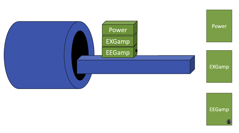
Figure 1. Schematic of the
positioning of the EEG amplifiers and power supply, behind the RF coil. A fiber optic temperature sensor was placed
between the EEGamp and theEXGamp, indicated by the 6, where previous
measurements have shown that EPI acquitions caused maximum heating.
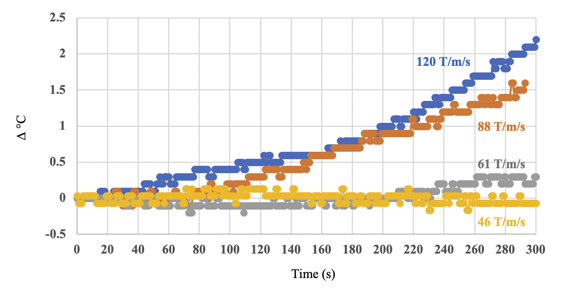
Figure 2. Observed
temperature rise at the location between the EEG amps indicated in Figure
1. Temperature rise was measured in four
phantom experiments, using four different slew rates for the EPI readout
gradients. After 5 minutes of continueous scanning, the slew rate of 120 T/m/s
caused an increase of more than 2 °C between tha amps. The slew rate of 61 T/m/s only caused a 0.25
°C temperature rise.
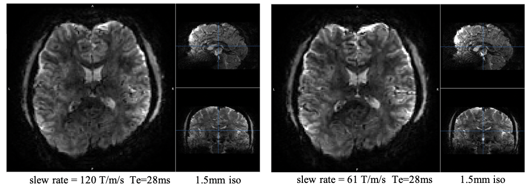
Figure 3. Example EPI volumes
acquired with the slew rates of 120 & 61 T/m/s. Ramp sampling minimized the impact on the overall acquisition time. These images show
that the image quality with the two slew rates is almost identical.
DOI: https://doi.org/10.58530/2024/3729