3721
In-vivo Validation of a Workflow to Predict Heating Around a Deep Brain Stimulation contacts1Center for Magnetic Resonance Research (CMRR), University of Minnesota, Minneapolis, MN, United States
Synopsis
Keywords: Safety, Safety
Motivation: To develop a reliable method to predict heating around deep brain stimulation implants as a safety assessment prior to scanning patients.
Goal(s): To investigate the accuracy of a heating prediction workflow in-vivo.
Approach: We surgically inserted a DBS electrode into swine brains and predicted the heating around DBS contacts, with and without perfusion. Our heating prediction workflow uses a new MR-based current measurement, proposed for this work, as well as quasi-static electromagnetic and thermal simulations to predict heating around the electrical contacts of DBS electrodes.
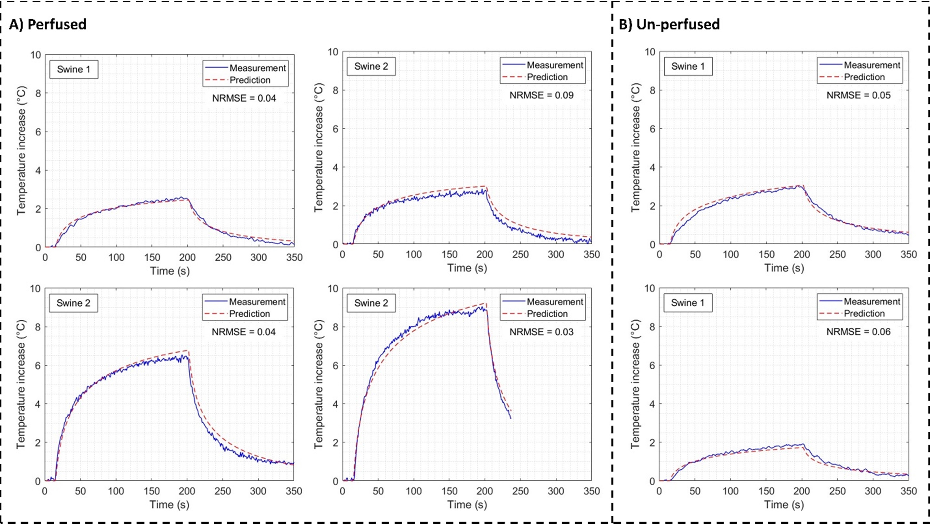
Results: The predicted temperature around electrical contacts agreed with the measurements (NRMSE ≤ 0.09).
Impact: Our workflow predicts heating around the electrical contacts in-vivo without complex modeling and simulations. The results demonstrate the reliability of the workflow to assess heating risk before scanning patients with DBS implants.
Introduction
Radiofrequency (RF) heating is a serious safety concern for patients with deep brain stimulation (DBS) implants during MRI scans1. A reliable and accurate heating prediction (pre-scan) is essential to assess the safety of patients with DBS implants2. Sadeghi-Tarakameh et al3 proposed a workflow where the heating around the electrical contacts of DBS electrodes was predicted using an MR-based current measurement4 method and quasi-static simulations3 (electromagnetic and thermal). In this study, we validated the workflow by predicting and measuring the heating around DBS contacts in swine, in-vivo and post-sacrifice. To generate RF heating and conduct imaging in the swine brain, a toroidal transmit-receive coil5 is used.Method
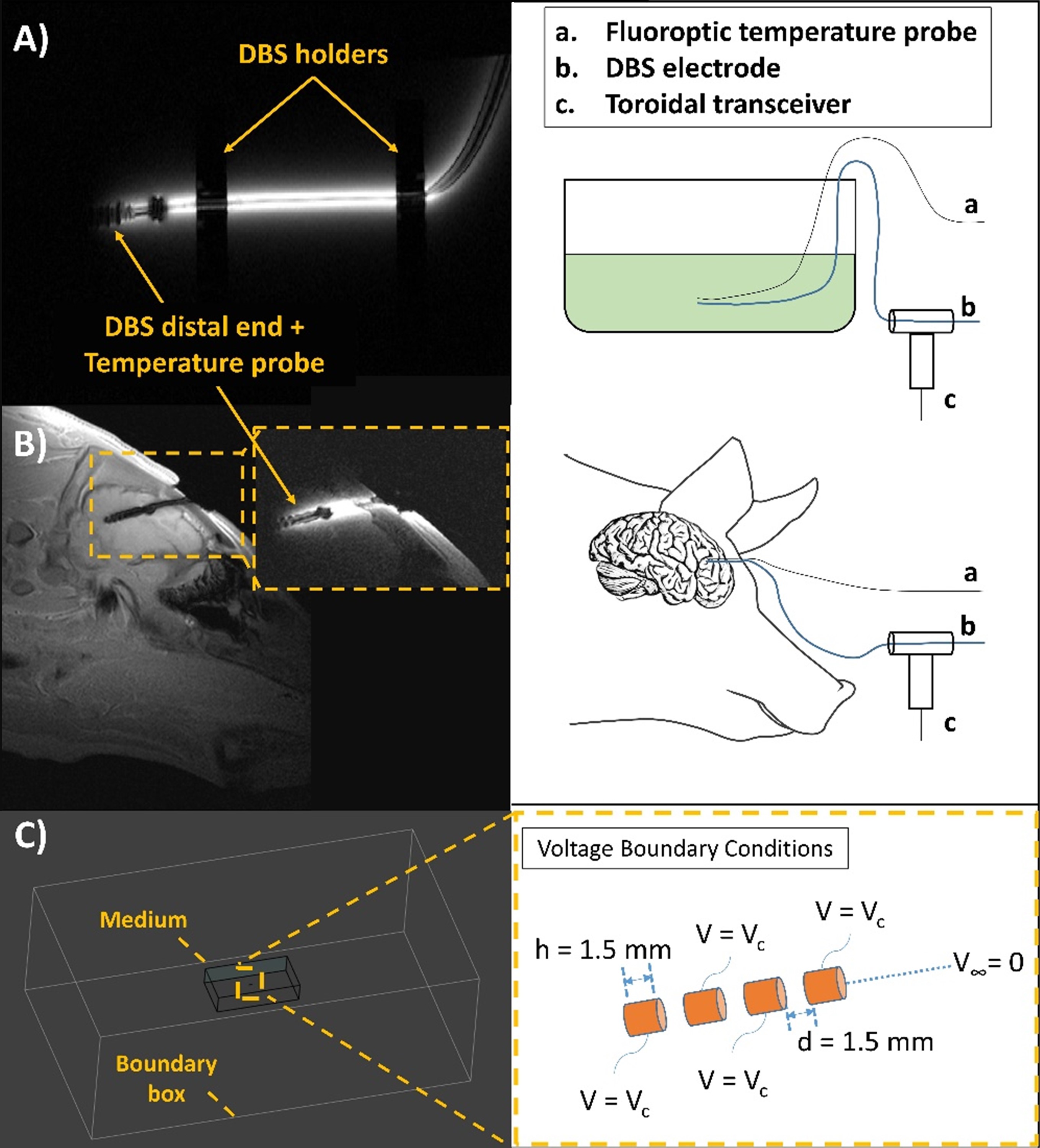
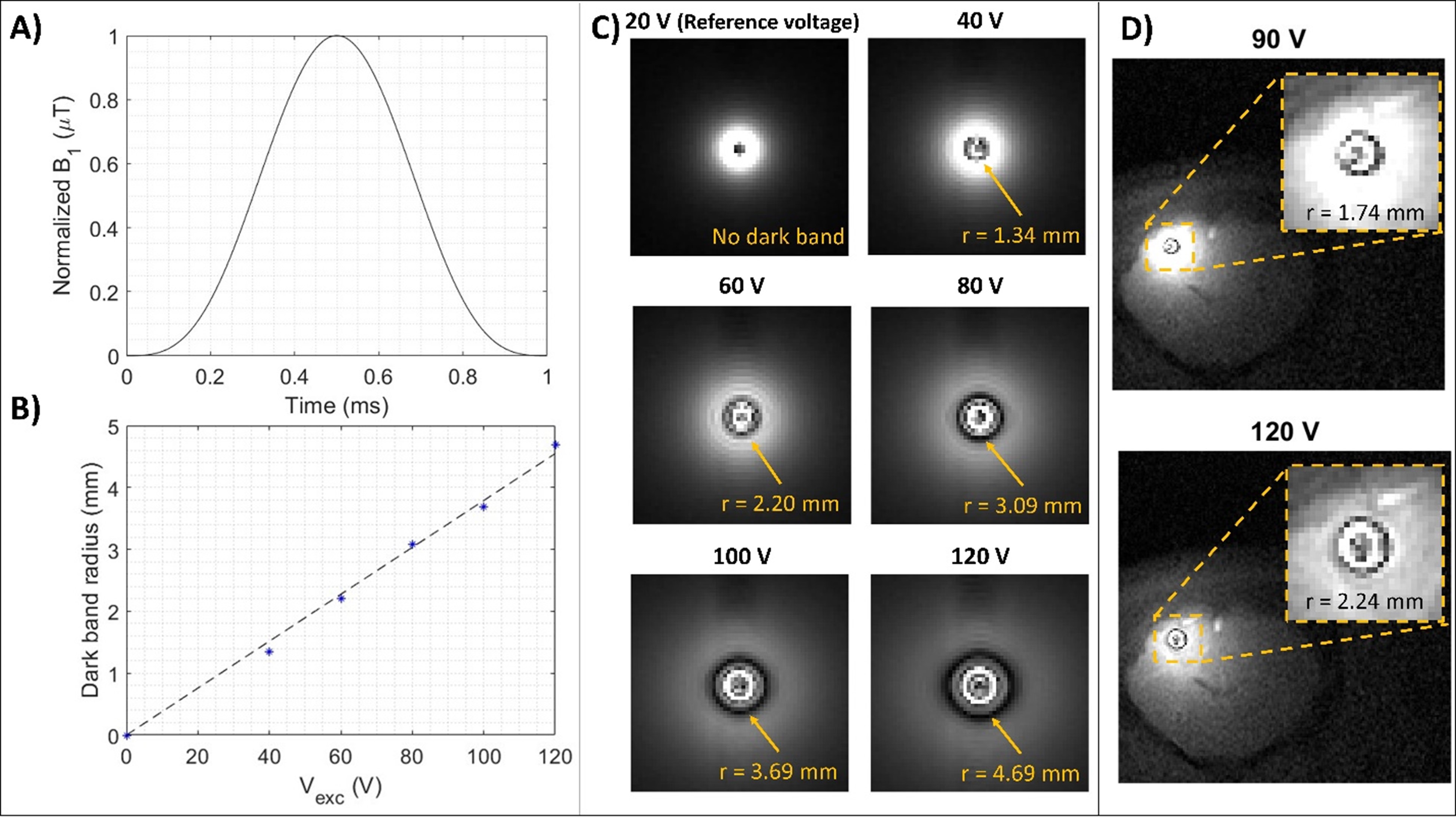
The experimental setups for calibrating electrode transimpedance, a parameter vital for temperature prediction, and heating experiments are shown in Figure 1. The proximal portion of a DBS (model 6173, Abbott Laboratories, Chicago, IL) was inserted through the cavity of a toroidal transceiver for imaging in a 3T Magnetom Prisma scanner (Siemens Healthineers, Erlangen, Germany). The electrode was exposed to a 3D-TSE sequence (TR/TE = 300/3.06 ms, echo train length = 15, FA = 150°, in-plane resolution = 0.5 mm, slice thickness = 1.25 mm) and fluoroptic temperature probes (Lumasense Technologies, CA, USA) measured the heating near the most distal contact.The equivalent transimpedance, RDBS, was calculated as the ratio of the voltage generated at the electrical contacts, Vc, and the induced current along the electrode shaft, Is. Vc was estimated in quasi-static EM and thermal simulations by determining the voltage boundary condition that simulated a heating curve matching the measured heating3. A new current measurement method was designed to measure Is. When the excitation voltage of a 2D-GRE sequence (TR/TE = 300/103 ms, in-plane resolution = 0.34 – 0.5 mm) surpasses the reference voltage, a circular dark band appears around the DBS electrode. This artifact corresponds to spins experiencing a 180° flip angle. The B1+ experienced by these spins is expressed as
$$B_{1,perVolt}^{+}=\frac{1}{{2}\gamma\cdot{V_{excit}}\int_{0}^{t_p}rf(t)dt}$$
where Vexc is the excitation voltage, rf(t) denotes the time-dependent variation of the RF pulse, and tp represents the pulse duration of the 2D-GRE sequence. Figure 2 shows the linear relationship between Vexc and rdark band. The current along the shaft can be calculated as follows:
$$I_s=\frac{B_{1,perVolt}^{+}\cdot4\pi{r_{darkband}}}{\mu}\cdot{V_{sequence}}$$
where rdark band is the radius of the dark band and Vsequence is the root-mean-square (RMS) voltage applied during the heating MRI sequence.
RDBS for average swine brain tissue was calibrated before the swine experiments, in a rectangular phantom filled with the following homogenous mixtures of different conductivity and permittivity values:
1) A hydroxyethylcellulose (HEC) gel (14 g/L), CuSO4 (0.25 g/L), and varying NaCl concentration.
2) A polyvinylpyrrolidone (PVP) gel (920 g/L), NaCl (40 g/L) and 0.5% agarose concentration.
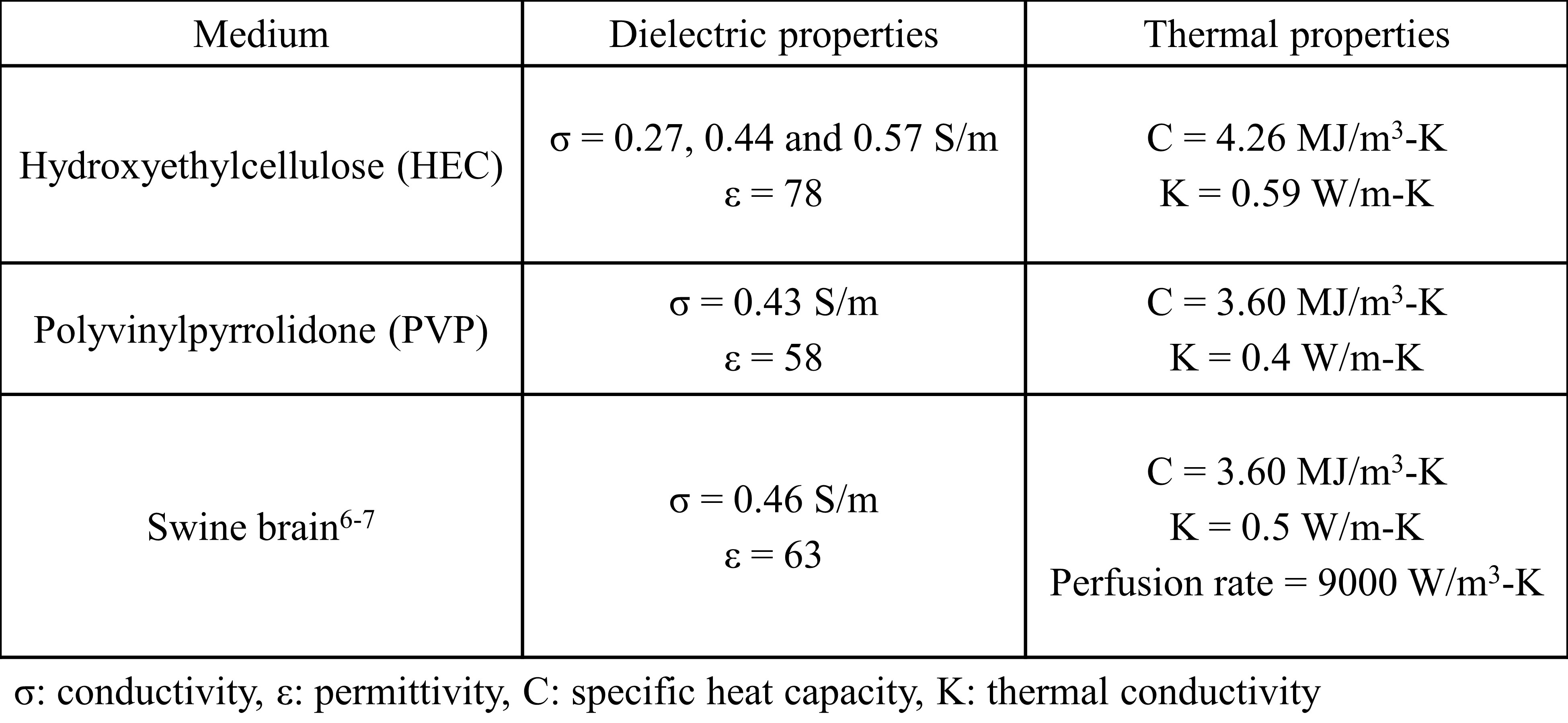
The values similar to the dielectric properties of grey and white matter were used in the calibration6. Table 1 summarizes the dielectric and thermal properties used in all experiments6-7. Two temperature probes were used for calibration in HEC. Conversely, a single temperature probe was used for calibration in PVP but the DBS insertion length was varied to investigate its effect on RDBS.
All swine experiments were compliant with a UMN IACUC-approved protocol. A 4-5 cm distal portion of the DBS electrode and a fluoroptic temperature probe were surgically implanted into the swine brain after anesthesia. Four in-vivo heating experiments were conducted with two animals, and two heating experiments were conducted post-sacrifice with one animal. The Vc for each heating experiment was calculated by multiplying the measured Is with the calibrated RDBS and used as an input to the EM and thermal simulations to predict heating around the contacts.
Results
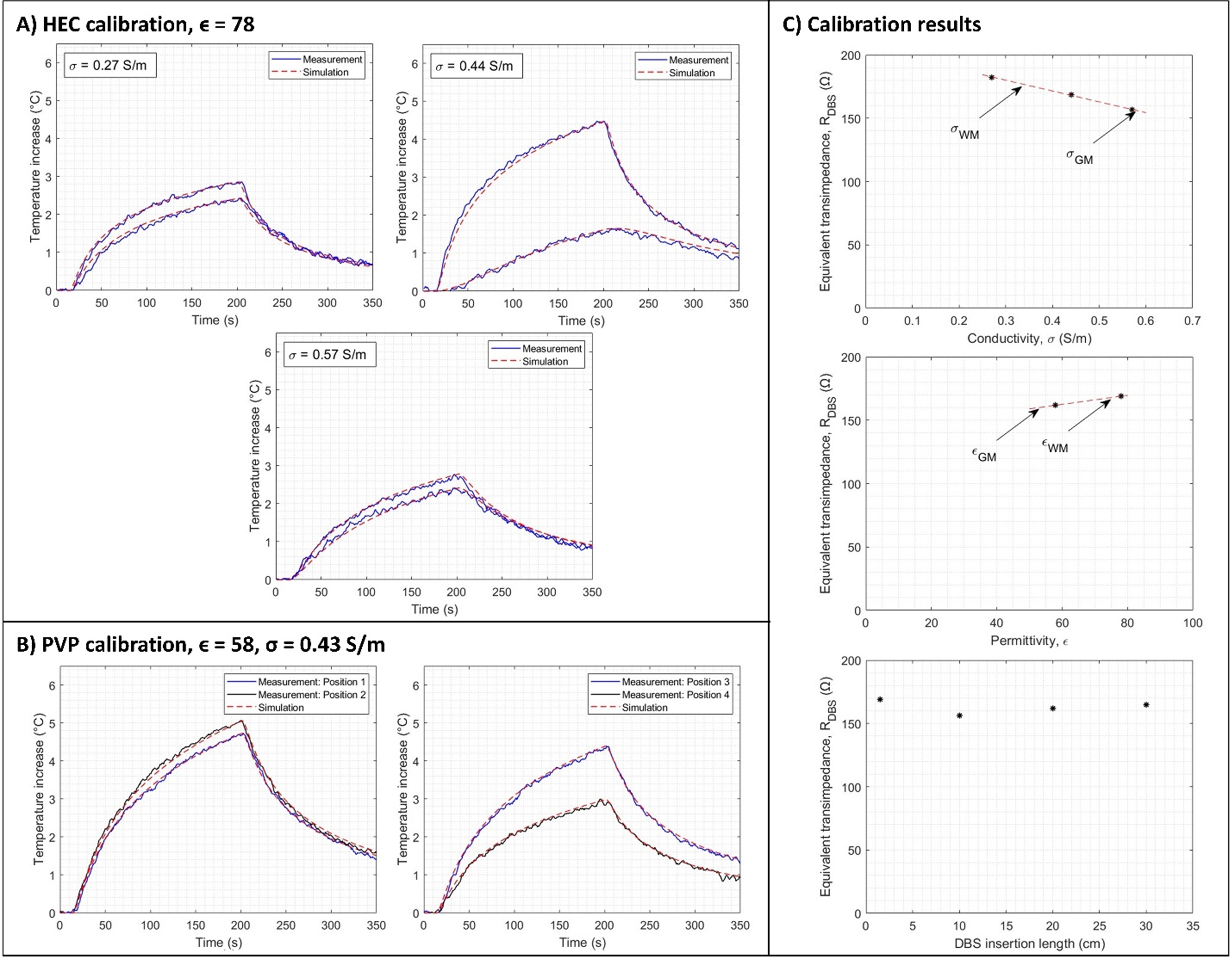
Figure 3 shows the temperature matching obtained with RDBS values calibrated for different dielectric properties. RDBS for swine brain tissue was calculated as 161 Ω using linear interpolation. Notably, Figure 3 also shows the minimal variation RDBS (< 3%) with respect to DBS insertion length in the toroidal transceiver. Figure 4 shows the good agreement between the heating predictions and measurements in swine with NRMSE ≤ 0.09.Discussion
Calibrating RDBS for average swine brain tissue demonstrated robust and accurate heating predictions that agreed with measured heating around DBS contacts for both in-vivo and post-sacrifice studies. In the future, comprehensive sensitivity analyses will be conducted to quantify a safety margin that accounts for different uncertainties in temperature prediction.Conclusion
A previously proposed temperature prediction workflow was validated with swine experiments. The equivalent transimpedance was calibrated for dielectric properties approximating the average swine brain tissue. The interpolated transimpedance resulted in heating predictions that agree with the measured heating with ≤ 0.09 NRMSE.Acknowledgements
This work was supported by NIBIB P41 EB027061, S10OD017974-01, and NINDS R01NS115180. We extend sincere appreciation to Lindsay Knoll, Rhianna Golden, Shannon Wilks, Dr. Whitney McGee, and Dr. Giuseppe Dell'Anna of Research Animal Resources (RAR) for their support and assistance during the swine experiments.References
- Henderson JM, Tkach J, Phillips M, Baker K, Shellock FG, Rezai AR. Permanent neurological deficit related to magnetic resonance imaging in a patient with implanted deep brain stimulation electrodes for Parkinson's disease: case report. Neurosurgery. 2005;57(5):E1063. doi:10.1227/01.neu.0000180810.16964.3e
- Boutet A, Chow CT, Narang K, et al. Improving Safety of MRI in Patients with Deep Brain Stimulation Devices. Radiology. 2020;296(2):250-262. doi:10.1148/radiol.2020192291
- Sadeghi-Tarakameh A, Zulkarnain NIH, He X, Atalar E, Harel N, Eryaman Y. A workflow for predicting temperature increase at the electrical contacts of deep brain stimulation electrodes undergoing MRI. Magn Reson Med. 2022;88(5):2311-2325. doi:10.1002/mrm.29375
- Eryaman Y, Kobayashi N, Moen S, et al. A simple geometric analysis method for measuring and mitigating RF induced currents on Deep Brain Stimulation leads by multichannel transmission/reception. Neuroimage. 2019;184:658-668. doi:10.1016/j.neuroimage.2018.09.072
- Etezadi-Amoli M, Stang P, Kerr A, Pauly J, Scott G. Interventional device visualization with toroidal transceiver and optically coupled current sensor for radiofrequency safety monitoring. Magn Reson Med. 2015;73(3):1315-1327. doi:10.1002/mrm.25187
- Andreuccetti D, Fossi R and Petrucci C: An Internet resource for the calculation of the dielectric properties of body tissues in the frequency range 10 Hz - 100 GHz. IFAC-CNR, Florence (Italy), 1997. Based on data published by C.Gabriel et al. in 1996. [Online]. Available: http://niremf.ifac.cnr.it/tissprop/
- Shrivastava D, Abosch A, Hughes J, et al. Heating induced near deep brain stimulation lead electrodes during magnetic resonance imaging with a 3 T transceive volume head coil. Phys Med Biol. 2012;57(17):5651-5665. doi:10.1088/0031-9155/57/17/5651
Figures