3718
Towards rapid T2 mapping using geometry in bSSFP imaging1Physics, University of Washington, Seattle, WA, United States, 2Radiology, University of British Columbia, Vancouver, BC, Canada, 3Radiology and Biomedical Imaging, University of California, San Francisco, San Francisco, CA, United States
Synopsis
Keywords: Quantitative Imaging, Quantitative Imaging, T2 quantitative mapping relaxometry, bSSFP, ellipse, elliptical signal model
Motivation: Quantitative T2 mapping is useful for indication of neuropathological states such as multiple sclerosis, but current methods are time consuming and can be challenging at low T2 values needed to indicate pathology.
Goal(s): To compute T2 maps efficiently and analytically.
Approach: T2 maps are computed by exploiting the geometry of the bSSFP signal. The algorithm is enhanced with added regularization, linearization, and solution combination, and is evaluated in simulations and phantom images.
Results: The improved algorithm demonstrates precision in simulations and a phantom, especially for lower T2 values. Reconstructed phantom T2 values were realistic, indicating its promise as a diagnostic tool.
Impact: Standard quantitative T2 mapping requires significant scan time for adequate fitting; here an analytical T2 mapping method is demonstrated that doesn’t require additional scan time beyond the associated artifact-free bSSFP images generated, inspiring further exploration.
Introduction
Quantitative T2 maps have the potential to serve as biomarkers for neuropathological states1. However, standard methods for T2 mapping employ fitting algorithms, demanding large scan times for adequate precision. Recently the SNR-efficient balanced steady state free precession (bSSFP) MRI sequence has been demodulated and unlocked using the geometric solution (GS) of the elliptical signal model (ESM)2. Tissue parameters have since been mapped by fitting the ESM using 6-10 phase-cycled acquisitions3. We recently proposed the 1st analytic T2 mapping method based on the ESM4; we now refine the technique and extend it to MR imaging. Regularization and linearization during the ellipse-to-circle transformation is added, and the multi-step optimal weighted average (OWA) is simplified to a single step, reducing solution noise, singularities, and complexity. Neurologically-relevant low T2 regions are investigated in simulations and a phantom, and compared with standard T2 mapping. Results indicate promise for a robust T2 mapping solution, warranting further study.Methods
Theory:The bSSFP ESM is defined in Eq.(1), where parameters $$$M_0,a,b$$$ are expressed in terms of the equilibrium magnetization $$$M_0$$$, flip angle $$$\alpha,TR$$$, and relaxation times $$$T_1$$$ and $$$T_2$$$. Here $$$\theta$$$ and $$$\phi$$$ respectively denote the off-resonant phase accumulation at $$$TR$$$ and $$$TE$$$, and $$$\psi$$$ denotes the RF phase cycling increment:$$\begin{aligned}I(\theta,\psi)&=M\frac{1-ae^{i(\theta+\psi)}}{1-b\cos(\theta+\psi)}e^{-i\phi},\quad(1)\\E_1:&=\exp(-TR/T_1)\\E_2:&=\exp(-TR/T_2)\\a:&=E_2\\M:&=\frac{M_0(1-E_1)\sin\alpha}{1-E_1\cos\alpha-E_2^2(E_1-\cos\alpha)}\\b:&=\frac{E_2(1-E_1)(1+\cos\alpha)}{1-E_1\cos\alpha-E_2^2(E_1-\cos\alpha)}\end{aligned}$$
Previous work transformed the elliptical bSSFP signal I to the “J-circle”:$$J(\theta,\psi)=I(\theta,\psi)(1-b\cos(\theta+\psi))\equiv\,M\,e^{-i\phi}(1-a\,e^{i(\theta+\psi)}),\quad(2)$$
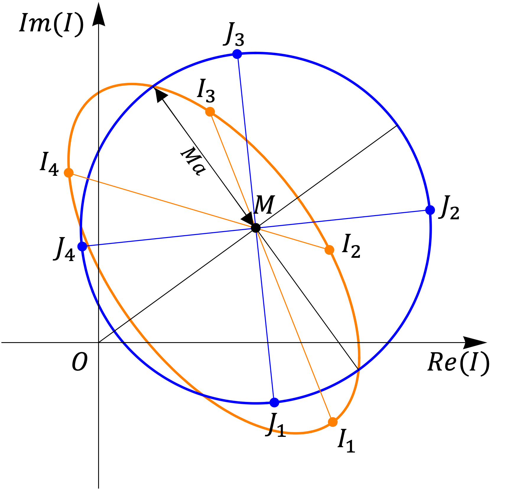
Figure 1 indicates that the J-circle’s radius is $$$Ma$$$, allowing direct calculation of T2. Here we regularize the $$$b\cos\theta,b\sin\theta$$$ transformation terms in a manner similar to GS linearization (LGS)2 by minimizing the regional energy $$$E$$$ of weighted solutions from the cross-point M:$$E=\sum_{\text{region}}\left|I_w-Me^{-i\phi}\right|^2,\quad(3)$$
where weighted solutions are formed from linear pairs of phase-cycled images $$$I_1,I_2,I_3,$$$ and $$$I_4$$$ via weights $$$w_{13},w_{24}$$$:$$\begin{aligned}I_{w13}&=I_1w_{13}+I_3(1-w_{13}),\\I_{w24}&=I_2w_{24}+I_4(1-w_{24}),\quad(4)\end{aligned}$$
$$$b\cos\theta,b\sin\theta$$$ can then be solved based on their direct relations to these weights:$$b\cos\theta=1-2w_{13},\quad\,b\sin\theta=2w_{24}-1,\quad(5)$$
and algebraic manipulation permits computation of $$$a$$$ and T2:$$a=\left(1-\frac{e^{i\phi}J(\theta,\psi)}{M}\right)e^{-i(\theta+\psi)},\quad(6)$$$$T_2=-TR/\log a$$
Four $$$T_2$$$ solutions corresponding to the four bSSFP acquisitions are combined using OWA:$$\text{sol}=\sum_j\,w_j\text{sol}_j,\quad\,w_j=\frac{1/V_j}{\sum_k1/V_k},\quad(7)$$
Here $$$V_j$$$ denotes the regional noise variance computed from the difference between solution $$$\text{sol}_j$$$ and the estimated ground truth.
Validation:
Four phase-cycled bSSFP images with $$$\psi=0^\circ,90^\circ,180^\circ,$$$ and $$$270^\circ$$$ respectively are generated for simulations and experimental MRI.
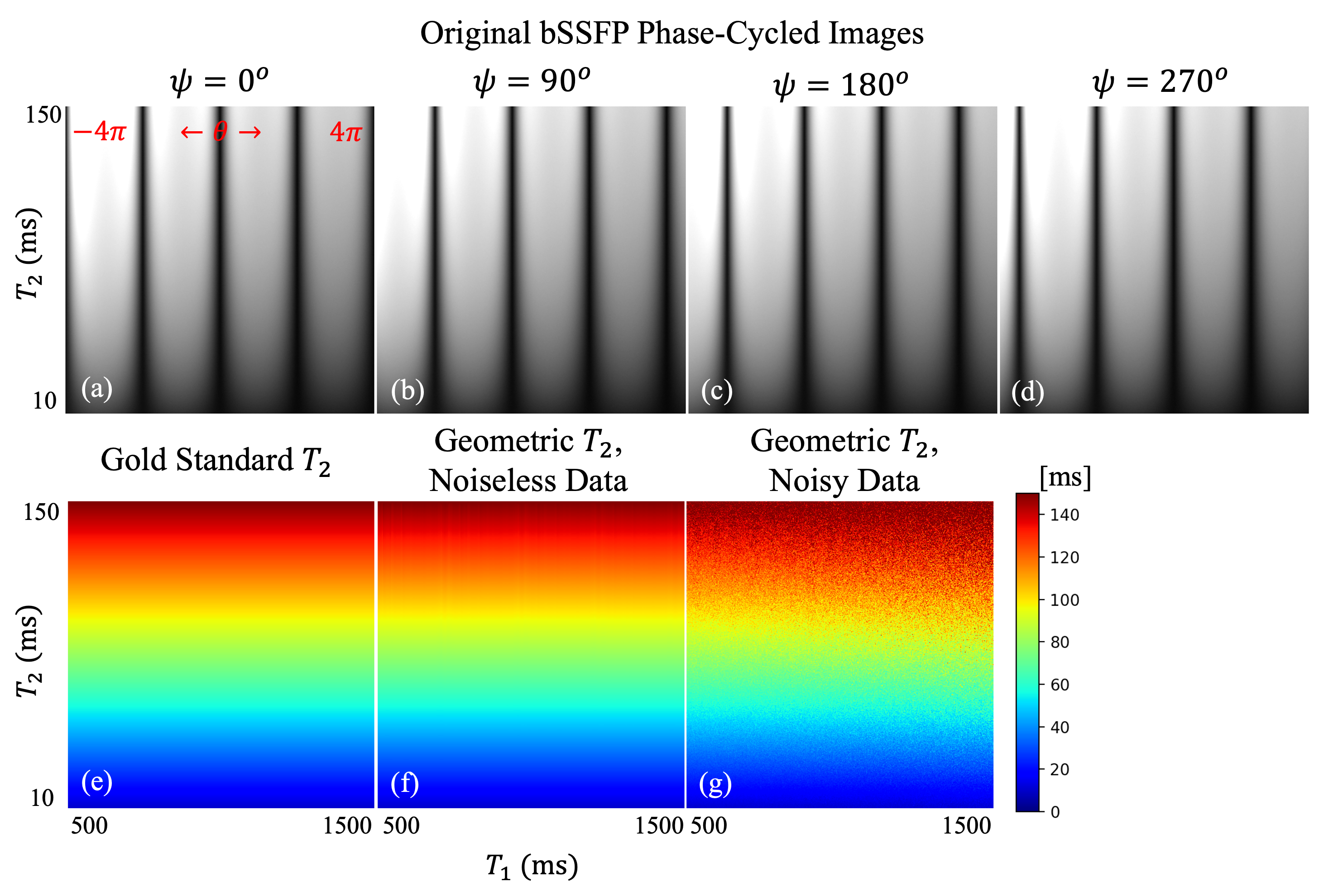
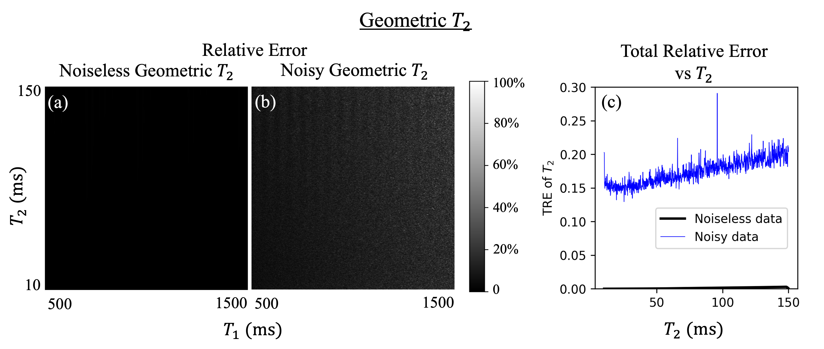
Simulations employed $$$\alpha=30^\circ$$$, $$$TR=10\,\text{ms}$$$, T2 varied vertically from $$$10\,\,\text{to}\,\,150\,\text{ms} $$$, and T1 (500 to 1500 ms) and $$$\theta(-4\pi\,\,\text{to}\,\,4\pi)$$$ varied horizontally. Bivariate Gaussian noise at 2% of the mean signal intensity is added. The OWA solution for T2 is then computed pixel-wise5 and the total relative error (TRE) evaluated6.
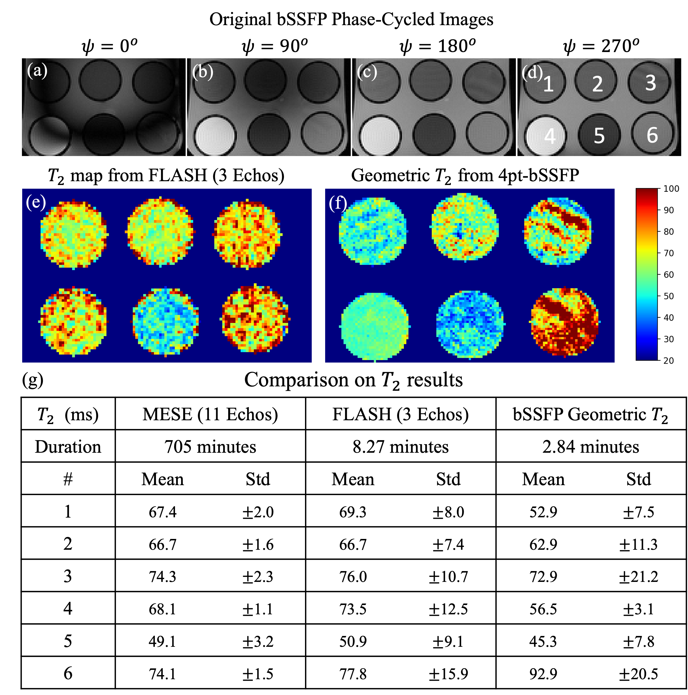
MRI experiments of a phantom containing vials of variable T2 surrounded by agar gel were performed on a 3T Vida scanner (Siemens Healthineers, Erlangen, Germany). 3D bSSFP data were acquired using $$$\alpha/TR/TE=45^\circ/4.9\,\text{ms}/2.4\,\text{ms}$$$, and required 2.8 minutes. Coil elements and T2 solutions were combined using OWA with the Sum of Squares of contributions as an estimate of ground truth. This T2 solution were compared with a T2 map obtained using a 3-echo FLASH sequence acquired with $$$\alpha/TR/TE1/TE2/TE3=12^\circ/276\,\text{ms}/10\,\text{ms}/35\,\text{ms}/55\,\text{ms}$$$ requiring 8.3 minutes, and a high fidelity multi-echo spin echo (MESE) sequence with $$$\alpha/TR=90^\circ/10\,\text{ms}$$$ and 11 echoes spanning $$$TE=5\,\text{ms}$$$ to $$$250\,\text{ms}$$$ that required near 12 hours of scan time.
Results
Figure 2 and Figure 3 show that our T2 solution is exact and accurate in noiseless scenarios, and exhibits realistic accuracy in low T2 regions with noise present, experiencing a slight increase in TRE as T2 increases.Figure 4 shows that 3-echo FLASH and 11-echo MESE T2-fitting sequences achieve similar results in the phantom, while geometric T2 deviates slightly but exhibits good precision, especially for lower T2 values.
Discussion
An analytic T2 map method is demonstrated and evaluated in simulation and phantom images. Simulations show that the exact nature of the T2 computation in noiseless scenarios is leveraged to yield faithful results in noise. Phantom results demonstrate the precision and efficiency of our T2 solution that required 2.8 minutes (compared with 11-echo/3-echo fitting methods that required 705/8.3 minutes respectively). This inspires future utilization of the method given that the geometric bSSFP approach also generates artifact-free images1. The quality for low T2 regions is especially promising given that classical fitting methods are constrained by achievable echo times in their ability to characterize this parameter range.Conclusion
We extend analytical T2 mapping using bSSFP to real data, showing its capacity for characterizing low T2 values. Its good accuracy and rapid acquisition time suggests its suitability in clinical quantitative tasks.Acknowledgements
No acknowledgement found.References
[1] Bottomley PA, Hardy CJ, Argersinger RE, Allen-Moore G. A review of 1H nuclear magnetic resonance relaxation in pathology: are T1 and T2 diagnostic? Med Phys 1987;14(1):1–37.
[2] Xiang Q-S, Hoff MN. Banding Artifact Removal for bSSFP Imaging with an Elliptical Signal Model. Magn Reson Med, 2014; 71(3):927:933, doi: 10.1002/mrm.25098.
[3] Shcherbakova, Y., van den Berg, C.A.T., Moonen, C.T.W. and Bartels, L.W. (2018), PLANET: An ellipse fitting approach for simultaneous T1 and T2 mapping using phase-cycled balanced steady-state free precession. Magn. Reson. Med, 79: 711-722.
[4] Dong Y, Xiang Q-S, Hoff MN. Towards analytical T2 mapping using the bSSFP elliptical signal model. In: Proc. ISMRM, Vancouver, 2021, Abstr# 4709.
[5] Xiang Q-S, Henkelman RM. Weighted Average and Its application in Ghost Artifact Reduction. In: 9th SMRI proceedings. ; 1991. p. 222.
[6] Chang Z, Xiang Q-S. Highly accelerated MRI by skipped phase encoding and edge deghosting with array coil enhancement (SPEEDACE). Med Phys 2006;33:3758.
Figures