3717
Optimizing and Evaluating Fast 3D MulTiPlex (MTP) Imaging for Infant Brain Imaging1School of Biomedical Engineering, ShanghaiTech University, Shanghai, China, 2United Imaging Healthcare, Shanghai, China, 3United Imaging Healthcare America, Houston, TX, United States, 4United Imaging Intelligence, Shanghai, China, 5Shanghai Clinical Research and Trial Center, Shanghai, China
Synopsis
Keywords: Quantitative Imaging, Quantitative Imaging, Multiparametric Imaging, Infant Brain Imaging
Motivation: The novel MulTiPlex (MTP) technique can generate high-resolution, multi-contrast, qualitative/quantitative images, but its reliability and potential for infant study have not been investigated.
Goal(s): To optimize MTP parameters and evaluate its reliability and validity for infant study.
Approach: n this study, we systematically assessed MTP’s test-retest reliability, compared MTP with varied accelerating rates using AI-assisted Compressed Sensing (ACS), followed by a comprehensive appraisal of its applicability in infant brain imaging, with both qualitative and quantitative methods.
Results: The findings indicate that MTP exhibits commendable reliability and holds significant promise for building large infant MRI database.
Impact: MTP generates assorted images with varied spectrum of contrasts and precious quantitative images that take hours to acquire conventionally. Our established MTP protocol combines these merits with fast acquisition, warranting its significant usefulness in infant development and clinical studies.
Introduction
The MulTiPlex (MTP)1 technique features a dual-TR, dual-flip angle (FA), and multi-echo design. Within each TR, a number of gradient echoes are acquired, providing different levels of susceptibility weighting effects. In total, a single MTP scan generates multiple sets of qualitative and quantitative images, including aT1w2, QSM, T1, T2*, PD maps, etc. Since these attribute maps provide valuable insights into different facets of the developing brain, MTP holds great potential in infant brain research. While quantitative accuracy of MTP has been well-established, its performance regarding reproducibility and applicability in infant brain imaging remains unexplored, partially due to its long acquisition time. Recently, an innovative deep learning acceleration technique, AI-assisted Compressed Sensing (ACS)3,4 was used to accelerate MTP scan for more practical uses. As such, the acceleration factor of MTP should be optimized to balance scanning time and image quality, before its wide application to infant development and disease studies.Methods
Ten healthy volunteers (ages 19-24) were scanned with a 64-channel head coil using a research-dedicated 3.0T scanner (uMR890, United Imaging) equipped with MTP and ACS. Each volunteer was scanned twice on two separate days seven days apart with MTP plus ACS: TR=9.09/38.61ms, TE= 3.64/9.65/13.29/19.3/22.94/28.95ms, flip angles=4o/16o, voxel size=0.8×0.8×1.6 mm3, field of view=230×190×144 mm3, and acceleration factor=4. The subjects were also scanned with T1w FSP images (voxel size=0.8×0.8×0.8 mm3) for brain region-of-interest (68 cortical and 18 subcortical ROIs according to the DK atlas and Freesurfer aseg atlas5) segmentation6,7 and registration. After T1w images were registered to aT1w images, mean T1, T2*, PD, and QSM values were extracted from the ROIs and compared using intra-class correlation coefficients (ICC) to evaluate the measurement reliability.To better validate the ACS-accelerated MTP, we further compared multiple MTP images from the same subject acquired with different ACS acceleration factors: 4.4, 5, 5.5, and 6. For qualitative evaluation, the aT1w images were compared. For quantitative assessment, various imaging attributes values in the subcortical ROIs were extracted in the same way as mentioned above and compared. After optimized ACS acceleration rate was chosen, several infants and children (ages 0-5) were scanned using MTP with the optimized ACS acceleration rate (4.4) and all the attributes maps were evaluated by experienced radiologists.
Results
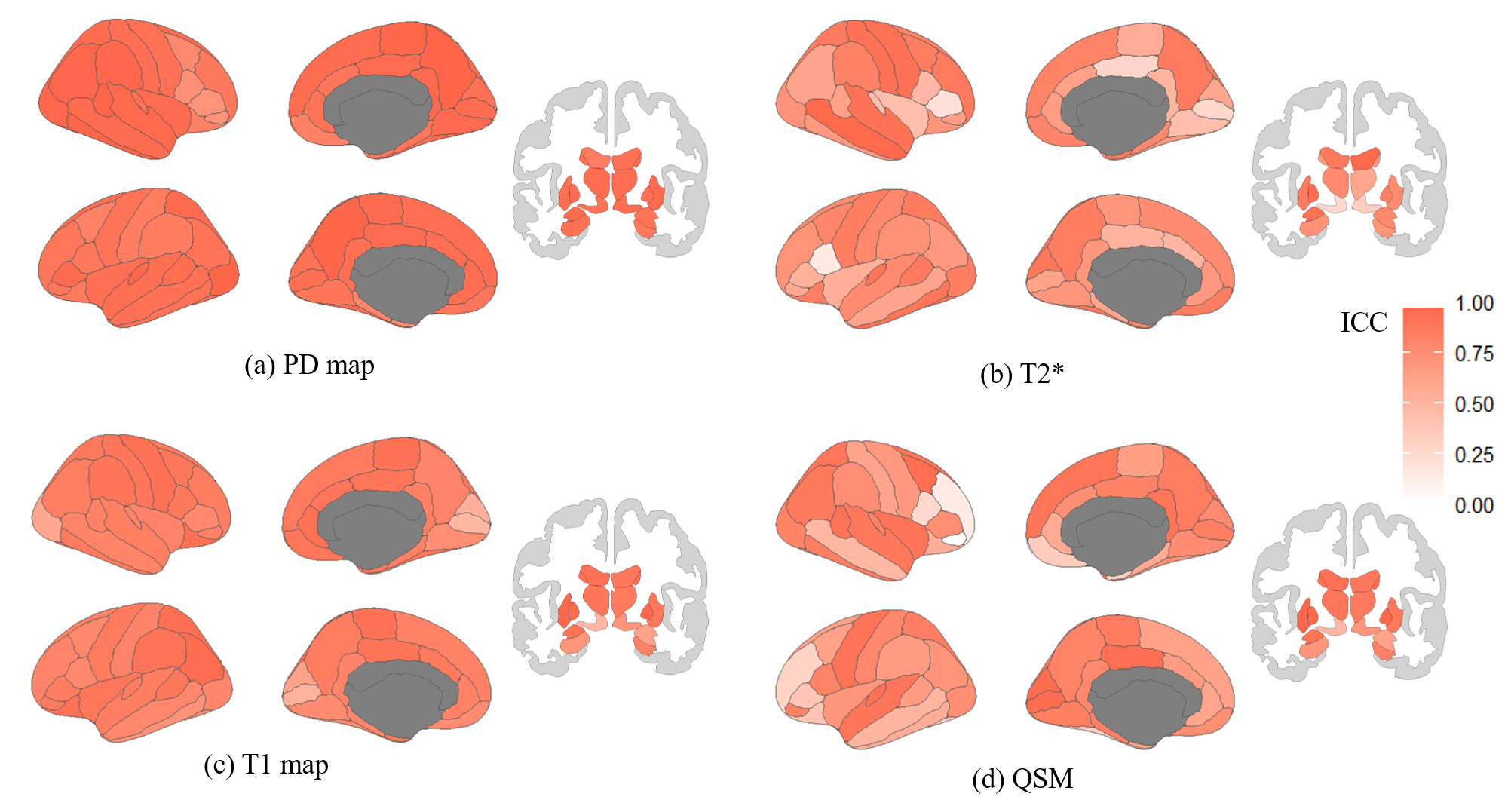
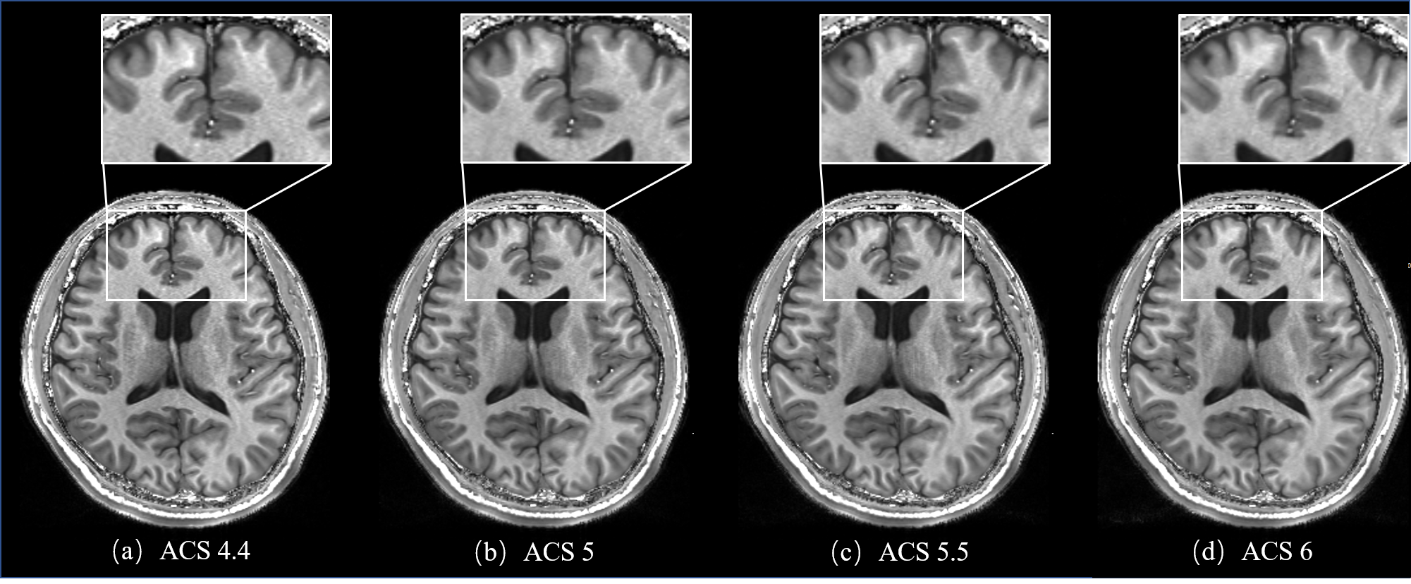
Good reliability (ICC≥0.75) was observed in 94.1%, 44.1%, 89.7%, and 47.0% of the cortical ROIs for PD map, T2* , T1 map and QSM respectively (Fig. 1). Poor reliability (ICC<0.4) was observed only in 4 and 11 out of 68 ROIs for T2* and QSM. For subcortical regions, ICC below 0.4 was found only in 4 subcortical ROIs for T2*.Regarding aT1w images, there was no notable difference observed among them. However, with ACS rate increasing, aT1w images tend to have increased smoothness (Fig. 2).
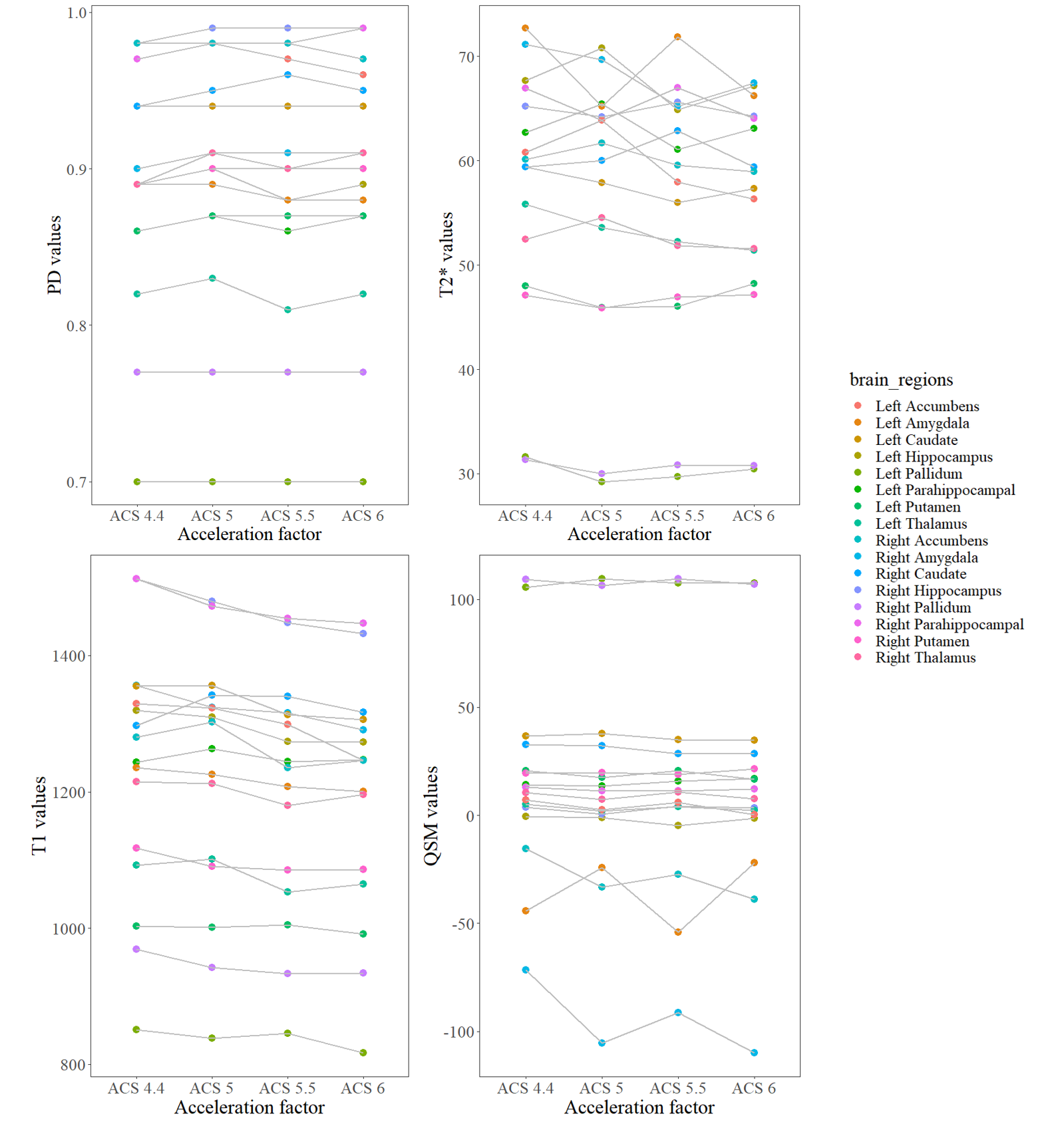
Fig. 3 illustrates that MTP values exhibit little variation across various acceleration factors for most of the brain regions, however, T2* and QSM values display fluctuations in several regions as the acceleration factor changes.
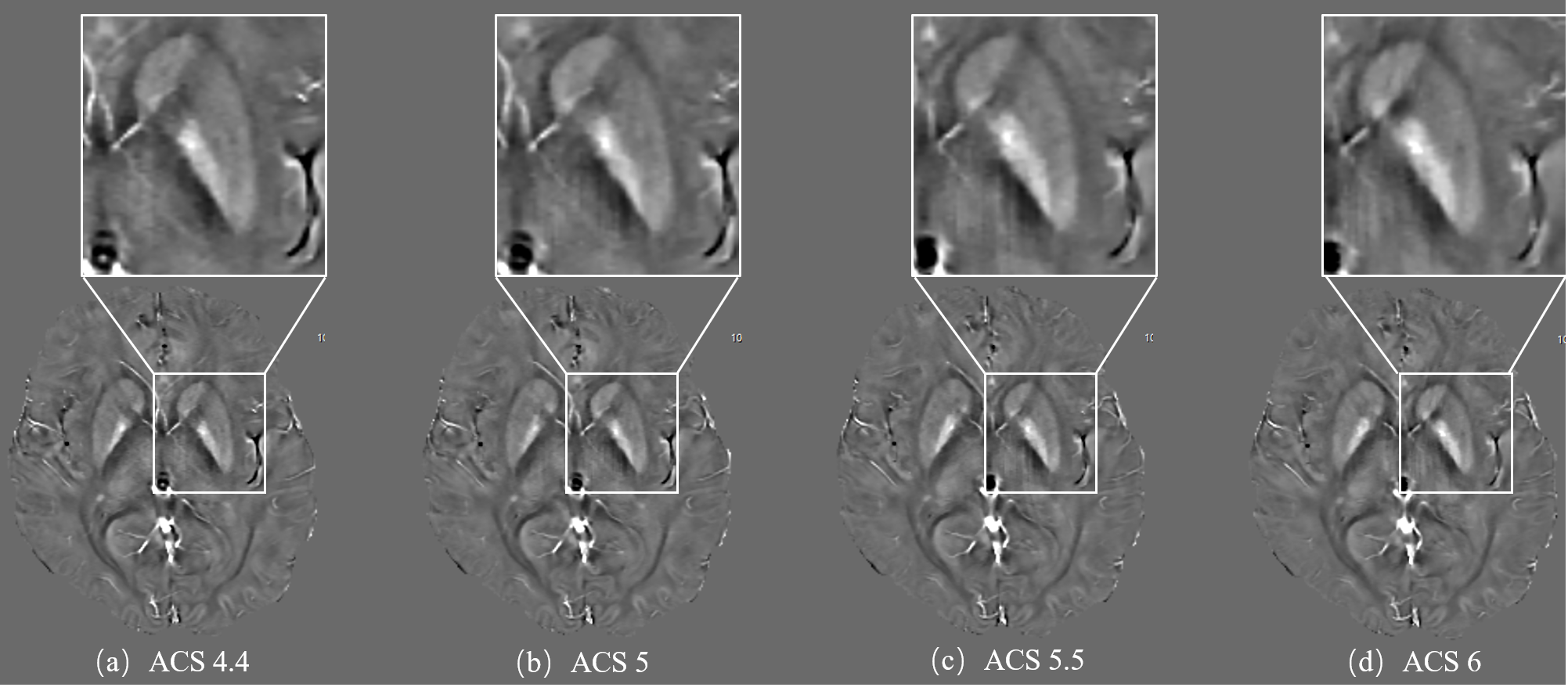
Noticeable ringing artifacts appeared in the highly accelerated images such as ACS6 (Fig. 4b-d), which is considered as a negative effect caused by the relatively high acceleration factor considering the current resolution settings, i.e. higher acceleration performs better with higher resolution. No such artifacts were visible in images with ACS4.4 (Fig. 4a).
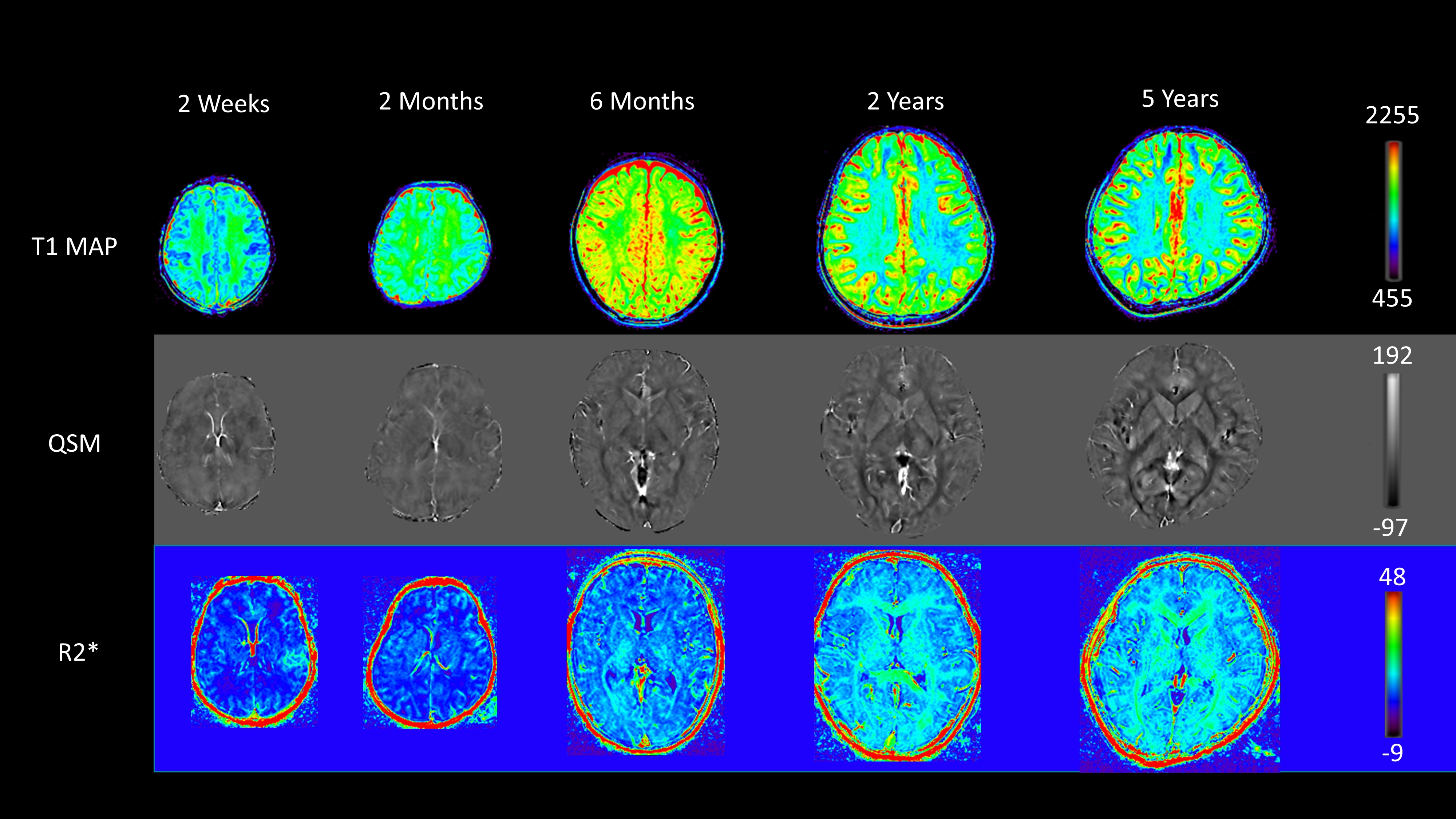
Fig. 5 presents the first application of MTP with ACS for the acquisition of multi-faceted infant brain images. All of the MTP images have very good quality and reflect age-specific patterns that are distinct from adults.
Discussion
All attribute maps of MTP exhibit fair-to-good test-retest reliability. For QSM, the values may be affected by results of brain extraction in the preprocessing steps8. The difference in positioning, which affects the B0 field, may also contribute to the inconsistencies. Besides qualitative observations, quantitative analysis indicated no discernible trends in MTP image variations as acceleration factors are adjusted. The ringing artifacts in QSM images, particularly at higher factors, are likely attributed to the overly under-sampled k-space center. By considering acquisition time, artifact levels, sharpness, and robustness together, we recommended ACS acceleration factor of 4.4. The implementation of optimized MTP on infant scans for early developmental study was considered successful.Conclusion
This study validated and optimized the integration of MTP with ACS, and initiated its application on infant brain imaging. Compared to conventional MR imaging, MTP offers multiple qualitative and quantitative maps with acceptable SNR, reliability, accuracy, and robustness without sacrificing acquisition/reconstruction efficiency. The flexible design of MTP signal acquisition allows specific contrast mechanisms to cater to the various research needs. With optimized parameters, promising future of MTP in infant brain imaging is warranted.Acknowledgements
This work is partially supported by the STI 2030—Major Project (2022ZD0209000, 2021ZD0200516), Shanghai Pilot Program for Basic Research—Chinese Academy of Science, Shanghai Branch (JCYJ-SHFY-2022-014), Open Research Fund Program of National Innovation Center for Advanced Medical Devices (NMED2021ZD-01-001), Shenzhen Science and Technology Program (No. KCXFZ20211020163408012), and Shanghai Pujiang Program (No. 21PJ1421400).References
1. Y. Ye, J. Lyu, Y. Hu, Z. Zhang, J. Xu, and W. Zhang, "MULTI‐parametric MR imaging with fLEXible design (MULTIPLEX)," Magnetic Resonance in Medicine, vol. 87, no. 2, pp. 658-673, 2022.
2. Y. Ye et al., "Augmented T1‐weighted steady state magnetic resonance imaging," NMR in Biomedicine, vol. 35, no. 8, p. e4729, 2022.
3. Eric Z. Chen et al. "Accelerating 3D MULTIPLEX MRI Reconstruction with Deep Learning," Joint Annual Meeting ISMRM-ESMRMB & ISMRT Annual Meeting, 2021
4. R. Zhai et al., "Intelligent incorporation of AI with model constraints for MRI acceleration," in Proceedings of the 29th Annual Meeting of ISMRM, 2021.
5. B. Fischl, "FreeSurfer," Neuroimage, vol. 62, no. 2, pp. 774-781, 2012.
6. J. Wei, F. Shi, Z. Cui, Y. Pan, Y. Xia, and D. Shen, "Consistent Segmentation of Longitudinal Brain MR Images with Spatio-Temporal Constrained Networks," in International Conference on Medical Image Computing and Computer-Assisted Intervention, 2021: Springer, pp. 89-98.
7. L. Wang et al., "LINKS: Learning-based multi-source IntegratioN frameworK for Segmentation of infant brain images," NeuroImage, vol. 108, pp. 160-172, 2015.
8. Y. Ye et al., "Dynamic streaking artifact regularization for QSM," in Proceedings of the 27th Scientific Meeting, International Society for Magnetic Resonance in Medicine, 2019: ISMRSM Montreal.
Figures