3716
Ultra-Fast Submillimeter Whole Brain QSM Using Highly Accelerated 3D Echo-Planar Imaging with a Complex Convolutional Neural Network1Department of Neurology, Cedars-Sinai Medical Center, Los Angeles, CA, United States, 2Biomedical Imaging Research Institute, Cedars-Sinai Medical Center, Los Angeles, CA, United States, 3Siemens Healthineers, Brisbane, Australia, 4Siemens Healthineers, CA, United States, 5Department of Imaging, Cedars-Sinai Medical Center, Los Angeles, CA, United States
Synopsis
Keywords: Quantitative Imaging, Quantitative Susceptibility mapping
Motivation: To restore image quality of highly accelerated QSM for data interpretation.
Goal(s): To evaluate the feasibility of a denoising convolutional neural network for generating high-quality submillimeter isotropic QSM acquired in 2-3 minutes at 3-Tesla.
Approach: A previously developed network for denoising complex-valued MRI data was applied to accelerated 3D-EPI scans (TA: 2min and 3min). Image quality was evaluated and QSM values obtained with and without denoising were compared against reference non-accelerated non-denoised 3D-EPI (TA: 6min).
Results: SNR and structural measures demonstrated improved image quality when denoising the accelerated data. Similar QSM values were observed for both highly accelerated denoised 3D-EPI and reference 3D-EPI.
Impact: Our approach for denoising complex-valued 3D-EPI brain images shows the feasibility of producing high-quality, whole-brain, submillimeter isotropic QSM acquired in 2-3 minutes at 3-Tesla, facilitating its clinical adoption.
Introduction
Three-dimensional echo planar imaging (3D-EPI) enables isotropic submillimeter imaging of the whole brain at 3T in a few minutes.1 The use of 3D-EPI has already been proposed for quantitative susceptibility mapping (QSM), i.e., a post-processing technique to non-invasively quantify the magnetic susceptibility properties in brain tissues.2 Recent developments combining the 3D-EPI sequence with Controlled Aliasing In Parallel Imaging Results In Higher Acceleration (CAIPIRINHA) were demonstrated to efficiently reduce the scan time down to 2 minutes.3 However, this reduction in scan time is achieved at the cost of reduced signal-to-noise ratio (SNR). To address this issue, we recently developed a deep learning-based complex-valued image denoiser (CDnCNN)4,5 to restore SNR while maintaining high structural fidelity. In this study, we evaluated for the first time the feasibility of generating ultra-fast submillimeter isotropic QSM from highly accelerated 3D-EPI brain images after denoising with our CDnCNN model.Methods
We previously developed a Complex-valued Denoising Convolutional Neural Network (CDnCNN) to denoise MRI data in the spatial domain as an extension of the Denoising Convolutional Neural Network (DnCNN).6 In our CDnCNN model, we modified the convolutional layers and rectified linear unit activation functions to process complex values, allowing for both magnitude and phase images to be reconstructed after denoising. In this study, we applied it to denoise an MRI dataset of 5 subjects scanned prospectively and evaluated the subsequently reconstructed QSM images (Figure 1). These 5 subjects underwent 3T imaging at Cedars-Sinai Medical Center using a 3D-EPI acquisition without and with CAIPIRINHA undersampling applied at two acceleration factors: R=3 (TA: ~3 minutes) and R=4 (TA: ~2 minutes). Main sequence parameters were: TE = 35ms, TR = 64ms, matrix size = [384 x 312 x 256], voxel size = 0.65 x 0.65 x 0.65 mm, and bandwidth = 394Hz. After image acquisition, image denoising was performed offline on the highly accelerated 3D-EPI data (R=3 and R=4). We first reconstructed the denoised T2*-weighted magnitude and phase images, and then generated QSM from these denoised images using the total generalized variation QSM (TGV-QSM) method.2To evaluate the quality of the denoising, we compared the peak signal-to-noise ratio (PSNR) and structural similarity index measure (SSIM) of the T2*-weighted magnitude and phase images with and without denoising. For evaluating the effect of denoising on QSM, we compared regions of interest (ROIs) calculated on QSM maps obtained from T2*-weighted magnitude and phase images with and without denoising. For this purpose, tissue segmentation was performed on T1 MPRAGE scans acquired during the same session and registered to the 3D-EPI scans using the FreeSurfer7 software. Additionally, automated lesion segmentation was performed on registered T2-FLAIR and registered T1-MPRAGE using the MIMoSA algorithm.8,9 Statistical analysis was performed using paired t-tests to compare QSM values of ROIs with and without denoising against reference values obtained for non-accelerated non-denoised images.
Results
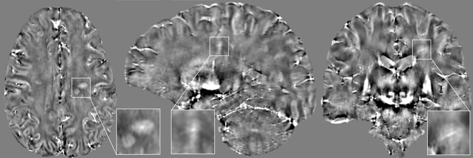
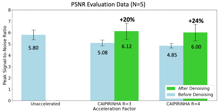
The average PSNR values substantially increased when denoising for both acceleration factors: 20% increase for R=3 and 24% increase for R=4 when comparing the same images with and without denoising; slightly higher PSNR values were also observed when comparing denoised R=3 (6.12) and R=4 (6.00) against the reference non-accelerated non-denoised images (5.80) (Figure 2). SSIM values measured remained high for both acceleration factors after denoising (mean ± SD: 0.888 ± 0.011 for R=3; 0.878 ± 0.010 for R=4). Statistical testing showed that there were no statistically significant differences between QSM values with or without acceleration and denoising (Figure 3). Finally, representative examples of T2*-weighted magnitude, phase, and QSM images of the 3D-EPI scans acquired with and without acceleration are shown with and without denoising using our CDnCNN model (Figure 4). A multiplanar view is also shown for a submillimeter isotropic QSM generated using 2-minute denoised 3D-EPI with CAPIRINHA acceleration (R=4) (Figure 5).Discussion
Our CDnCNN model was able to efficiently denoise the highly accelerated 3D-EPI scans while preserving anatomical and pathological details on all three susceptibility-based imaging modalities. More importantly, denoising with our model had no effects on the QSM values computed for the different brain ROIs and were consistent with those generated from the reference non-accelerated non-denoised scans. We did observe a slight blurring in the sagittal and coronal planes of the QSM images after denoising which we are currently investigating (Figure 5).Conclusion
We showed, for the first time, the feasibility of generating ultra-fast submillimeter isotropic QSM from highly accelerated 3D-EPI brain images after denoising with our CDnCNN model. Future work will be aimed at developing a deep learning based super-resolution method to remedy the slight blurring observed on QSM after denoising.Acknowledgements
We acknowledge the National MS Society (NMSS) RG-2110-38526, National Institutes of Neurological Disorder and Stroke (NINDS) 1U01NS116776-01, and Department of Defense, and Erwin Rautenberg Foundation for research support.References
1. Sati P, Thomasson DM, et al. Rapid, high-resolution, whole-brain, susceptibility-based MRI of multiple sclerosis. Multiple Sclerosis Journal. vol. 20. October 2014.
2. Langkammer C, Bredies K, Poser B, et al. Fast Quantitative Susceptibility Mapping using 3D EPI and Total Generalized Variation. NeuroImage. vol. 111. May 2015.
3. Nair S, et al. Feasibility of ultra-fast, high-resolution, T2*-weighted brain imaging using 3D echo-planar imaging and CAIPIRINHA [abstract]. 2023 ISMRM & ISMRT Annual Meeting & Exhibition. Abstract no. 2838.
4. Quah B, Nair S, et al. Denoising highly accelerated T2* weighted brain MRI using a deep learning convolutional neural network [abstract]. 2023 ISMRM & ISMRT Annual Meeting & Exhibition. Abstract no. 4188.
5. Quah B, Nair S, et al. Complex Convolutional Neural Networks for Denoising Accelerated Submillimeter Magnetic Susceptibility Brain MRI [abstract]. 109th Scientific Assembly and Annual Meeting of Radiological Society of North America 2023. Abstract no. 442.
6. Zhang K, Zuo W, Chen Y, Meng Deyu, Zhang L. Beyond a Gaussian Denoiser: Residual Learning of Deep CNN for Image Denoising. IEEE Transactions on Image Processing. vol. 26, no. 7, 2017.
7. Fischl, B. Freesurfer. NeuroImage, 2012;62(2):774-781.
8. Valcarcel AM, Linn KA, Vandekar SN, Satterthwaite TD, Muschelli J, Calabresi PA, Pham DL, Martin ML, Shinohara RT. MIMoSA: an automated method for intermodal segmentation analysis of multiple sclerosis brain lesions. Journal of Neuroimaging. 2018;28(4):389-98.
9. Avants BB, Tustison NJ, Stauffer M, Song G, Wu B, Gee JC. The Insight ToolKit image registration framework. Frontiers in Neuroinformatics. 2014;8:44.
Figures
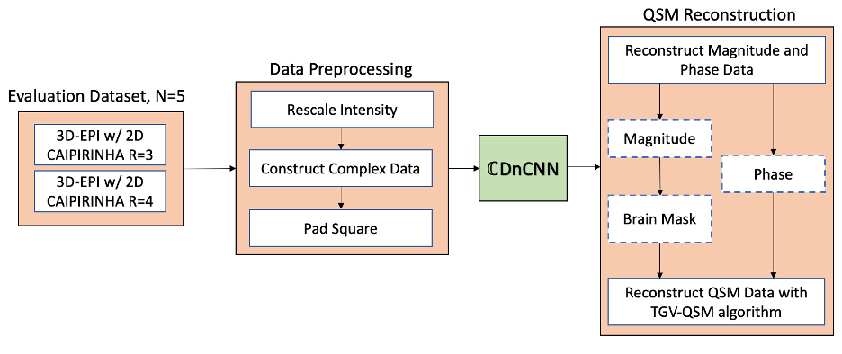
An evaluation dataset of 5 subjects acquired at two different acceleration factors (R=3 & 4; TA: 3 min & 2 min) was used. The data underwent standard preprocessing steps before denoising with CDnCNN. QSM images were generated from the data with and without denoising to compare their differences.
R: Acceleration Factor; TA: Time of Acquisition; CDnCNN: Complex Denoising Convolutional Neural Network. TGV: Total Generalized Variation; QSM: Quantitative Susceptibility Mapping.
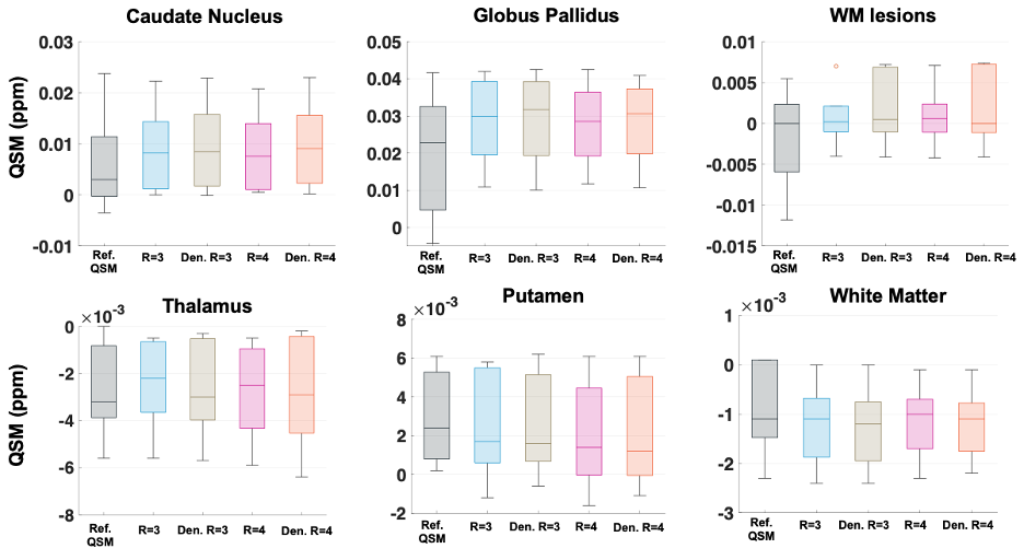
Boxplot diagram showing the ranges of QSM values within different regions of interest. There were no statistically significant differences in QSM values between the reference 3D-EPI without CAIPIRINHA acceleration and 3D-EPI with CAIPIRINHA acceleration after denoising (no p-values lower than 0.05).
Column Order (Left-to-Right): Reference QSM from unaccelerated 3D-EPI; Undenoised 3D-EPI with CAIPIRINHA R=3; Denoised 3D-EPI with CAIPIRINHA R=3; Undenoised 3D-EPI with CAIPIRINHA R=4; Denoised 3D-EPI with CAIPIRINHA R=4.
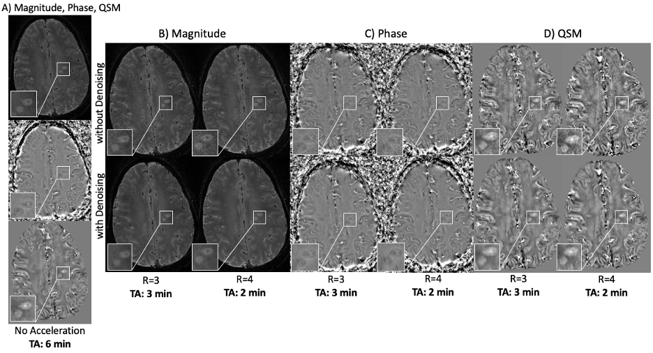
(A) T2* weighted magnitude, phase and QSM images acquired and reconstructed using 3D-EPI without CAIPIRINHA acceleration (TA: ~6 mins). (B) & (C) & (D) T2* weighted magnitude, phase and QSM images acquired and reconstructed using 3D-EPI with CAIPIRINHA at acceleration factors: R=3 (AT: ~3 min), and R=4 (AT: ~2 min). Brain images are shown with and without denoising using our deep learning CDnCNN model. A MS lesion is depicted within the zoomed-in figures.
EPI: Echo planar imaging; TA: Time of Acquisition; CDnCNN: Complex Denoising Convolutional Neural Network.