3714
Quantitative multi-parametric mapping of human subcortex at ultrahigh field1Department of Neurophysics, Max Planck Institute for Human Cognitive and Brain Sciences, Leipzig, Germany, 2GIGA-CRC in vivo imaging, University of Liège, Liège, Belgium, 3Full brain picture Analytics, Leiden, Netherlands, 4HUN-REN-PTE Clinical Neuroscience MR Research Group, Pécs, Hungary, 5Department of Neurology, University of Pécs, Pécs, Hungary, 6Integrative Model-based Cognitive Neuroscience Research Unit, University of Amsterdam, Amsterdam, Netherlands, 7Felix Bloch Institute for Solid State Physics, Faculty of Physics and Earth System Sciences, Leipzig University, Leipzig, Germany, 8Wellcome Centre for Human Neuroimaging, Institute of Neurology, University College London, London, United Kingdom
Synopsis
Keywords: Quantitative Imaging, Quantitative Imaging, Brain, Neuro, Subcortex
Motivation: Many neurodegenerative diseases affect subcortical nuclei early. Quantitative MRI (qMRI) offers a unique non-invasive tool for detection of neurodegeneration at its early stage, paving the way for development of potential therapies.
Goal(s): Ultra-high resolution multi-modal cartography of human subcortex capable of detecting subtle longitudinal changes in macro- and microstructure of subcortical nuclei.
Approach: We combined high resolution multi-parametric mapping using 7T, pTx and advanced reconstruction with automated segmentation of subcortical structures. Performance was tested across two sites.
Results: While qMRI repeatability varied strongly by structure, automated parcellation was highly repeatable, making the protocol a promising candidate for further studies.
Impact: We present a method for multi-contrast quantification of subcortical microstructure. Our high-resolution MRI acquisition and analysis protocol aims to enable longitudinal multi-center studies to detect subcortical neurodegeneration at its early stage.
Introduction
Ultra-high resolution quantitative MRI offers a unique opportunity to characterize multiple small nuclei in the human subcortex1,2. Particularly in the context of neurodegenerative diseases like Parkinson’s disease it offers the possibility for early detection of neurodegeneration of small subcortical structures where neurodegeneration starts. However, in order to be able to detect the subtle changes in small structures on a longitudinal basis high quality reproducible ultra-high resolution quantitative data are necessary, along with dedicated processing pipelines. Moreover, since MR contrast mechanisms in the subcortex are complex and far from understood, multi-contrast approaches are needed in order to visualize and quantify different nuclei. Here we present an ultra-high resolution multi-parametric qMRI protocol optimized for the cartography of the human subcortex. We assessed the performance of this acquisition in test–retest experiments across two imaging sites. We present data quality and test–retest reproducibility of subcortical morphometry and microstructure.Methods
Four healthy adult participants (1 female, mean age 45.25±6.3 years), each took part in at least two scanning sessions at 7T (MAGNETOM Terra, Siemens Healthineers, Erlangen, Germany) using parallel transmission (pTx) and an 8 transmit-/32 receive-channel radiofrequency head coil (Nova Medical, Wilmington, USA). Scanning was performed across two sites: MPI CBS in Leipzig, Germany and GIGA-Institute in Liège, Belgium.Multi-parametric acquisition consisted of whole-brain 3D multi-echo GRE with pTx kt-points excitation at an isotropic resolution of 0.6 mm (TR 22.4 ms, FA PDw/T1w 7/22°, six equispaced echoes TE 3.00 .. 15.6 ms, CAIPIRINHA R=2x2, TA 8:25 per contrast) and B1 mapping for transmit field correction3. The total acquisition time including shimming was around 24 minutes.
Images were reconstructed with AC-LORAKS4,5 and MCPC-3D-S6 followed by LCPCA denoising7. The images of one participant were strongly affected by motion artifacts and excluded. The remainder were processed with the open source hMRI toolbox8 (https://hMRI.info), yielding R2*, PD and R1 maps. Subcortical structures were automatically parcellated by Multi-contrast Anatomical Subcortical Structures Parcellation (MASSP)9 using all three contrasts as input. QSM was additionally calculated by SEPIA10 (PDF11 for background field removal, Star-QSM12 for dipole inversion) using MPM-QSM (https://github.com/fil-physics/MPM_QSM) on the PD-weighted images. To assess the repeatability of subcortical segmentation and quantification on the remaining participants, the dilated Dice overlap coefficient and mean distance between boundary delineations were computed9, together with qMRI parameters within each structure.
Results
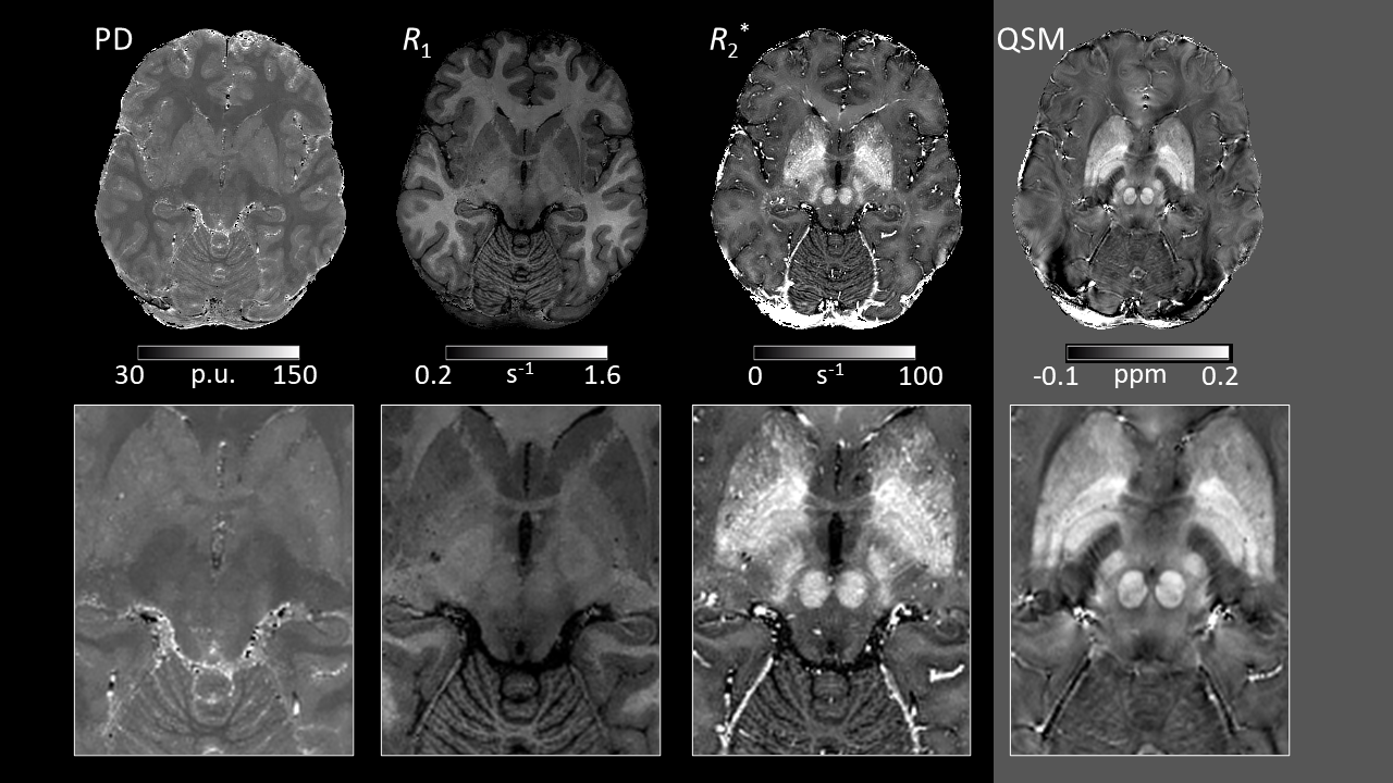
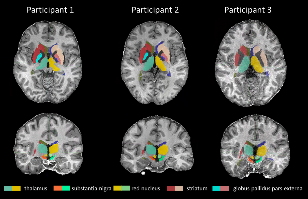
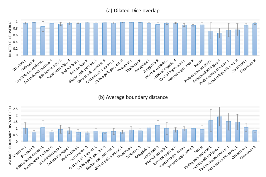
High quality, ultra-high resolution, intrinsically co-aligned maps of qMRI parameters R2*, PD, R1, and QSM (Fig. 1) were obtained in three out of four participants. The 0.6 mm resolution multi-parameter mapping (MPM) delineated cortical and subcortical anatomical structures with a high contrast-to-noise ratio (CNR) across the brain. MASSP parcellations shown in Fig. 2 demonstrate robust parcellation of subcortical structures across participants.As measured by dilated Dice overlap, Fig. 3 shows that all segmented structures retained at least 65% overlap between repeated scanning sessions with most structures above 95%. The average boundary distance confirmed the high overlap with values typically between one and two voxels.
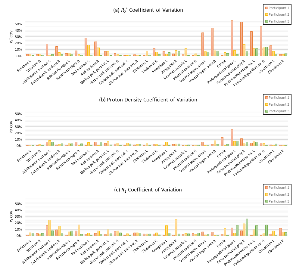
The differences in quantitative values between repeated sessions are reported in Fig. 4. Coefficients of variation were generally less than 10% but highly variable between participants and regions, with repeatability lower especially in smaller structures (e.g. subthalamic nucleus (STN), periaqueductal gray (PAG), pedunculopontine nucleus (PPN)) with less robust segmentation and fewer voxels.
Discussion and conclusion
In this study, we investigated the reproducibility of a subcortical quantitative multi-parameter mapping protocol. In contrast to other traveling head studies13,14, our aim was to enable studies primarily of subcortical structures between testing sites. While initial data on inter-session variability are promising, heralding the detection of 10% changes in myelin and iron content or more, further work is required to distinguish inter- from intra-site bias. MASSP performed well with the provided parameter maps and could be extended to additionally use QSM for higher accuracy. The combination of high resolution, efficient acquisition times, and multi-parametric contrasts makes this protocol a promising candidate for detailed studies of the subcortex in health and disease.Acknowledgements
The research leading to these results has received funding from JPND/ZonMW (grant 1051006211003), JPND/Federal Ministry of Education and Research under support code 01ED2210, the National Research, Development and Innovation Office, Hungary (2019-2.1.7-ERA-NET-2022-00046), JPND2021-650-301 and JPND/FRS-FNRS Belgium (Grants R.8011.21 & T.0238.23).References
1. Forstmann B , Hollander G et al. Towards a mechanistic understanding of the human subcortex. Nat. Rev. Neurosci., 2017; 18(1):57-65.
2. Keuken M, Isaacs B et al. Visualizing the Human Subcortex Using Ultra-high Field Magnetic Resonance Imaging. Brain Topogr., 2018; 31(4):513-545.
3. Pine K, Gross-Weege N et al. Parallel transmit (pTx) kT-points pulses improve 500µm resolution quantitative multi-parameter mapping (MPM) at 7T. Proc. Intl. Soc. Magn. Reson. Med. 2023.
4. Kim T, Haldar J. LORAKS Software Version 2.0: Faster Implementation and Enhanced Capabilities. University of Southern California, Los Angeles, CA, Technical Report USC-SIPI-443, May 2018.
5. Haldar J. Autocalibrated LORAKS for fast constrained MRI reconstruction. Proc. IEEE Int. Symp. Biomed. Imag., 2015; 910-913.
6. Dymerska B, Eckstein et al. Phase Unwrapping with a Rapid Opensource Minimum Spanning TreE AlgOrithm (ROMEO). Magn. Reson. Med, 2020; 85(4):2294-2308.
7. Bazin P-L, Alkemade A et al. Denoising High-Field Multi-Dimensional MRI With Local Complex PCA. Front Neurosci., 2019; 13:1066.
8. Tabelow K, Balteau E et al. hMRI – A toolbox for quantitative MRI in neuroscience and clinical research. Neuroimage, 2019; 194:192-210.
9. Bazin P-L, Alkemade A et al. Multi-contrast anatomical subcortical structures parcellation. eLife, 2020; 9:e59430.
10. Chan K-S, Marques J. SEPIA—Susceptibility mapping pipeline tool for phase images. Neuroimage, 2021; 227:117611.
11. Liu T, Khalidov I et al. A novel background field removal method for MRI using projection onto dipole fields (PDF). NMR Biomed., 2011; 24:1129-1136.
12. Wei H, Dibb R et al. Streaking artifact reduction for quantitative susceptibility mapping of sources with large dynamic range. NMR Biomed., 2015; 28:1294-1303.
13. Sherif S, Aghaeifar A et al. Repeatability of ultra-high-resolution Multi-Parametric Mapping across five 7T sites. Proc. Intl. Soc. Magn. Resn. Med. 2022.
14. Voelker M, Kraff O et al. The traveling heads 2.0: Multicenter reproducibility of quantitative imaging methods at 7 Tesla. Neuroimage, 2021; 232:117910.
Figures