3710
Spin and gradient echo multiple overlapping echo detachment (SAGE-MOLED) for high-fidelity T2-T2* mapping and simultaneous DSC-DCE imaging1Department of Electronic Science, Xiamen University, Xiamen, China, 2Department of Magnetic Resonance Imaging, The First Affiliated Hospital of Zhengzhou University, Zhengzhou University, Zhengzhou, China, 3Clinical & Technical Support, Philips Healthcare, Shenzhen, China
Synopsis
Keywords: Quantitative Imaging, DSC & DCE Perfusion, Spin and gradient echo, Deep learning
Motivation: Conventional spin and gradient echo EPI (SAGE-EPI) sequence has limited spatial resolution and suffers from geometric distortion.
Goal(s): Our goal was to develop a high-fidelity simultaneous T2 and T2* mapping technique used for dynamic MR imaging.
Approach: We developed a single-shot spin and gradient echo multiple overlapping-echo detachment acquisition (SAGE-MOLED) method with deep learning reconstruction. The proposed method was tested in phantom and in vivo human brains, and a single-dose contrast dynamic imaging was performed on a clinical case.
Results: SAGE-MOLED achieves distortion-free high-precision T2 and T2* mapping compared to gold-standard methods and was successfully used in simultaneous DSC-DCE imaging.
Impact: Rapid high-fidelity T2 and T2* mapping can be achieved using our proposed SAGE-MOLED technique, which further enables leakage-corrected simultaneous DSC and DCE imaging at 1.7×1.7×4.0 mm3 spatial resolution and 1.9 s temporal resolution.
Introduction
Dynamic susceptibility contrast (DSC) and dynamic contrast enhanced (DCE) imaging are widely used for MRI-based assessment of brain tissue abnormalities.1 A spin- and gradient-echo echo-planar imaging (SAGE-EPI) sequence has been developed for simultaneous quantification of T2 and T2* and directly to eliminate the error caused by T1-leakage effects in dynamic parameters estimation.2,3 However, SAGE-EPI trends to have poor image quality with low spatial resolution (~3×3×5 mm3), due to its limited and repeated EPI readout modules. Additionally, the EPI readout of SAGE-EPI results in B0-induced geometric distortions, which may introduce additional errors in perfusion parameters calculation. To address these challenges, we proposed a method that utilizes multiple overlapping-echo detachment (MOLED) technique4-6 to increase the number of accessible echoes without sacrificing spatial resolution, termed SAGE-MOLED. The proposed method was validated to achieve simultaneous DSC-DCE imaging with a single-dose contrast injection at 1.7×1.7×4.0 mm3 spatial resolution and 1.9 s temporal resolution.Methods
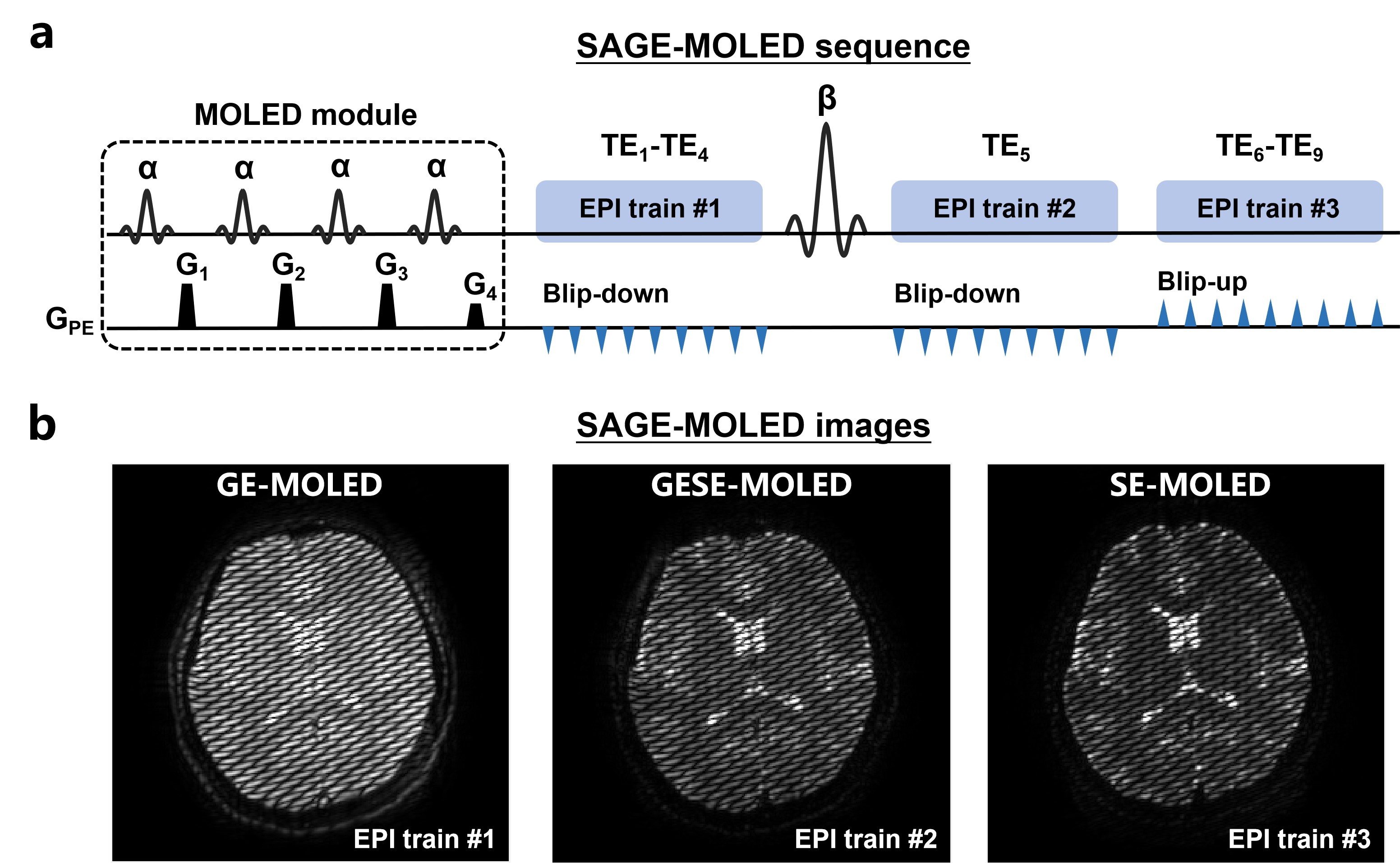
Pulse sequence: Figure 1(a) shows the sequence diagram of SAGE-MOLED with three EPI trains. As for MOLED module, four excitation pulses with the same flip angle α = 30° and echo-shifting gradients G1-G4 are used to prepare spin and gradient echoes with different echo times. EPI train #1-2 and EPI train #3 use different blip polarities, resulting in opposite geometric distortion to achieve distortion-free mapping, as shown in Figure 1(b).Phantom and in vivo experiments: All data were acquired on a 3T whole-body MRI system with a 32-channel head coil (Ingenia CX; Philips Healthcare). A custom phantom containing 9 small tubes with different concentrations of MnCl2 solutions was used for validation. The acquisition parameters of SAGE-MOLED were as follows: FOV = 22×22 cm2, resolution = 1.7×1.7×4.0 mm3, TE = 7.5, 21.1, 34.8, 48.6, 57.6, 72.6, 86.2, 100.0, 113.8 ms, TR = 6.0 s, slice number = 21, SENSE acceleration factor = 3. Reference T2 and T2* maps were obtained using single-echo SE with TE = 20, 40, 60, 90, 120 ms and multi-echo GRE with TE = 8, 16, 24, 32, 40, 48 ms. For DSC-DCE imaging, the SAGE-MOLED sequence was incorporated in a clinical protocol and was conducted during a single dose contrast injection, using TR = 1.9 s and dynamic scan number = 110.
Deep learning parametric mapping and analysis: A five-level UNet was trained to perform end-to-end parametric mapping (T2, T2*, M0, ∆B0 and B1+) from complex-valued SAGE-MOLED images. A total of 4,000 pairs of synthetic data with distortion-free labels were generated for network training through Bloch simulation.7 For DSC-DCE analysis, the dynamic SAGE-MOLED T2 and T2* maps are susceptibility-sensitive and were therefore used to calculate the cerebral blood volume and flow (CBV and CBF), while the SAGE-MOLED M0 maps reflect the T1 signal change and were therefore used to calculate the transfer constant (Ktrans).
Results
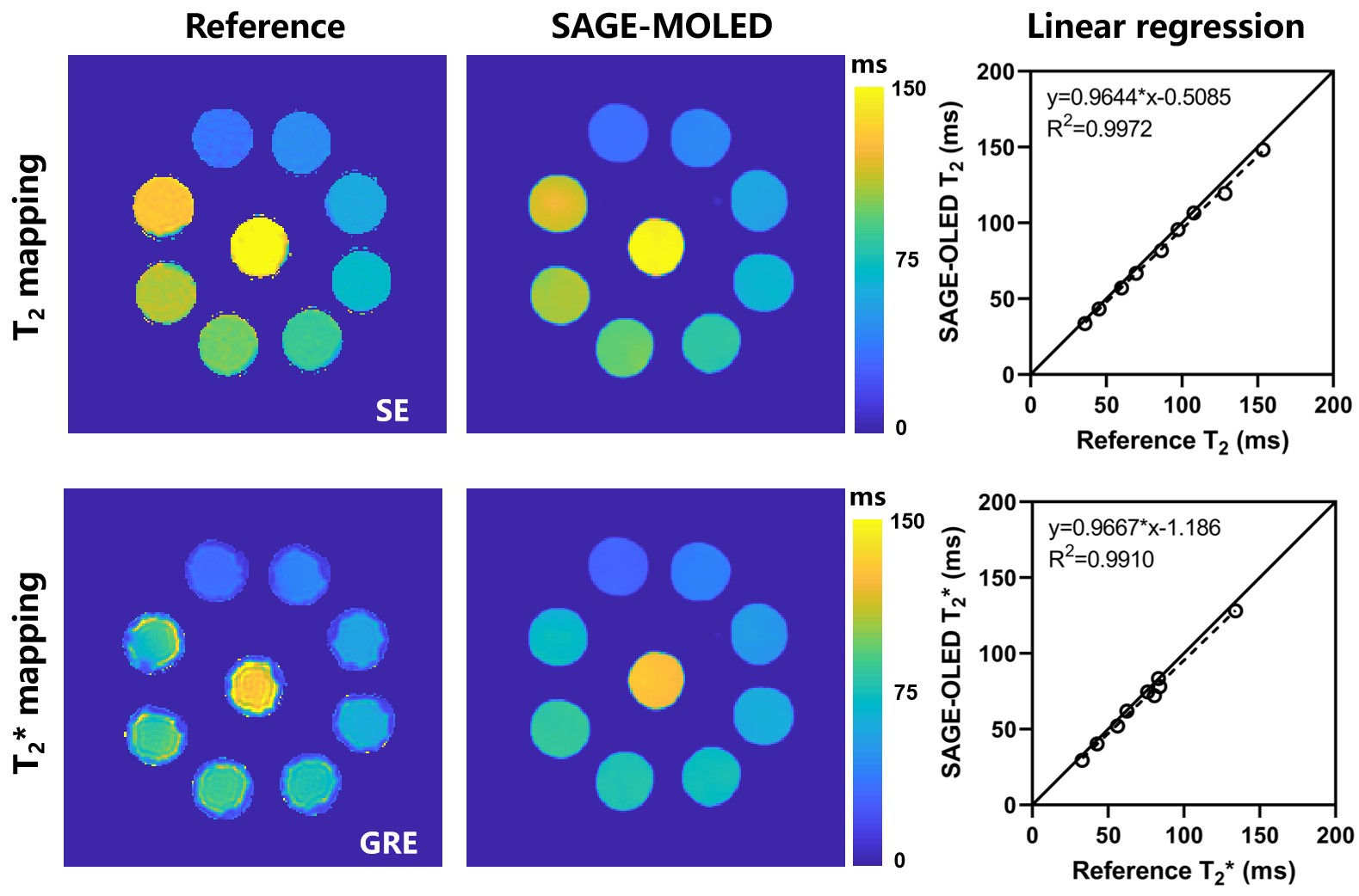
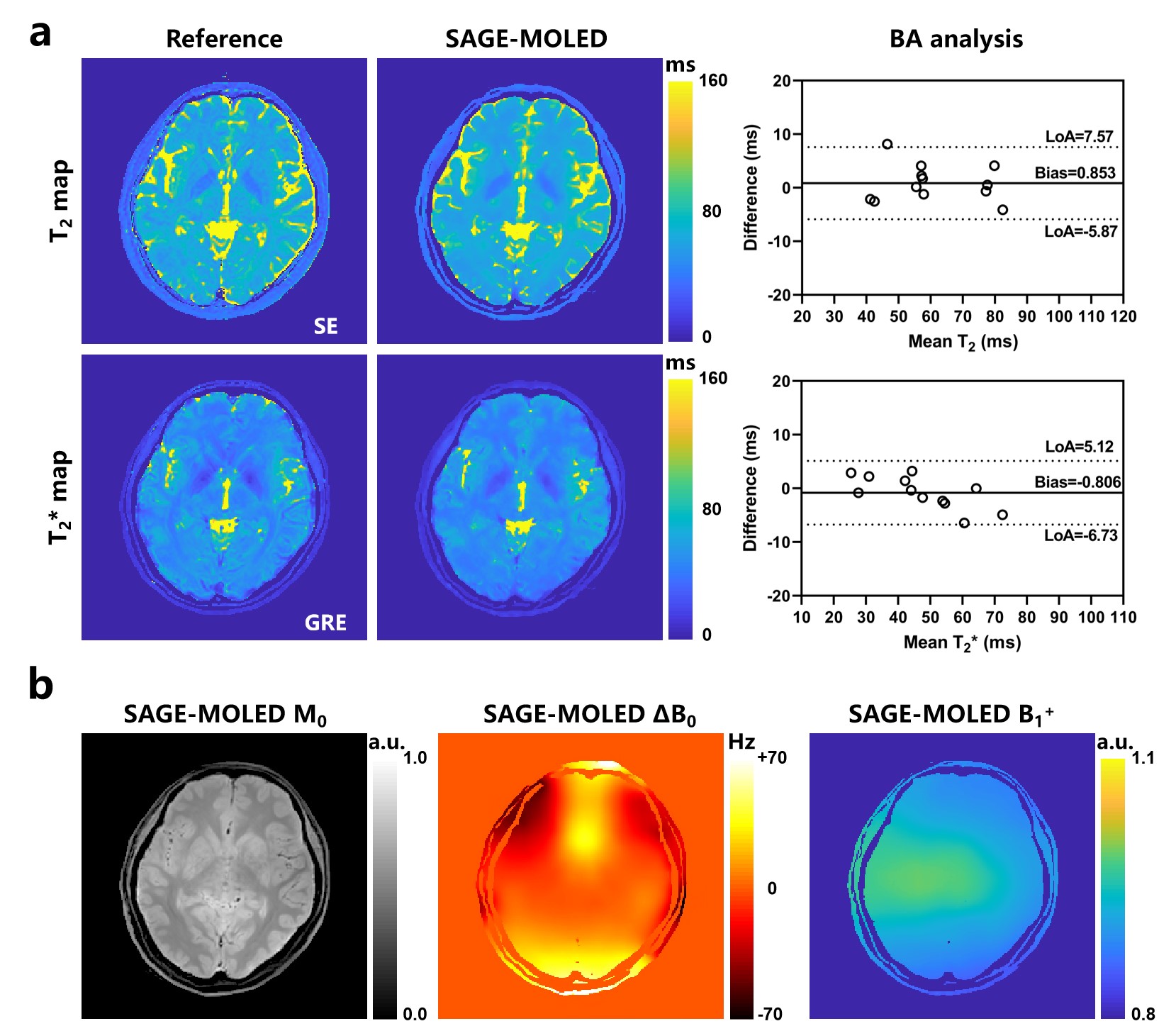
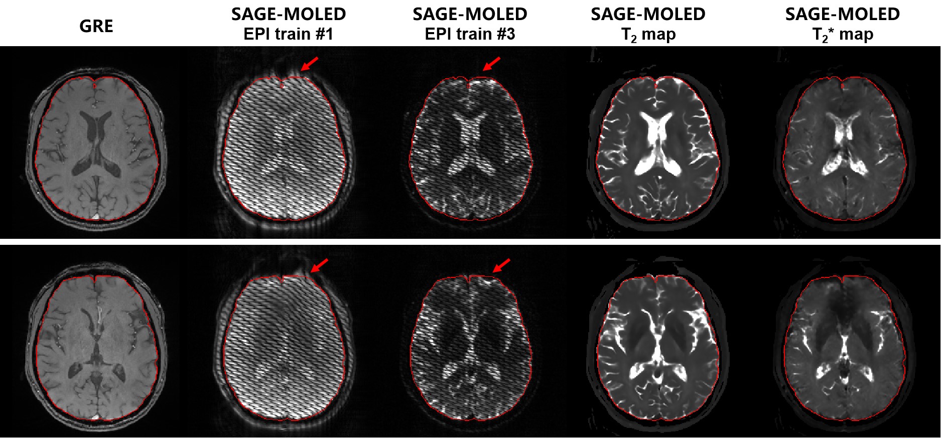
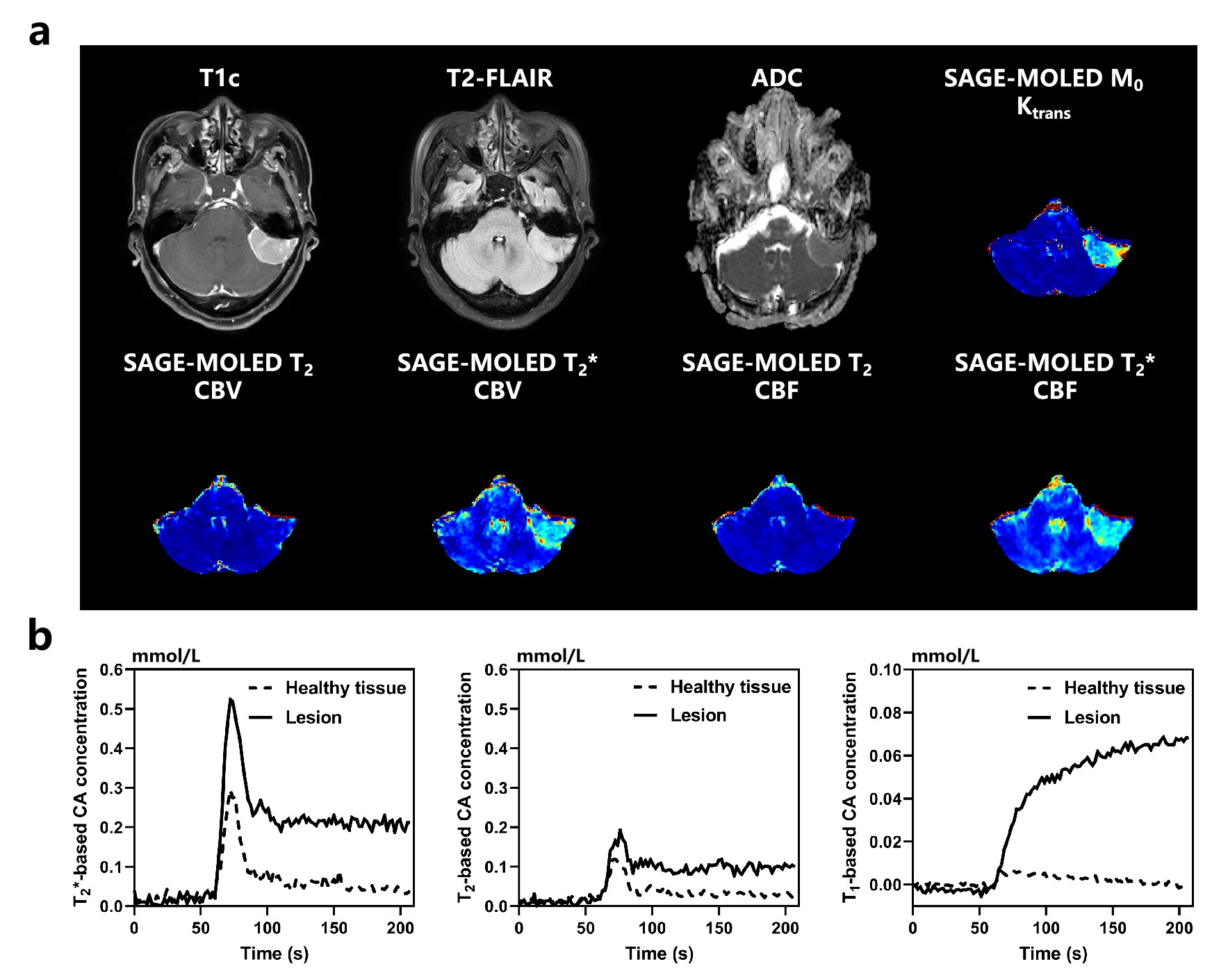
The results of the phantom experiments are shown in Figure 2. Good agreement is seen between SAGE-MOLED and reference methods. The linear regression analysis indicates a strong positive correlation between the two methods with Pearson’s correlation coefficient (PCC) = 0.998 for T2 and with PCC = 0.995 for T2*. The human brain experiments show the same trend as the phantom experiments in that the parametric maps from SAGE-MOLED are consistent with the reference methods with the bias of 0.853 ms for T2 and 0.806 ms for T2* (Figure 3a). In addition, the reconstructed M0, ∆B0 and B1+ maps of SAGE-MOLED from the same slice can be seen in Figure 3(b). Figure 4 illustrates the robustness of SAGE-MOLED to B0-induced geometric distortion. Compared with the original MR images, the parametric maps are distortion-free and comparable to GRE images. A representative clinical case is shown in Figure 5. From the perfusion maps of T2* and M0, we can see that the regions with high CBV, CBF and Ktrans agree well with the tumor regions on the postcontrast T1-weighted image. The tumor shows a faster T1/T2*-based CA concentration increase than healthy tissue, and the T2*-based concentration subsequently recovered above baseline. However, there is no significant signal change in dynamic T2 maps.Discussion and conclusion
The proposed SAGE-MOLED sequence shows excellent performance in quantifying brain T2 and T2* values, and its accuracy is close to the lengthy gold standard methods. In dynamic SAGE-MOLED imaging, perfusion and permeability parameters analysis benefits from quantitative MRI and can avoid T1 or T2* leakage effects, as reported by previous works.6,8 As a proof-of-concept, this work has not yet combined SAGE-MOLED with advanced acceleration techniques such as simultaneous multislice acquisition, and future work will further enhance the temporal resolution of SAGE-MOLED to improve its volume coverage.Acknowledgements
This work was supported in part by the National Natural Science Foundation of China under grant numbers 82071913, 12375291 and 22161142024.References
1. Quarles C, Bell L, Stokes A, Imaging vascular and hemodynamic features of the brain using dynamic susceptibility contrast and dynamic contrast enhanced MRI, Neuroimage 2019; 187:32-55.
2. Schmiedeskamp H, Straka M, Newbould R, et al. Combined spin- and gradient-echo perfusion-weighted imaging. Magn Reson Med. 2012; 68: 30-40.
3. Schmiedeskamp H, Andre J, Straka M, et al. Simultaneous perfusion and permeability measurements using combined spin- and gradient-echo MRI. J Cerebr Blood F Met. 2013; 33: 732-743.
4. Cai CB, Wang C, Zeng YQ, et al. Single-shot T2 mapping using overlapping-echo detachment planar imaging and a deep convolutional neural network. Magn Reson Med. 2018;80:2202-2214.
5. Yang QQ, Ma L, Zhou Z, et al. Rapid high-fidelity T2* mapping using single-shot overlapping-echo acquisition and deep learning reconstruction. Magn Reson Med. 2023;89:2157-2170.
6. Ma LC, Wu J, Yang QQ, et al. Single-shot multi-parametric mapping based on multiple overlapping-echo detachment (MOLED) imaging. Neuroimage. 2022;263:119645.
7. Yang QQ, Lin YH, Wang JC, et al. Model-based synthetic data-driven learning (MOST-DL): Application in single-shot T2 mapping with severe head motion using overlapping-echo acquisition. IEEE Trans Med Imaging. 2022;41:3167-3181.
8. Hu Z, Ghristodoulou A, Wang N, et al. MR multitasking-based dynamic imaging for cerebrovascular evaluation (MT-DICE): Simultaneous quantification of permeability and leakage-insensitive perfusion by dynamic T1/T2* mapping. Magn Reson Med. 2023;89:161-176.
Figures