3707
Transient-state CSF suppression for time-efficient, high-resolution 3T whole-brain 3D relaxometry1Computational Imaging Group for MR diagnostics & therapy, Center for Image Sciences, University Medical Center Utrecht, Utrecht, Netherlands
Synopsis
Keywords: Quantitative Imaging, Quantitative Imaging
Motivation: Cerebrospinal fluid (CSF) pulsation artifacts using 3D unbalanced gradient-echo sequences hinder the accuracy of whole-brain relaxometry measurements.
Goal(s): To develop a CSF-suppressed 3D MR-STAT sequence and eliminate CSF pulsation artifacts, allowing for fast, high-resolution whole-brain relaxometry with enhanced image quality.
Approach: The time-varying RF flip-angle train is optimized with an additional CSF suppression constraint to achieve both high quantitative parameter encoding capability and low CSF signal intensity.
Results: The new CSF-suppressed 3D MR-STAT sequence has been validated on healthy volunteers, demonstrating effective mitigation of CSF ghosting artifacts, and allowing whole-brain relaxometry acquisition within 5.5-minute scan time.
Impact: The proposed MR-STAT sequence with CSF suppression requires a short scan time (less than 6 minutes) and shows robustness in whole-brain 1mm3 isotropic relaxometry, and therefore shows the ability to adopt quantitative imaging in broad neuroscientific and clinical applications.
Introduction
Three-dimensional unbalanced GRE (gradient-echo) sequences are known to be highly sensitive to the pulsatile flow of fluids[1,2]. In 3D quantitative imaging, cerebrospinal fluid (CSF) pulsation artifacts can be observed when using either steady-state or transient-state SSFP protocols[3,4,5]. The CSF pulsation leads to ghosting artifacts which originate from cerebral ventricles and affect surrounding tissues. These artifacts are more conspicuous when CSF signal intensity is comparable or higher to that of the surrounding tissues[6].In this work, we propose a new method to mitigate the CSF pulsation artifacts when using transient-state, gradient-spoiled GRE sequences. The method is inspired by a recently developed steady-state T2 mapping protocol[7], which uses the proper combination of RF amplitudes and quadratic RF phases to suppress the CSF signal and the related ghosting artifacts.
In this abstract, we show that a similar CSF signal suppression strategy can be also applied to eliminate CSF artifacts in a transient-state 3D unbalanced multi-parametric protocol. Specifically, we show that a smooth RF flip-angle train can be optimized for a 3D MR-STAT (Magnetic Resonance Spin Tomography in Time-Domain) framework[8], to simultaneously achieve low CSF signal intensity and high T1/T2 encoding capability. The proposed MR-STAT protocol allows for a five-minute, 1mm3 isotropic whole brain scan which is much more robust to CSF flow artifacts than base-line 3D protocols. The new 3D MR-STAT sequence is preliminarily tested on a gel/water phantom and on 3 healthy volunteers.
Theory
- Base-line Sequence:
- Proposed CSF-suppressed flip-angle train optimization:
In order to suppress the CSF ghosting artifacts, we add an additional term to the minimization objective function $$$F_{Cramer-Rao}(\alpha, \phi'')$$$, shown as below
$$F(\alpha, \phi'')= F_{Cramer-Rao}(\alpha, \phi'')+\lambda \frac{\parallel S_{CSF}(\alpha, \phi'') \parallel_2}{\parallel S_{WM}(\alpha, \phi'') \parallel_2}. (1)$$
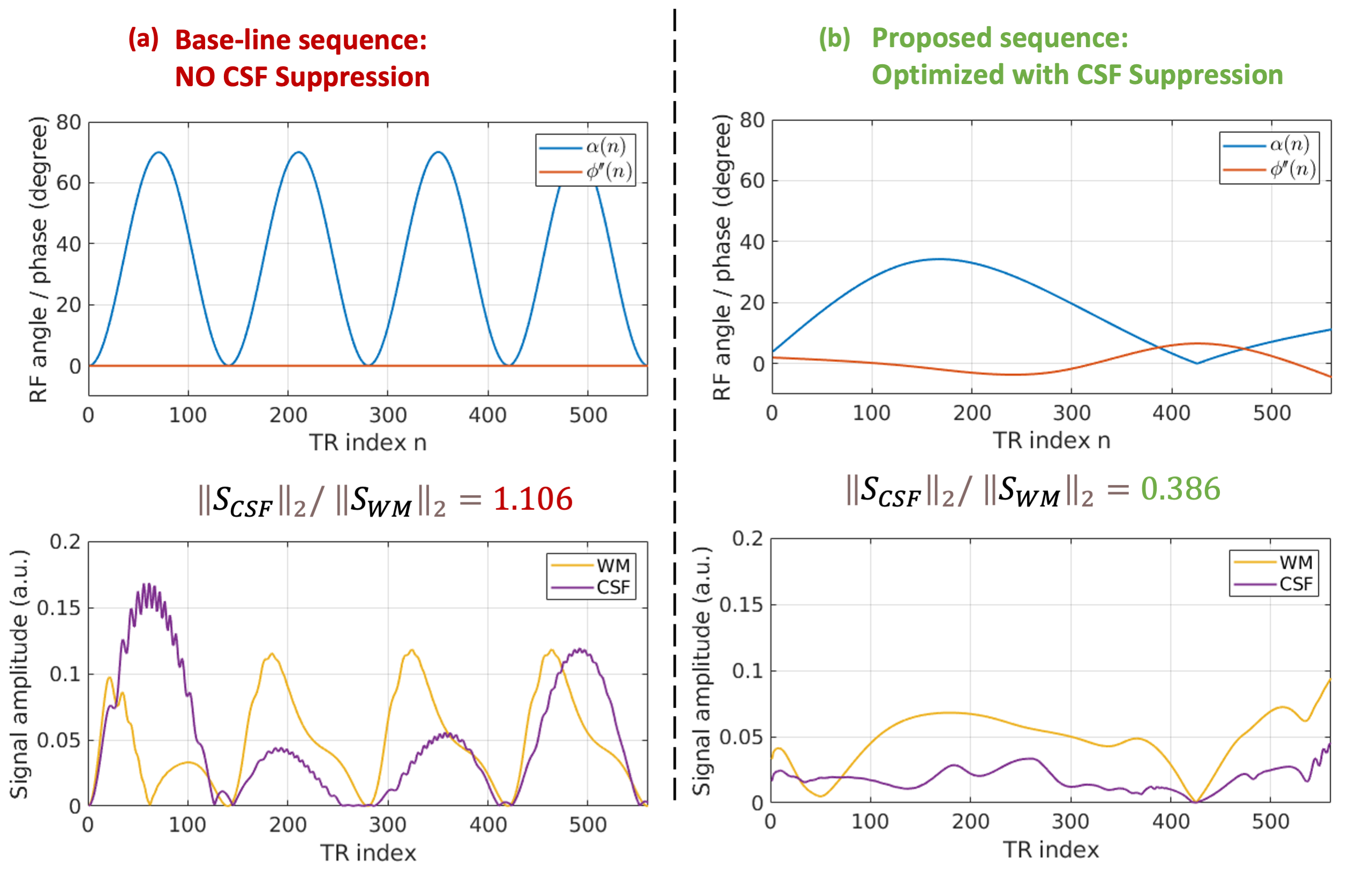
Here, $$$S_{CSF}(\alpha, \phi'')$$$ and $$$S_{WM}(\alpha, \phi'')$$$ are temporal MR signals for different tissue parameter values, namely $$$T_1=4s, T_2=2s$$$ for the CSF signal ($$$S_{CSF}$$$) and , $$$T_1=0.9s, T_2=0.05s$$$ for the white matter signal ($$$S_{WM}$$$). In this way, the CSF signal is minimized and therefore the related artifacts will be mitigated.
Method
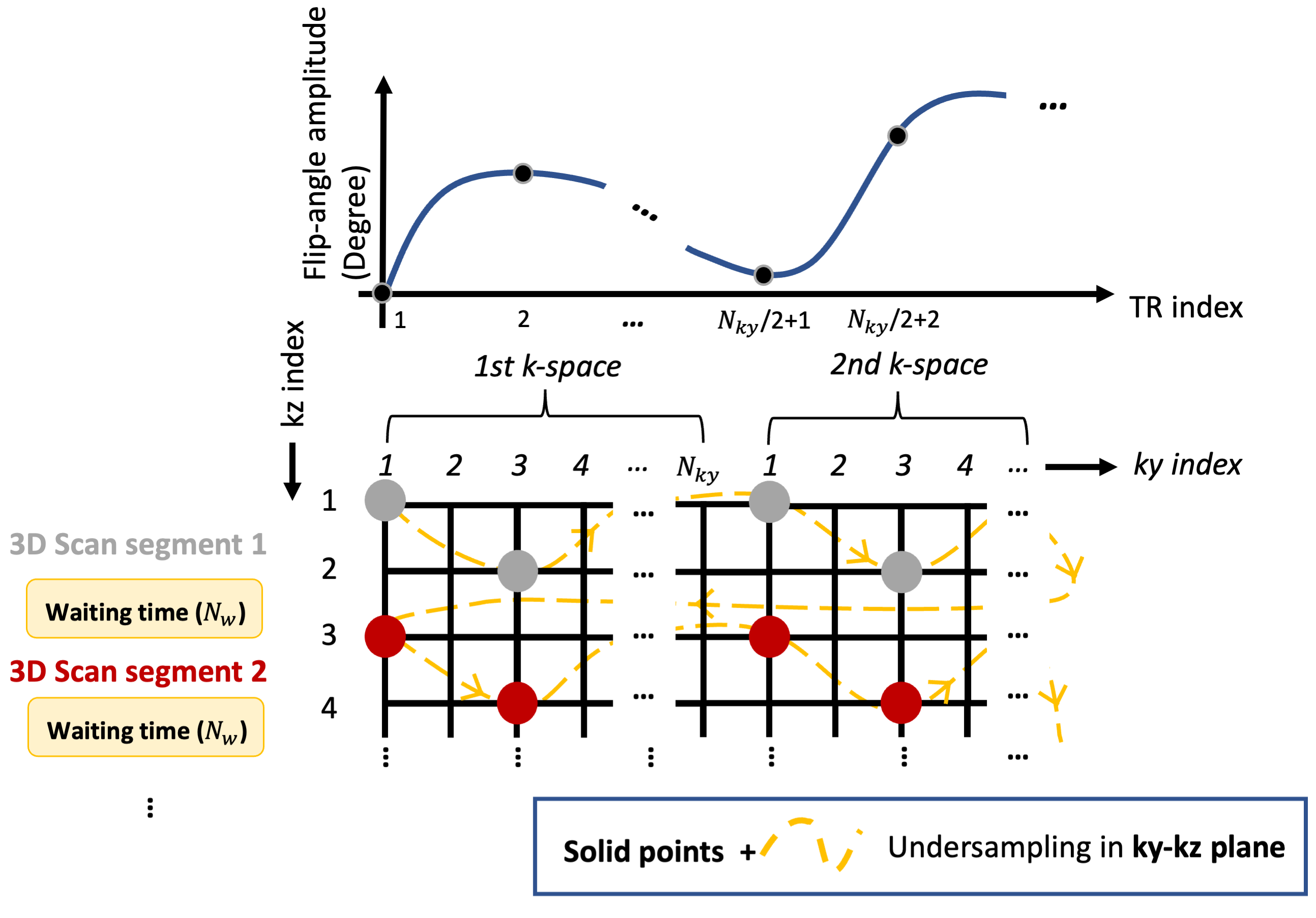
The proposed 3D MR-STAT protocol (Fig.1) was implemented on a 3T Philips Elition system. Both the base-line sequence and the proposed sequence were used for 3D scans of a phantom (9 Eurospin gel tubes and 1 tap-water tube) and in-vivo (on 3 healthy volunteers) using a 32-channel receive head coil. The total scan time for the proposed sequence is 5min37sec for 1mm3 whole brain coverage.A two-step reconstruction strategy described previous literature[5] was used for all reconstructions.
Results
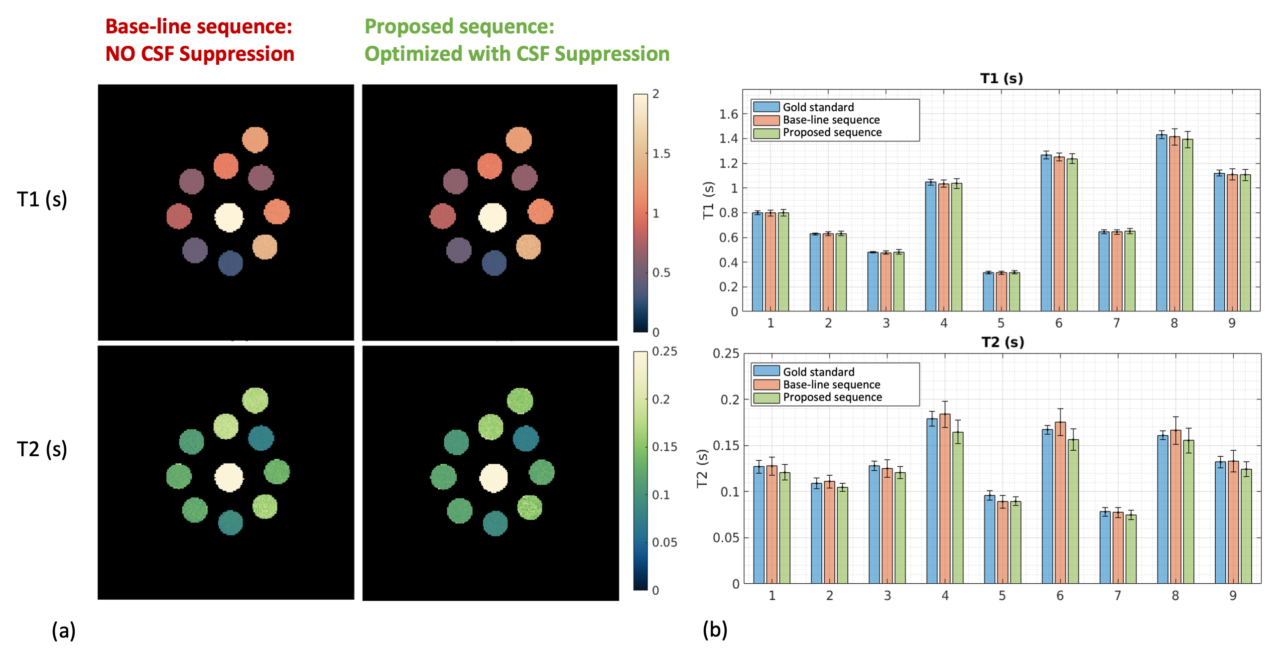
Fig.2 shows the flip-angle trains of (a) the base-line sequence[5] and (b) the proposed CSF-suppressed sequence. The simulated MR signals computed for the two sequences are plotted in the second row, showing that the proposed protocol leads to a much lower CSF-to-WM signal ratio.Fig.3 shows the results of the phantom experiment. Mean T1 and T2 values computed from both sequences show high agreements with the gold standards, and the precision (standard deviations) is similar for the two sequences.
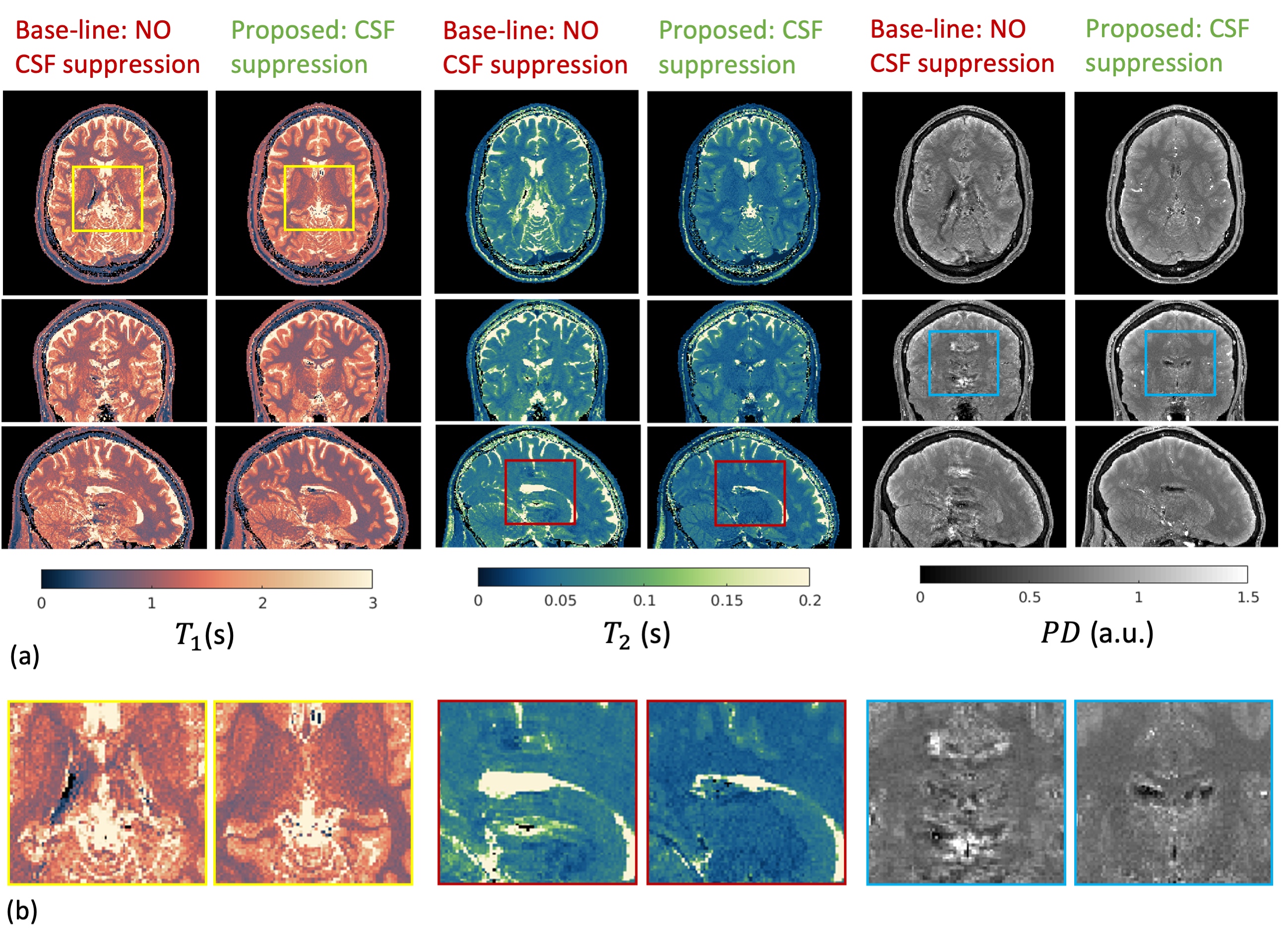
Fig. 4 shows the in-vivo results for one healthy volunteer. The CSF ghosting artifacts contaminate the image quality in surrounding regions for the baseline sequence. The proposed CSF-suppressed sequence shows instead greatly improved image quality for all quantitative maps.
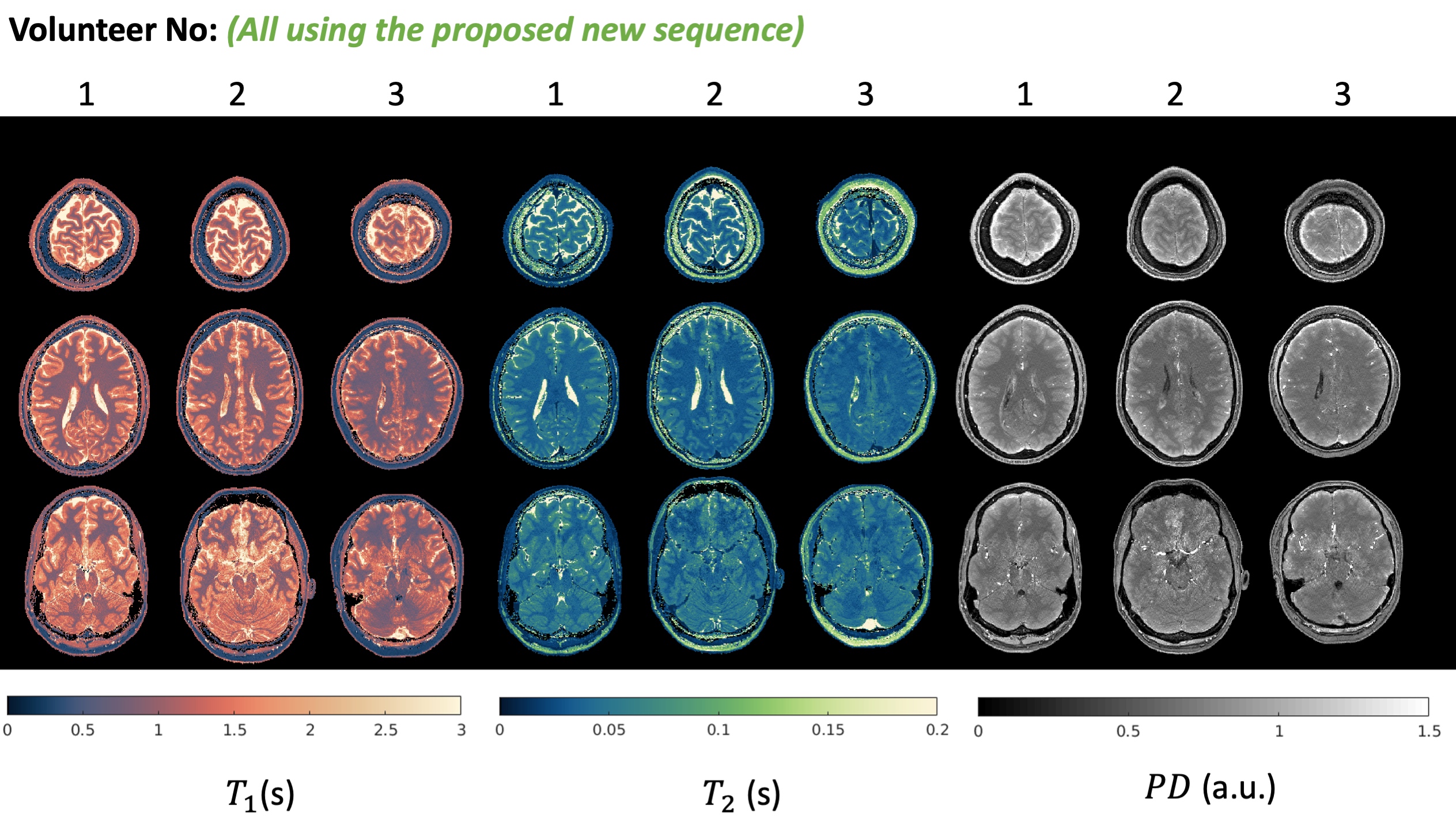
Fig.5 shows the in-vivo results from all three healthy volunteers using the new optimized sequence, proving overall high robustness to CSF artifacts and consistent relaxometry quality.
Conclusion and Discussion
We demonstrated the efficiency of the CSF suppression technique on healthy volunteers. The 3D MR-STAT sequence with CSF suppression has been validated for acquiring high quality, 1mm3 whole-brain relaxometry within 5.5-minute scan time. This robust, fast protocol for isotropic whole-brain relaxometry will potentially broaden the application of quantitative imaging in different clinical work-flow. Other CSF artifact mitigation methods, such as phase-encoding pattern optimization[1,12], will be also investigated in the future.Acknowledgements
No acknowledgement found.References
[1] Anderson, Christian E., et al. "Regularly incremented phase encoding–MR fingerprinting (RIPE‐MRF) for enhanced motion artifact suppression in preclinical cartesian MR fingerprinting." Magnetic resonance in medicine 79.4 (2018): 2176-2182.
[2] Seginer, Amir, and Rita Schmidt. "Messing up to clean up: Semi‐randomized frequency selective space‐filling curves to suppress physiological signal fluctuations in MRI." Magnetic Resonance in Medicine (2023).
[3] Palma, Giuseppe, et al. "A novel multiparametric approach to 3D quantitative MRI of the brain." PloS one 10.8 (2015): e0134963.
[4] Pirkl, Carolin M., et al. "Accelerated 3D whole-brain T1, T2, and proton density mapping: feasibility for clinical glioma MR imaging." Neuroradiology (2021): 1-21.
[5] Liu, Hongyan, et al. "A three‐dimensional Magnetic Resonance Spin Tomography in Time‐domain protocol for high‐resolution multiparametric quantitative magnetic resonance imaging." NMR in Biomedicine (2023): e5050.
[6] Lisanti, Christopher, et al. "Normal MRI appearance and motion-related phenomena of CSF." American Journal of Roentgenology 188.3 (2007): 716-725.
[7] Seginer, Amir, and Rita Schmidt. "Phase-based fast 3D high-resolution quantitative T2 MRI in 7 T human brain imaging." Scientific reports 12.1 (2022): 14088.
[8] Sbrizzi, Alessandro, et al. "Fast quantitative MRI as a nonlinear tomography problem." Magnetic resonance imaging 46 (2018): 56-63.
[9] Breuer, Felix A., et al. "Controlled aliasing in volumetric parallel imaging (2D CAIPIRINHA)." Magnetic Resonance in Medicine: An Official Journal of the International Society for Magnetic Resonance in Medicine 55.3 (2006): 549-556.
[10] Fuderer, Miha, et al. "Efficient performance analysis and optimization of transient‐state sequences for multiparametric magnetic resonance imaging." NMR in Biomedicine 36.3 (2023): e4864.
[11] Fuderer, Miha, et al. "Multi-parametric quantitative MRI without inversion pulses by optimized RF phase modulation." Proc Intl Soc Mag Reson Med. Vol. 31. 2023.
[12] Seginer, Amir, and Rita Schmidt. "Messing up to clean up: Semi‐randomized frequency selective space‐filling curves to suppress physiological signal fluctuations in MRI." Magnetic Resonance in Medicine (2023).
Figures