3705
Intravoxel incoherent motion analysis after preoperative endovascular embolizationvascular embolization in supratentorial meningioma1Division of Health Sciences, Graduate School of Medical Sciences, Kanazawa University, Kanazawa, Japan, 2Nagoya City University Hospital, Nagoya, Japan
Synopsis
Keywords: Tumors (Pre-Treatment), Diffusion/other diffusion imaging techniques, Meningioma, Embolization, IVIM
Motivation: Some studies have reported decreased tumor volume and blood flow after preoperative endovascular embolization in meningioma, but it remains unclear how the meningioma and peritumoral edema are altered after endovascular embolization, and a practical evaluation method has not been established.
Goal(s): We investigated IVIM analysis to evaluate perfusion and diffusion simultaneously before and after endovascular embolization in supratentorial meningioma.
Approach: We assessed IVIM parameters (FD*, F, D*, and D) before and after endovascular embolization in supratentorial meningioma.
Results: FD* and F were significantly lower after endovascular embolization in meningioma than those before the endovascular embolization.
Impact: The IVIM analysis makes it possible to evaluate perfusion and diffusion simultaneously after preoperative endovascular embolization in supratentorial meningiomas in a short time.
INTRODUCTION
Meningiomas are the most frequent primary brain tumors, and preoperative embolization may be performed for intraoperative bleeding reduction and tumor softening.1 Some studies have reported decreased tumor volume and blood flow after endovascular embolization in meningioma.2,3 However, it remains unclear how the meningioma and peritumoral edema are altered after endovascular embolization, and a practical evaluation method has not been established. The study aim was to investigate intravoxel incoherent motion (IVIM) analysis using diffusion magnetic resonance imaging (MRI) to evaluate perfusion and diffusion simultaneously before and after preoperative endovascular embolization in supratentorial meningioma.METHODS
Six patients with supratentorial meningiomas (mean age, 68.3 ± 10.2 years) were included in the study. On a 3.0-T MRI, single-shot diffusion echo planar imaging was performed with the smallest number of b-values (0, 200, and 1000 s/mm2) to shorten the scan time (within 93 seconds) before and after (1–2 days) preoperative endovascular embolization in meningiomas. Next, a biexponential IVIM model was used to perform voxel-wise estimations of the perfusion-related diffusion coefficient (D*), perfusion fraction (F), multiplication of D*and F (FD*), and restricted diffusion coefficient (D). Then, F, D*, FD*, and D in the meningioma and peritumoral edema were compared before and after preoperative endovascular embolization. P values of <0.05 indicated statistical significance.RESULTS AND DISCUSSION
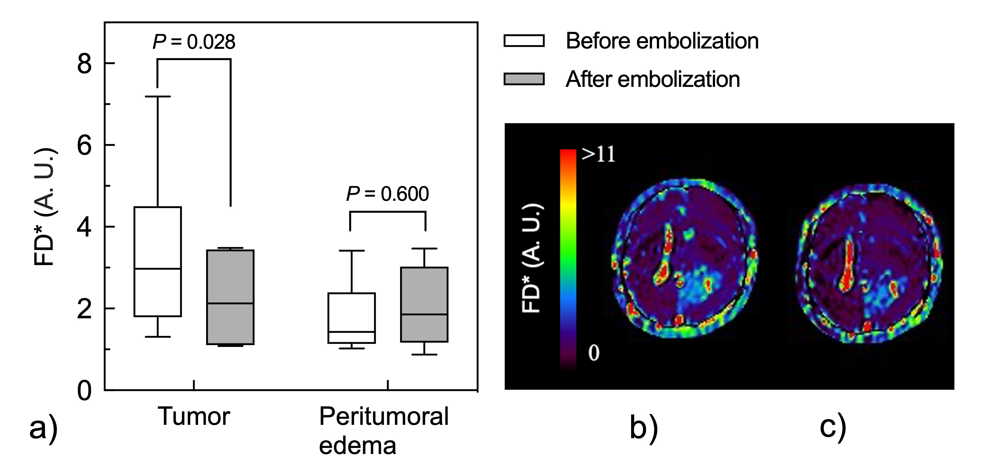
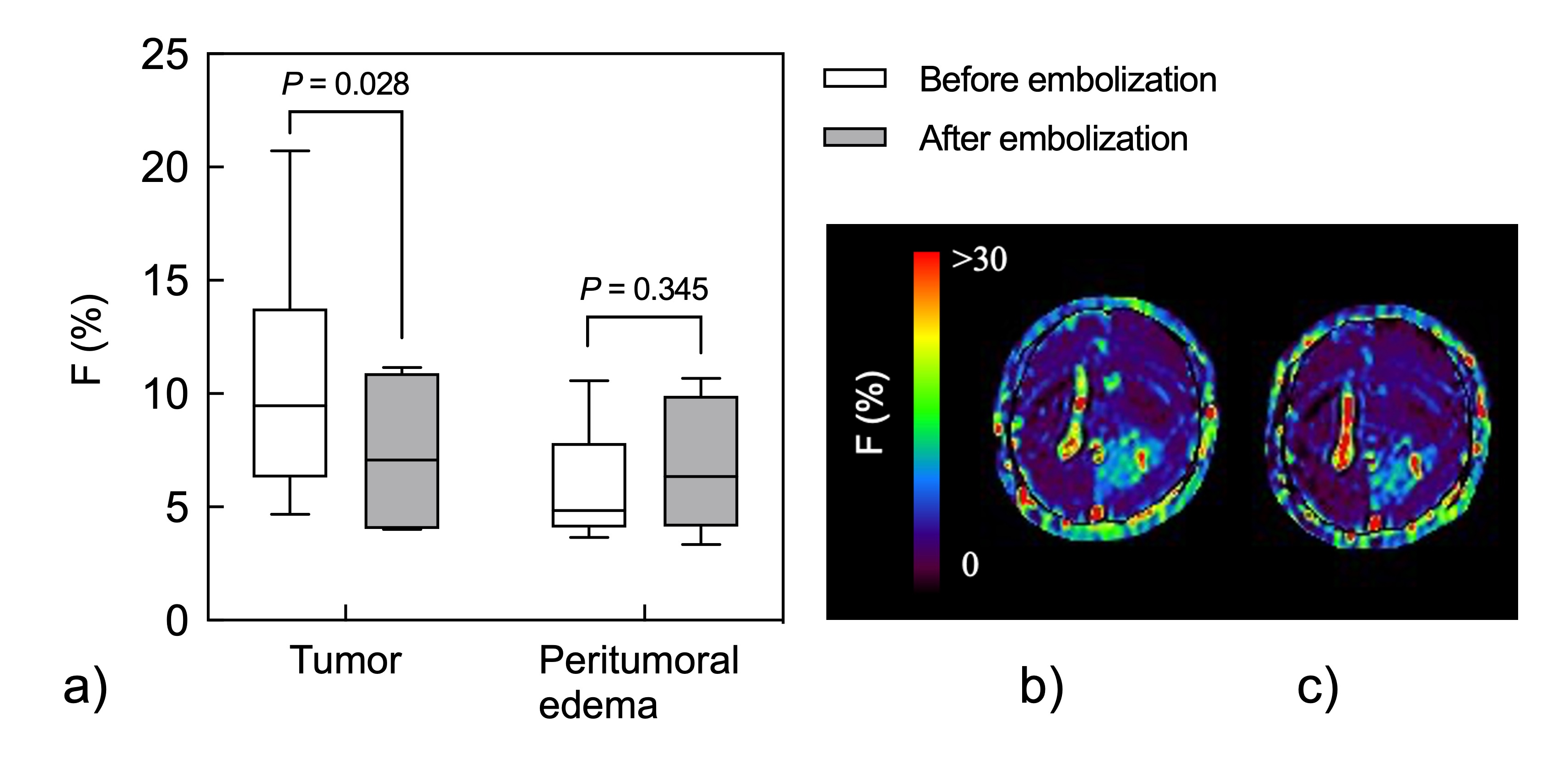
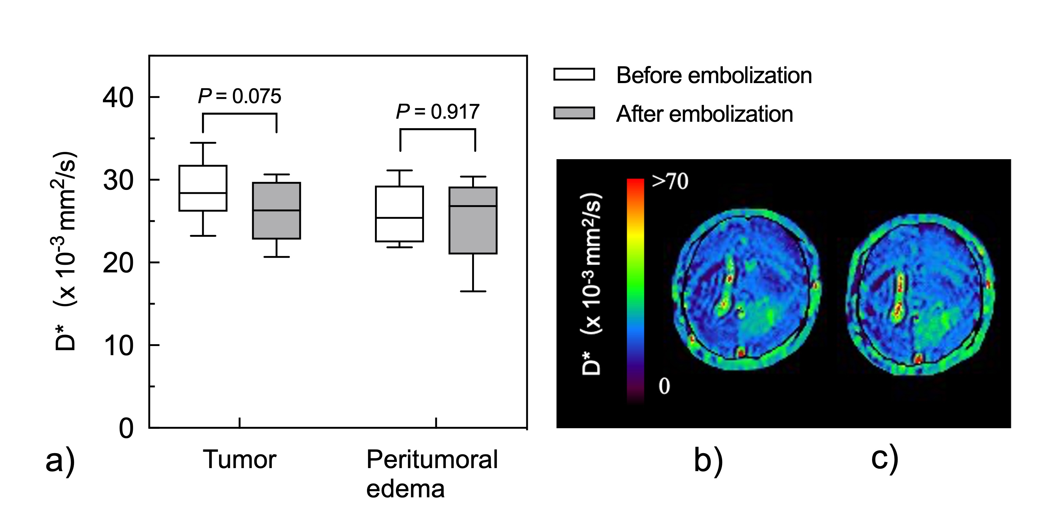
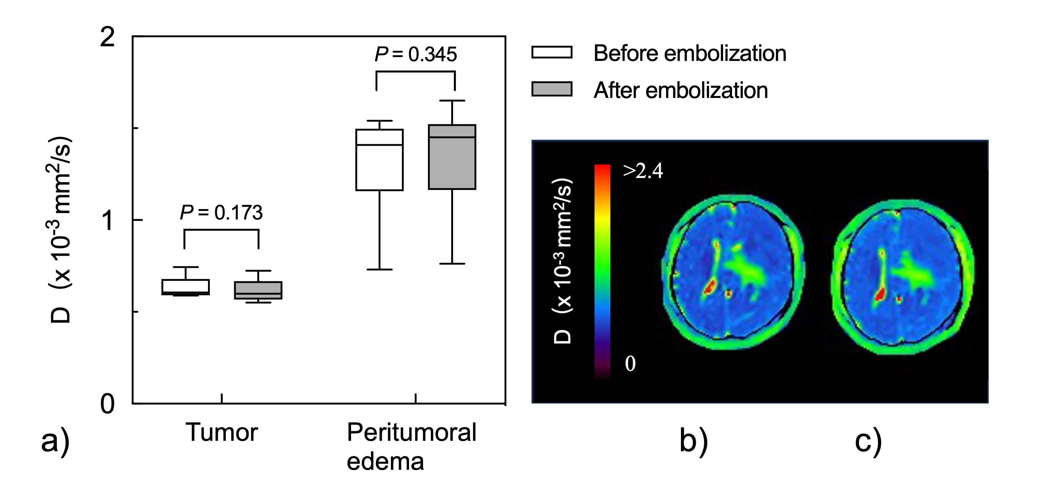
FD* and F, which depend on the cerebral blood flow and volume respectively,4,5 were significantly lower after endovascular embolization in meningioma than those before the endovascular embolization (reduction rates of FD* and F, 28.4 ± 21.4% and 26.0 ± 19.9%, respectively; P < 0.05 for both) (Figs. 1 and 2). These results were consistent with evaluations assessed using pseudocontinuous arterial spin labeling2 and dynamic susceptibility contrast MRI3 reported in literature. However, in peritumoral edema, there was no significant difference in FD* and F before and after endovascular embolization. Moreover, no significant difference was observed in D* (inverse proportion to mean transit time4,5) and D in meningioma and peritumoral edema (P ≥ 0.05 for both) (Figs. 3 and 4). These results suggested that preoperative endovascular embolization did not affect the perfusion and diffusion of peritumoral edema as well as water diffusion of a meningioma except in the necrotic areas.CONCLUSION
The IVIM analysis enabled simultaneous rapid evaluation of perfusion and diffusion after preoperative endovascular embolization in supratentorial meningiomas.Acknowledgements
No acknowledgement found.References
- Jumah F, AbuRmilah A, Raju B, et al. Does preoperative embolization improve outcomes of meningioma resection? A systematic review and meta-analysis. Neurosurg Rev. 2021; 44: 3151-3163.
- Wanibuchi M, Komatsu K, Akiyama Y, et al. Quantitative assessment of flow reduction after feeder embolization in meningioma by using pseudocontinuous arterial spin labeling. World Neurosurg. 2016; 93: 237-245.
- Gruber P, Schwyzer L, Klinger E, et al. imaging of tumor volume, diffusivity, and perfusion after preoperative endovascular embolization in supratentorial hemispheric meningiomas. World Neurosurg. 2018; 120: e357-e364.
- Le Bihan D and Turner R. The capillary network: a link between IVIM and classical perfusion. Magn Reson Med. 1992; 27: 171-178.
- Le Bihan D. What can we see with IVIM MRI? Neuroimage. 2019; 187: 56-67.
Figures