3703
MRI and tumor-infiltrating CD8+ T cell-based nomogram for predicting meningioma recurrence risk stratification1Lanzhou University Second Hospital, Lanzhou, China
Synopsis
Keywords: Tumors (Pre-Treatment), Tumor
Motivation: To investigate the efficacy of MRI features and CD8+ T cells in predicting risk stratification for meningioma recurrence.
Goal(s): To develop a reliable nomogram incorporating MRI features and CD8+ T cells to predict meningioma recurrence.
Approach: Conventional MRI features, ADC histogram parameters, and CD8+ T cells were recorded and compared. This model was the first to combine clinical, imaging, and TME data to predict meningioma recurrence.
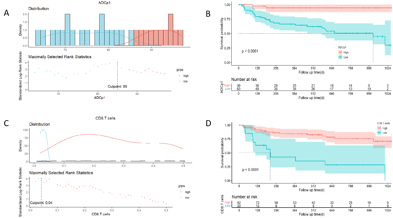
Results: The ADCp1 and CD8+ T cells as predictive variables for meningioma recurrence and patients with low ADCp1 or CD8+ T cell counts had higher recurrence rates than those with high ADCp1 or CD8+ T cell counts.
Impact: The findings will improve prognostic accuracy for patients with meningioma and potentially allow for targeted treatment of individuals who have the recurrent form.
Introduction
Meningiomas account for 39.0% of all central nervous system (CNS) tumors [1]. Although >80% of meningiomas are benign, a subset become aggressive and are categorized by the World Health Organization (WHO) as grade 2 to 3 meningiomas [2]. These are difficult to completely eradicate surgically, and patients experience early or multiple tumor recurrences. Unfortunately, the 10-year progression-free survival (PFS) rates for grades 1, 2, and 3 meningiomas are 75–90%, 23–78%, and 0%, respectively [3].The tumor microenvironment (TME) consists of numerous cell types and a variable extracellular matrix that enhance tumor immune tolerance, directly influencing cancer progression and recurrence [4]. Tumor-infiltrating lymphocytes (TILs) are important components of the meningioma TME and have a major effect on underlying tumor growth, proliferation, and overall prognosis [5]. In particular, CD8+ TILs exert antitumor effects [6], and their decrease in meningiomas suggests the presence of an immunosuppressive tumor microenvironment. A recent study [7] showed that WHO meningioma grades are negatively correlated with the proportion of CD4+, CD8+, and PD-1+ lymphocytes. For immunotherapy to become a realistic therapeutic approach, TILs in meningiomas must be thoroughly characterized, but few studies are available with the necessary data.
Currently, qualitative analyses are limited in predicting meningioma recurrence/progression and risk stratification. Apparent diffusion coefficient (ADC) histogram analysis is now widely used for meningioma grading, subtyping, and differential diagnosis [8]. However, there are little data regarding whether ADC histogram parameters can predict meningioma recurrence preoperatively. Therefore, this study aimed to investigate the prognostic value of preoperative MRI features and tumor-infiltrating CD8+ T cells in risk stratification for meningioma recurrence.
Methods

Clinical, pathological, and imaging data of 102 patients with pathologically confirmed meningiomas were retrospectively analyzed. Patients were divided into recurrence and non-recurrence groups based on follow-up. Tumor-infiltrating CD8+ T cells were quantitatively assessed with immunohistochemical staining. Preoperative ADC histogram parameters were quantified in MaZda. Next, independent risk predictors of meningioma recurrence were identified with a multivariate COX proportional hazards model. A visual nomogram was then constructed and the predicted recurrence probability was 1–2 years. Kaplan-Meier curves were plotted for meningioma recurrence-free survival.Results
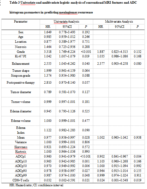
The risk factors for meningioma recurrence were ADCp1 (hazard ratio [HR] = 0.902, 95% confidence interval [95% CI]: 0.841~0.967, p = 0.004) and CD8+ T cells (HR = 0.024, 95%CI: 0.001~0.548, p = 0.019). The resultant nomogram had AUC values of 0.779 and 0.784 for 1- and 2-year predicted recurrence rates, respectively. The survival analysis revealed that patients with low ADCp1 or CD8+ T cell counts had higher recurrence rates than those with high ADCp1 or CD8+ T cell counts.Discussion
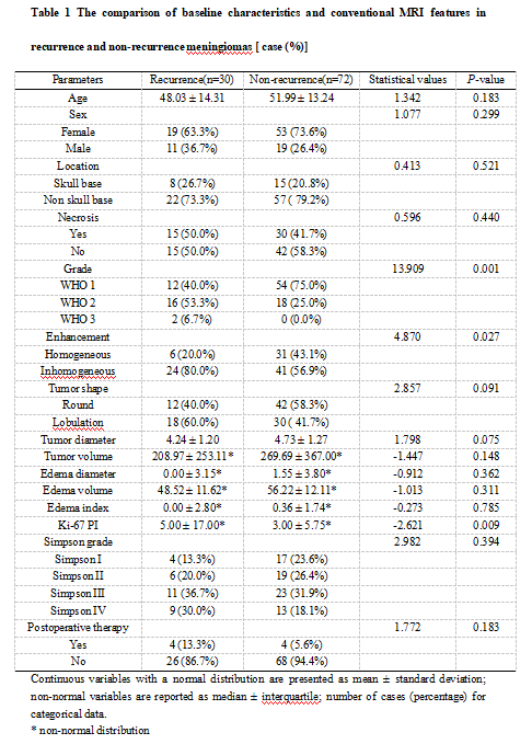
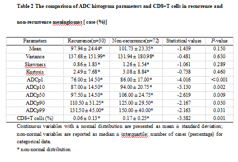
Previous study has found that meningioma recurrence is often associated with pathological grade, KI-67 PI, tumor diameter, location, necrosis, and peritumoral edema [9]. Similarly, we demonstrated that tumor grade and Ki-67 PI were risk factors for meningioma recurrence[10]. In addition, meningiomas are blood-rich tumors, and high-grade meningiomas often exhibit obvious intra-tumor heterogeneity that causes uneven enhancement, another important factor affecting meningioma recurrence.The results of comparing ADC histogram parameters revealed that they all differed significantly between two groups. This outcome is consistent with histopathological data showing that high-grade meningiomas exhibit higher mitotic activity and increased nuclear/cytoplasmic ratios of tumor cells. These characteristics in turn lead to lower ADC values and poor prognosis. Moreover, variance is valuable for assessing tumor heterogeneity, as it mainly responds to the degree of data dispersion in histogram parameters [11].
The presence of CD8+ TILs in meningiomas have a positive effect on survival [12]. We confirmed that the number of CD8+ T cells was significantly lower in the recurrence group than non-recurrence group. Likewise, Zhang et al. [13] found that high levels of TILs were associated with improved PFS. Similarly, an association between high CD8+ TIL levels and improved RFS has been observed in non-small cell lung carcinoma [14] and hepatocellular carcinoma [15]. Taken together, which illustrate the potential of CD8+ TILs as biomarkers for predicting meningioma recurrence.
Our model serves as an intuitive visual tool with demonstrable reliability and accuracy, while being an improvement to previous attempts. For example, a prior nomogram was built from clinical data to predict grade 2–3 meningiomas and measure meningioma prognosis [16-17]. In contrast, we included all meningioma grades to generate a model with broader adaptations. Most importantly, our nomogram incorporated TME data on CD8+ T cells, better reflecting the heterogeneity of meningiomas and thus increasing predictive accuracy.
Conclusion
In summary, ADCp1 and CD8+ T cells may be potential vivo biomarkers for predicting meningioma recurrence, providing clinicians a basis for guidance in personalised treatment and postoperative follow-up of meningioma patients.Acknowledgements
Not ApplicableReferences
1.Ostrom QT, Price M, Ryan K et al (2022) CBTRUS Statistical Report: Pediatric Brain Tumor Foundation Childhood and Adolescent Primary Brain and Other Central Nervous System Tumors
2.Diagnosed in the United States in 2014-2018. Neuro-oncology 24:iii1-iii38 Marastoni E, Barresi V (2023) Meningioma Grading beyond Histopathology: Relevance of Epigenetic and Genetic Features to Predict Clinical Outcome. Cancers. 2023;15(11)
3.Bi WL, Zhang M, Wu WW, Mei Y, Dunn IF (2016) Meningioma Genomics: Diagnostic, Prognostic, and Therapeutic Applications. Frontiers in surgery. 2016;3:40
4.Haslund-Vinding J, Møller JR, Ziebell M, Vilhardt F, Mathiesen T (2022) The role of systemic inflammatory cells in meningiomas. Neurosurgical review 45:1205-15
5.Turner CP, McLay J, Hermans IF et al (2022) Tumour infiltrating lymphocyte density differs by meningioma type and is associated with prognosis in atypical meningioma. Pathology 54:417-24
6.Butterfield LH (2015) Cancer vaccines. BMJ (Clinical research ed.) 350:h988
7.Du Z, Abedalthagafi M, Aizer AA et al (2015) Increased expression of the immune modulatory molecule PD-L1 (CD274) in anaplastic meningioma. Oncotarget 6:4704-16
8.Liu X, Huang X, Han T et al (2022) Discrimination between microcystic meningioma and atypical meningioma using whole-lesion apparent diffusion coefficient histogram analysis. Clinical radiology 77:864-9
9.Haddad AF, Young JS, Kanungo I et al (2020) WHO Grade I Meningioma Recurrence: Identifying High Risk Patients Using Histopathological Features and the MIB-1 Index. Frontiers in oncology 10:1522
10.Khanna O, Fathi Kazerooni A, Arif S et al (2023) Radiomic signatures of meningiomas using the Ki-67 proliferation index as a prognostic marker of clinical outcomes. Neurosurgical focus 54:E17
11.Han T, Liu X, Jing M et al (2023) ADC histogram parameters differentiating atypical from transitional meningiomas: correlation with Ki-67 proliferation index. Acta radiologica (Stockholm, Sweden : 1987):2841851231205151
12.Li YD, Veliceasa D, Lamano JB et al (2019) Systemic and local immunosuppression in patients with high-grade meningiomas. Cancer immunology, immunotherapy : CII 68:999-1009
13.Zhang Y, Wang X, Shi M, Song Y, Yu J, Han S (2022) Programmed death ligand 1 and tumor-infiltrating CD8(+) T lymphocytes are associated with the clinical features in meningioma. BMC cancer 22:1171
14.Huang B, Liu R, Wang P et al (2020) CD8(+)CD57(+) T cells exhibit distinct features in human non-small cell lung cancer. Journal for immunotherapy of cancer 8
15.Xu X, Tan Y, Qian Y et al (2019) Clinicopathologic and prognostic significance of tumor-infiltrating CD8+ T cells in patients with hepatocellular carcinoma: A meta-analysis. Medicine 98:e13923
16.Gao P, Kong T, Zhu X et al (2021) A Clinical Prognostic Model Based on Preoperative Hematological and Clinical Parameters Predicts the Progression of Primary WHO Grade II Meningioma. Frontiers in oncology 11:748586
17.Jia Z, Yan Y, Wang J et al (2021) Development and Validation of Prognostic Nomogram in Patients With WHO Grade III Meningioma: A Retrospective Cohort Study Based on SEER Database. Frontiers in oncology 11:719974
Figures