3701
Identification of Prognostic Imaging Biomarkers in H3 K27-Altered Diffuse Midline Gliomas in Adults: Impact of Tumor Oxygenation Biomarkers1Department of Radiology, Yonsei University College of Medicine, Seoul, Korea, Republic of
Synopsis
Keywords: Tumors (Pre-Treatment), DSC & DCE Perfusion
Motivation: The prognostic markers of H3 K27-altered diffuse midline gliomas (DMGs) in adults have yet to be studied.
Goal(s): To investigate prognostic markers for H3 K27-altered DMGs in adults, including clinical, qualitative, quantitative imaging phenotypes, and tumor oxygenation characteristics.
Approach: Clinical, qualitative, and quantitative imaging phenotypes were analyzed in 32 adults with H3 K27-altered DMGs. Cox analyses were conducted to determine predictors of overall survival in entire patients with H3 K27-altered DMG and subgroup of contrast-enhancing (CE) tumor.
Results: The presence of LM and higher rCMRO2 of CE tumor were independently associated with poor prognosis in adult patients with H3 K27-altered DMG.
Impact: Tumor oxygenation imaging biomarkers may provide valuable insight into the prognosis of H3 K27-altered diffuse midline gliomas, potentially reflecting higher metabolic activity in the tumor microenvironment.
Purpose
To investigate prognostic markers for H3 K27-altered diffuse midline gliomas (DMGs) in adults including clinical, qualitative, quantitative imaging phenotypes, and tumor oxygenation characteristics.Methods
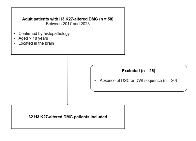
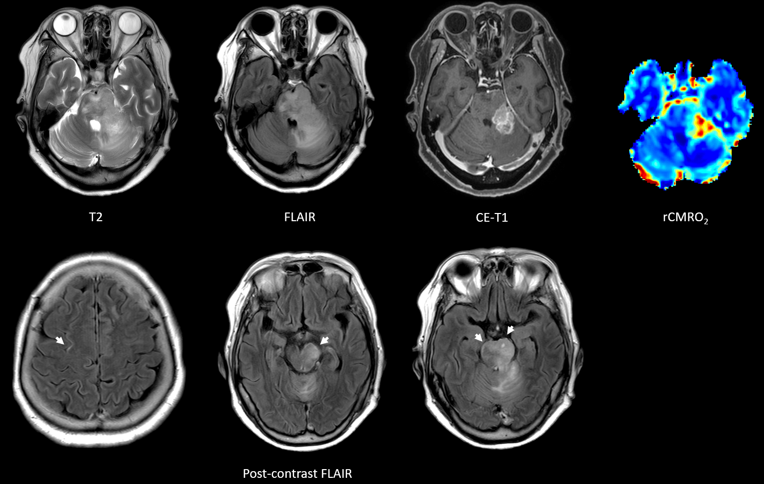
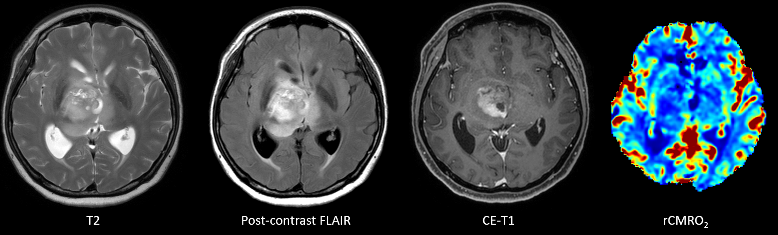
Retrospective chart and imaging reviews were conducted on 32 adult patients with H3 K27-altered DMGs between 2017 and 2023. Clinical and qualitative imaging characteristics were analyzed. Quantitative imaging assessment was performed from the tumor mask via automatic segmentation to calculate normalized cerebral blood volume (nCBV), capillary transit time heterogeneity (CTH), oxygen extraction fraction (OEF), relative cerebral metabolic rate of oxygen (rCMRO2) values, and mean ADC value. Leptomeningeal metastases (LM) was diagnosed with imaging. Cox analyses were conducted to determine predictors of overall survival (OS) in entire patients with H3 K27-altered DMG and subgroup of contrast-enhancing (CE) tumor.Results
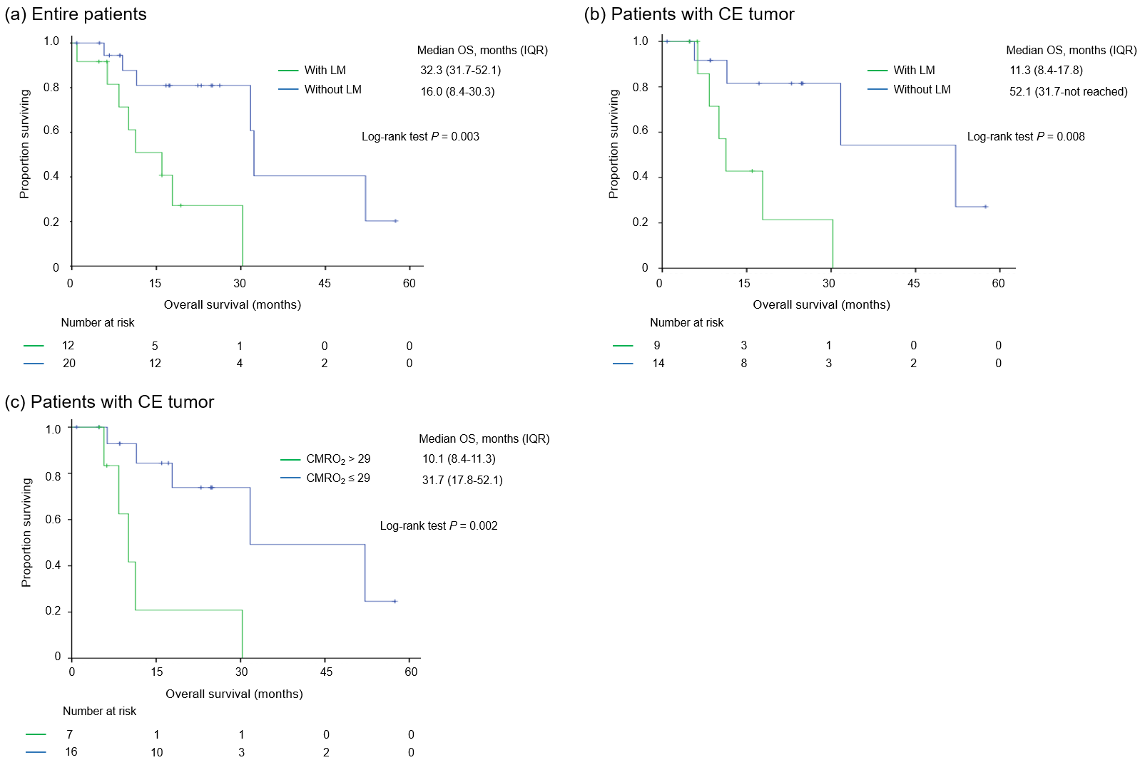
The median patient age was 40.5 years (range 19.9-75.7), with an OS of 30.3 months (IQR 11.3-32.3). On multivariable analysis on entire patients, the presence of LM was a predictor of OS (hazard ratio [HR] = 6.01, P = 0.009). In the subgroup of 23 (71.9%) patients with CE tumors, rCMRO2 of CE tumor (HR = 1.08, P = 0.019) and the presence of LM (HR = 5.92, P = 0.043) were independent predictors of OS.Conclusions
The presence of LM was independently associated with poor prognosis in adult patients with H3 K27-altered DMG. In patients with CE tumors, higher rCMRO2 of CE tumor, which may reflect higher metabolic activity in the tumor oxygenation microenvironment, may be a useful imaging biomarker to predict poor prognosis.Acknowledgements
NoneReferences
Persson ML, Douglas AM, Alvaro F, et al. The intrinsic and microenvironmental features of diffuse midline glioma: Implications for the development of effective immunotherapeutic treatment strategies. Neuro-Oncology 2022;24(9):1408-1422.Schulte JD, Buerki RA, Lapointe S, et al. Clinical, radiologic, and genetic characteristics of histone H3 K27M-mutant diffuse midline gliomas in adults. Neurooncol Adv 2020;2(1)::vdaa142.
Kickingereder P, Brugnara G, Hansen MB, et al. Noninvasive Characterization of Tumor Angiogenesis and Oxygenation in Bevacizumab-treated Recurrent Glioblastoma by Using Dynamic Susceptibility MRI: Secondary Analysis of the European Organization for Research and Treatment of Cancer 26101 Trial. Radiology 2020;297(1):164-175.
Park YW, Han K, Park JE, et al. Leptomeningeal metastases in glioma revisited: incidence and molecular predictors based on postcontrast fluid-attenuated inversion recovery imaging. Journal of Neurosurgery 2023;139(1):38-48.
López-Pérez CA, Franco-Mojica X, Villanueva-Gaona R, Díaz-Alba A, Rodríguez-Florido MA, Navarro VG. Adult diffuse midline gliomas H3 K27-altered: review of a redefined entity. J Neurooncol 2022;158(3):369-378.
Zheng L, Gong J, Yu T, et al. Diffuse Midline Gliomas With Histone H3 K27M Mutation in Adults and Children: A Retrospective Series of 164 Cases. The American Journal of Surgical Pathology 2022;46(6).
Vuong HG, Ngo TNM, Le HT, et al. Prognostic Implication of Patient Age in H3K27M-Mutant Midline Gliomas. Front Oncol 2022;12:858148.
Bin-Alamer O, Jimenez AE, Azad TD, Bettegowda C, Mukherjee D. H3K27M-Altered Diffuse Midline Gliomas Among Adult Patients: A Systematic Review of Clinical Features and Survival Analysis. World Neurosurgery 2022;165:e251-e264.
Figures