3699
3D Slip Interface Imaging on the basis of magnetic resonance elastography can preoperatively cauge the degree of meningioma–brain adhesion1Shengjing Hospital of China Medical University, Shenyang, China, 2Mayo Clinic College of Medicine, Rochester, MN, United States
Synopsis
Keywords: Tumors (Pre-Treatment), Tumor, magnetic resonance elastography, three dimensional slip interface imaging, tumor–brain adhesion, meningioma
Motivation: The adhesion of brain tumor to brain tissue is very important to evaluate the surgical risk and prognosis of patients
Goal(s): To investigate the ability of slip interface imaging (SII), based on a recently developed technique-magnetic resonance elastography (MRE), to predict the degree of meningioma–brain adhesion
Approach: slip interface imaging (SII), based on a recently developed technique-magnetic resonance elastography (MRE)
Results: SII agreed with the intraoperative assessment of the degree of tumor adhesion (medium agreement, k=0.688, 95% CI: 0.15-1)
Impact: Preoperative non-invasive evaluation of brain tumors and the degree of adhesion around the guidance of the surgeon to assess the risk of surgery and selection of surgical methods
Introduction
The adhesion of brain tumor to brain tissue is very important to evaluate the surgical risk and prognosis of patients.To investigate the ability of three dimensional slip interface imaging (3D SII), based on a recently developed technique-magnetic resonance elastography (MRE), to predict the degree of meningioma–brain adhesion.Methods
With Institutional Review Board approval and written informed consent,3D SII examinations were performed in five patients with meningiomas preoperatively.Each tumor was evaluated on six dimensions. All 3D SII examinations involved Intracranial shear motions were introduced using a soft, pillow-like head driver. The displacement field was acquired with an MRE on 3T MR scanners. The acquired shear motion data creates three dimensional slip interface imaging (3D SII).Two surgeon specialists in brain surgery primarily conducted all surgical resections, their intraoperative impressions of tumor adhesion serving as reference standards. The 3D SII findings were independently and blindly correlated with surgical findings of tumor adhesion by using the Cohen’s k coefficient.Results
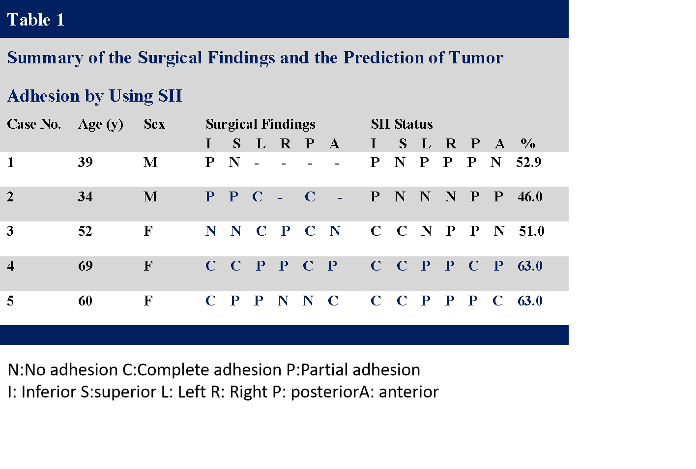
Among the five patients, 3D SII agreed with the intraoperative assessment of the degree of tumor adhesion in four patients (80%) cases (medium agreement, k=0.688, 95% CI: 0.15-1). Among the 26 dimensions, 3D SII agreed with 19 dimensions (73%) (medium agreement, k=0.592, 95% CI: 0.33-0.85).Discussion
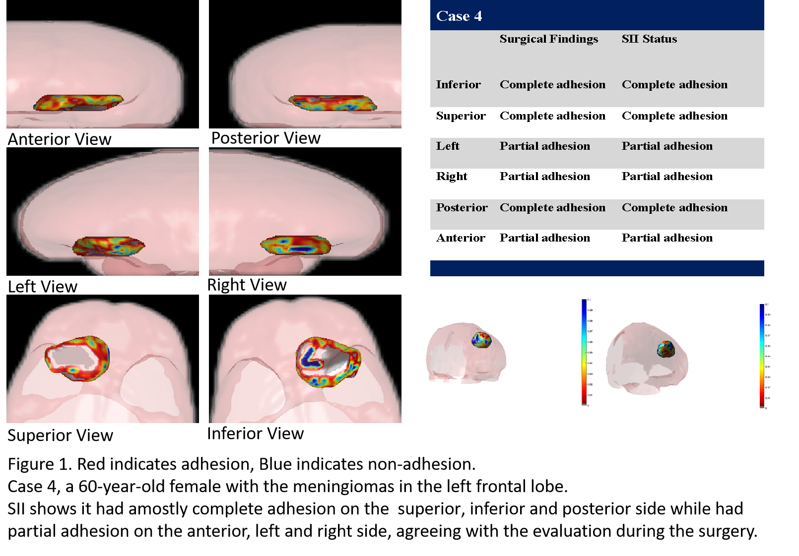
Case 1 had slight adhesions on one side of the foot and no adhesions on the head on the other side during the surgical procedure for the meningioma. Case 2 had adhesions at the origin of the temporal side of the occipital region, adhesions on the left ear, slight adhesions on the nasal side, slight adhesions on the other side of the ear, no adhesions in the cerebellum, and no adhesions on the top side during the meningioma surgery. Case3 had severe adhesions on the side of the cerebellum, adhesions on the nasal side, slight adhesions on the top side, slight adhesions on one side of the ear, no adhesions on the other side of the ear, and no adhesions on the occipital side during the meningioma surgery. Case 4 had severe adhesions on the side of the skull base, adhesions on the top side, adhesions on the occipital side, slight adhesions on one side of the ear, slight adhesions on the other side of the ear, and slight adhesions on the nasal side.had slight adhesion on one side of the foot and no adhesion on the head side during the surgery, agreeing with SII. Case 5 had severe adhesion on the side of the skull base, adhesion on the top side, adhesion on the occipital side, slight adhesion on one side of the ear, slight adhesion on the other side of the ear, and slight adhesion on the nasal side during the surgery, mostly agreeing with SII.Conclusion
3D SII, a noninvasive technique can be used to evaluates the degree of meningioma–brain adhesion preoperatively , to predict surgical risk and tumor resectability.Acknowledgements
No acknowledgement found.References
1. Yin Z, Hughes JD, Trzasko JD, et al. Slip interface imaging based on MR-elastography preoperatively predicts meningioma-brain adhesion. J Magn Reson Imaging 2017; 46:1007-1016.
2. Hughes JD, Fattahi N, Van Gompel J, et al. Higher-Resolution Magnetic Resonance Elastography in Meningiomas to Determine Intratumoral Consistency. Neurosurgery 2015; 77:653-658, 658-659.
3. Romani R, Tang WJ, Mao Y, et al. Diffusion tensor magnetic resonance imaging for predicting the consistency of intracranial meningiomas. Acta Neurochir (Wien) 2014; 156:1837-1845.
Figures