3698
Radiomics-based prediction of intraoperative bleeding rate during intracranial meningiomas surgery1Novosibirsk Neurosurgery Center, Novosibirsk, Russian Federation, 2Novosibirsk State University, Novosibirsk, Russian Federation
Synopsis
Keywords: Tumors (Pre-Treatment), Tumor
Motivation: The usefulness of radiomics in predicting intraoperative bleeding rate remains underestimated. Our objective was to examine the potential of radiomic characteristics to predict the intraoperative bleeding rate in patients with intracranial meningiomas.
Goal(s): Как To predict intraoperative bleeding rate in patients with intracranial meningiomas using radiomics, machine learning and regression methods.
Approach: Brain 3T MRI was performed with subsequent tumor segmentation and radiomics analysis.
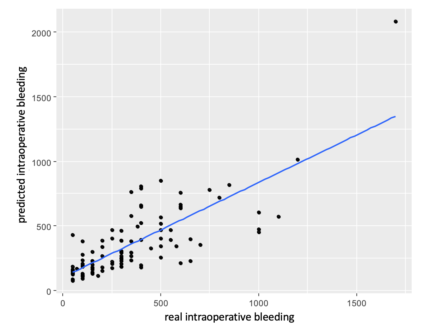
Results: The combination of six ADC- and ASL-based radiomics features allowed us to predict the intraoperative bleeding rate with raw residuals estimation -23 (-101; 68) (Me (1; 3 quantile)) in patients with intracranial meningiomas.
Impact: Our results provide an additional non-invasive tool for the evaluation of meningiomas, which potentially could impact the treatment tactic(for example, making a decision about performing a pre-surgicalembolization in cases with high-risk).
Introduction
Meningiomas are the most common primary tumor of the central nervous system, and surgery is the main treatment option for them [1]. However, meningioma resection is frequently accompanied by substantial bleeding, which is associated with an increased incidence of medical morbidities. Magnetic resonance imaging (MRI) has an essential role in preoperative tumor assessment. Furthermore, neuroimaging features, such as the radiomic characteristics of meningioma in different MRI sequences, could provide additional quantitative information on the tumor and be useful in predicting tumor grade, brain invasion, and recurrence [2].Nonetheless, the usefulness of radiomics in predicting intraoperative bleeding rate remains underestimated. Our objective was to examine the potential of radiomic characteristics to predict the intraoperative bleeding rate in patients with intracranial meningiomas.Methods
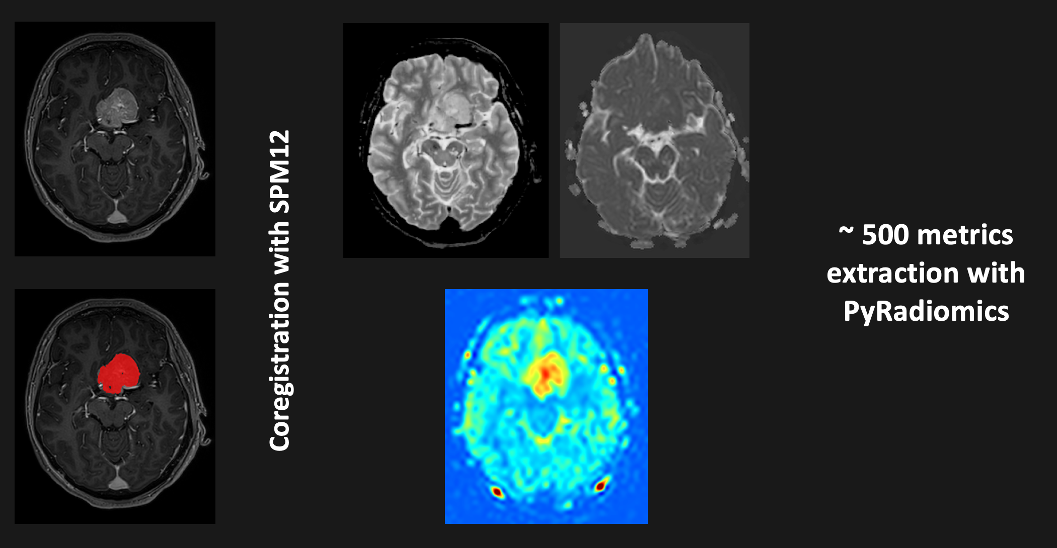
102 patients with primary diagnosed intracranial meningiomas were evaluated using high-resolution brain magnetic resonance imaging (MRI), which included the T1-weighted pre- and postcontrast, T2-weighted, diffusion-weighted sequences, as well as arterial spin labeling (ASL). 3T MRI data were analysed with ITK-Snap v4.1 [3], SPM12 (http://fil.ion.ucl.ac.uk/spm/), and PyRadiomicstoolbox [4], with semiautomatic tumor segmentation in postcontrast T1-weighted images, spatial coregistration between modalities, and subsequent extraction of radiomic features (Figure 1). The most significant predictors were determined by the 'random forest' machine learning-based method, with subsequent negative binomial regression analysis to model the dependency of selected metrics on intraoperative bleeding rate.Results
We found that the best predictors of intraoperative bleeding were the radiomic parameters of ASL and the apparent diffusion coefficient (ADC). Specifically, the combination of glszm_SizeZoneNonUniformity, ngtdm_Coarseness, glrlm_LowGrayLevelRunEmphasis metrics within ASL-based cerebral blood flow (CBF) maps and glrlm_GrayLevelNonUniformity, glrlm_RunEntropy, andglszm_ZoneEntropy metrics within ADC maps was the most effective (p < 0.01 for all factors). The resulting model allowed us to predict the intraoperative bleeding rate with raw residuals estimation -23 (- 101; 68) (Me (1; 3 quantile)) in patients with intracranial meningiomas (Figure 2). Tumor volume, localization and histological grade were much less significant predictors and did not improve the model.Discussion
In line with our hypothesis, radiomic features could provide valuable information for the presurgical evaluation of patients with intracranial meningiomas. The ADC and ASL were the two modalities to predict intraoperative bleeding rate in patients with intracranial meningiomas with high precision. Both diffusion and perfusion modalities provide the information about tissue structure, specifically, about tumor cellularity (ADC) and tumor microvasculature (ASL). On the other hand, all included in our model radiomic metrics generally reflect the signal inhomogeneity. Thus, according to our data, more heterogeneous tumors are more prone to bleeding. To our knowledge, this is the first attempt to predict theintraoperative bleeding rate with the radiomic method in patients with intracranial meningiomas. A previously published study demonstrated the association between tumour volume and intraoperative bleeding [5], which is expected and has been confirmed in our dataset.However, the combination of ADC and ASL-based radiomic metrics (as well as each metric independently) is much more effective in predicting bleeding rates. More studies with larger sample sizes are needed to prove our results and evaluate their utility in patient care.Conclusion
Our results provide an additional noninvasive tool for the presurgical evaluation of intracranial meningiomas, which could potentially affect the treatment tactic.Acknowledgements
No acknowledgement found.References
[1]R. A. Buerki, C. M. Horbinski, T. Kruser, P. M. Horowitz, C. D. James, and R. V. Lukas, “An overview of meningiomas,” Future Oncology, vol. 14, no. 21, p. 2161, Sep. 2018, doi: 10.2217/FON-2018-0006.
[2]H. Gu, X. Zhang, P. di Russo, X. Zhao, and T. Xu, “The Current State of Radiomics for Meningiomas: Promises and Challenges,” Front Oncol, vol. 10, p. 567736, Oct. 2020, doi: 10.3389/FONC.2020.567736/BIBTEX.
[3]P. A. Yushkevich, Y. Gao, and G. Gerig, “ITK-SNAP: an interactive tool for semi-automatic segmentation of multi-modality biomedical images,” Conf Proc IEEE Eng Med BiolSoc, vol. 2016, p. 3342, Oct. 2016, doi: 10.1109/EMBC.2016.7591443.
[4]J. J. M. Van Griethuysen et al., “Computational radiomics system to decode the radiographic phenotype,” Cancer Res, vol. 77, no. 21, pp. e104–e107, Nov. 2017, doi: 10.1158/0008-5472.CAN-17-0339/SUPPLEMENTARY-VIDEO-S2.
[5]S. Y. Hsu and Y. H. Huang, “Characterization and prognostic implications of significant blood loss during intracranial meningioma surgery,” Transl Cancer Res, vol. 5, no. 6, pp. 797–804, 2016, doi: 10.21037/TCR.2016.11.72.
Figures