3696
Identifying NF-2 Mutations in Meningiomas Based on Susceptibility Weighted Imaging for Patient Prognosis Using Machine Learning1Institute of Biomedical Engineering, Bogazici University, Istanbul, Turkey, 2Department of Radiology, Basaksehir Cam and Sakura City Hospital, Istanbul, Turkey, 3Department of Medical Pathology, Acibadem University, Istanbul, Turkey, 4Center for Neuroradiological Applications and Reseach, Acibadem University, Istanbul, Turkey, 5Electric and Electronic Engineering Department, Bogazici University, Istanbul, Turkey, 6Department of Neurosurgery, Acibadem University, Istanbul, Turkey, 7Department of Radiology, Acibadem University, Istanbul, Turkey
Synopsis
Keywords: Tumors (Pre-Treatment), Machine Learning/Artificial Intelligence
Motivation: Molecular markers, like neurofibromatosis type-2 (NF-2) mutations, highly impact patient outcomes in meningiomas, but they could only be assessed in excised tissue.
Goal(s): To develop a non-invasive approach for preoperatively identifying NF-2 mutations using susceptibility-weighted MRI (SWI) with radiomics and deep learning.
Approach: Preoperative SWI of 92 meningiomas with NF-2 status data were analyzed. Radiomics and deep learning were used to extract features of SWI, which were classified using traditional machine learning.
Results: Reduced tumor signal intensity, "en plaque" growth pattern, and intratumoral calcification were markers of NF2 mutation, which was identified with an accuracy of 74%.
Impact: This study employed SWI to predict NF-2 mutation through radiomics and deep learning features with 74% accuracy. Preoperative identification of NF-2 mutations might allow for personalized treatment planning resulting in better patient outcomes.
Introduction
Meningiomas are the most common primary intracranial tumors in adults, with diverse subtypes classified by the World Health Organization1,2. Accurate prognosis and treatment planning of meningiomas are crucial due to their variable aggressiveness, influenced by demographic, anatomical, histopathological, and clinical factors, along with molecular differences3. Genetic studies have shown that over two-thirds of meningiomas carry neurofibromatosis type 2 (NF-2) gene mutations or deletions, often associated with multiple tumors and a worse prognosis4-6. Identifying NF-2 mutations in meningiomas preoperatively is of interest for better treatment planning and outcome prediction. Susceptibility-weighted MRI (SWI), which could detect new blood vessel formation and intratumoral calcification, has been used for grading meningiomas7,8. This study aims to identify NF-2 mutations in meningiomas based on SWI using radiomics and deep learning features.Material-Methods
This study included 92 patients diagnosed with meningiomas, who underwent preoperative MRI scans and had available NF-2 mutation status. A neuropathologist assigned WHO grades. NF-2 allele loss was analyzed in tumor samples using Droplet digital polymerase chain reaction. All patients were scanned using a brain tumor imaging protocol at a 3T clinical MRI scanner (Siemens, Germany), including pre and post-contrast (Gd-DTPA) T1-weighted MRI (T1WI) (TR/TE = 589/10ms, FOV = 220mm, slice thickness = 3mm), fluid-attenuated inversion recovery (FLAIR) (TR/TE = 8320/92ms, TI = 2000ms, FOV = 220mm, slice thickness = 3mm), and SWI (TR/TE = 28/20ms, FOV = 220mm, slice thickness = 1.6mm). Morphological characteristics of the tumors were semi-quantitatively evaluated by a radiologist, including growth pattern, peritumoral edema, venous sinus invasion, hyperostosis, bone destruction, and intratumoral calcification. Tumor volumes were segmented on FLAIR using Slicer v4.8.1 (http://slicer.org/). After segmentation, image registration was carried out for each patient. This process involved aligning FLAIR images with high-resolution SWI images. The transformation matrices obtained from registration were utilized to align segmentation masks onto the SWI images, using the FMRIB Software (http://fsl.fmrib.ox.ac.uk/fsl/fslwiki/). Radiomics features of the FLAIR hyperintense region on SWI were extracted using Pyradiomics9. Univariate and multivariate logistic regression analyses were used to assess associations between textural features, morphological characteristics, age, and NF-2 mutational status. Additionally, deep learning features were extracted from FLAIR hyperintensity regions on SWI slices involving the largest tumoral area and the two adjacent slices. A pre-trained ResNet50 network was used for feature extraction10. Extracted features were used with classical machine learning models to identify NF2 mutation status. The model performance was evaluated through cross-validation, with accuracy, area under the curve (AUC), sensitivity, and specificity. The statistical analysis was conducted using R (v2022.12.0).Results
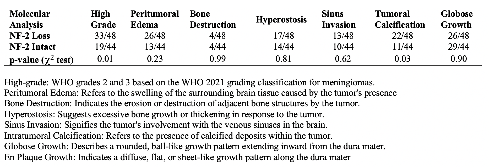
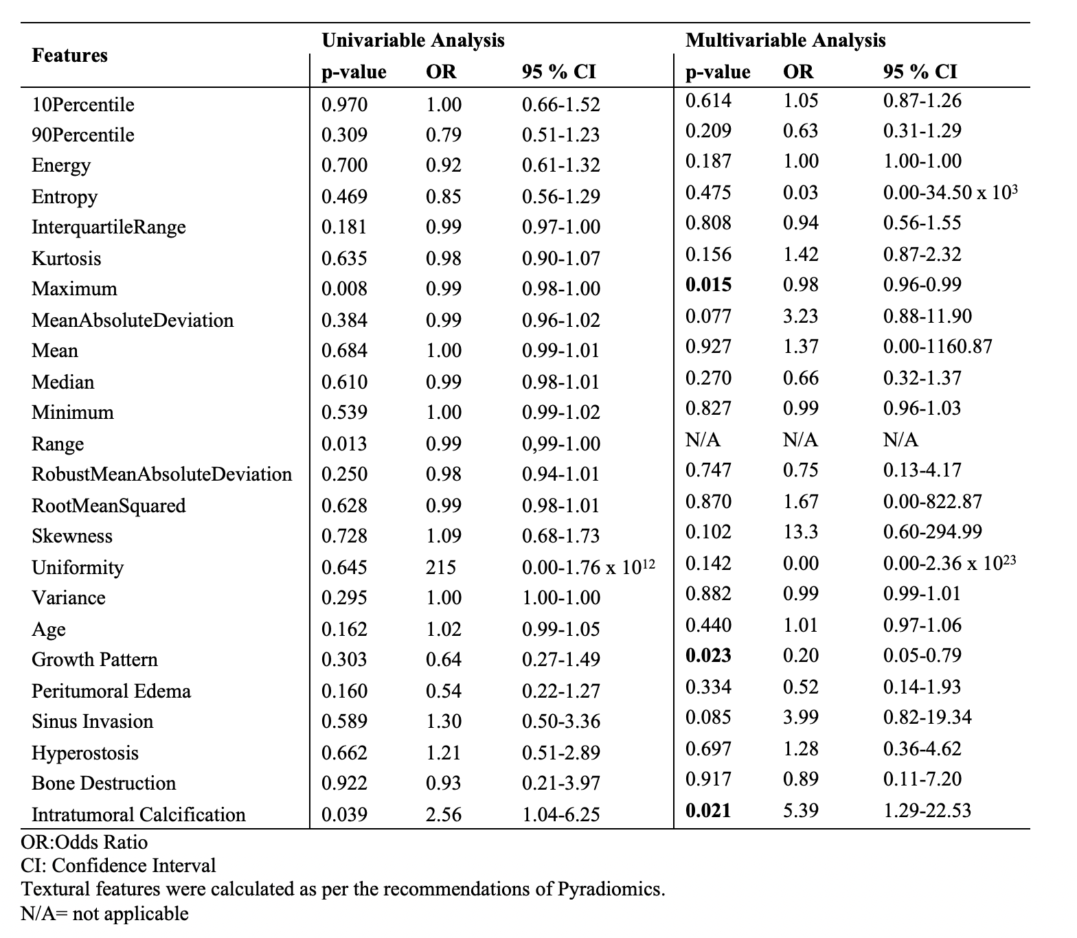
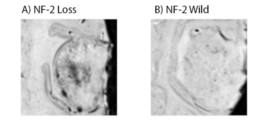
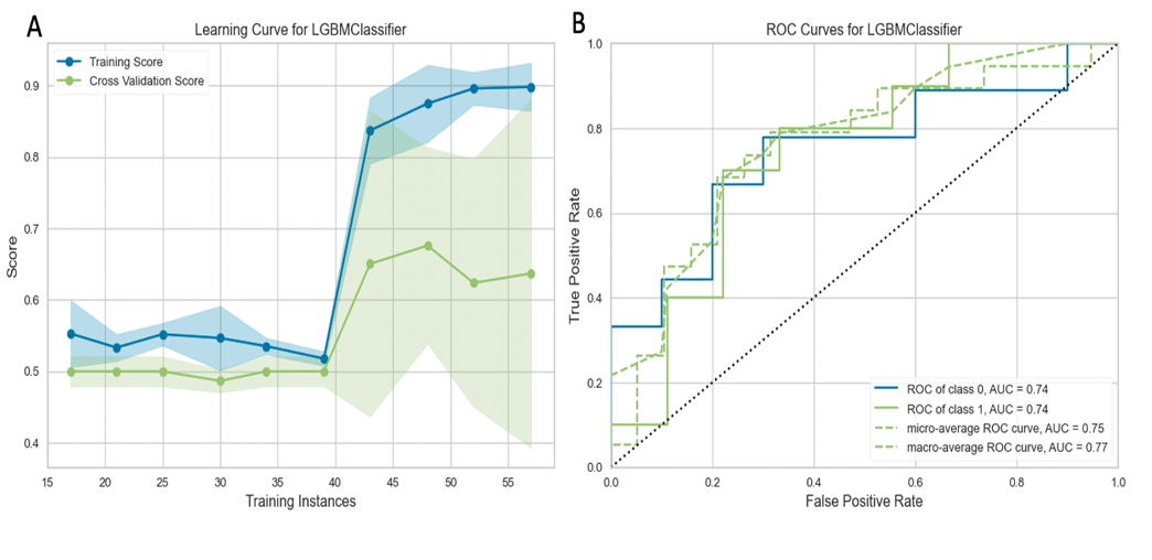
The mean age of the patient cohort was 52.9 ± 13.9 years. Among the 92 patients with available NF-2 mutation data, 48 exhibited NF-2 copy number loss (NF2-L), out of which 33 were categorized as high-grade tumors. On the other hand, 19 out of 44 meningiomas without NF-2 mutation (NF2-NL) were high-grade. There was a statistically significant difference between the NF2-L and N-NL groups in terms of the distribution of the tumor grade (p = 0.01, χ2 test) (Table 1). Multivariable logistic regression identified reduced maximum signal intensity within the tumor, the presence of an "en plaque growth" pattern, and the existence of intratumoral calcification as significant predictors of NF-2 copy number loss, with odds ratios of 0.98 (p = 0.015), 0.20 (p = 0.023), and 5.39 (p = 0.021), respectively (Table 2). Figure 1 shows SWI of the largest tumor region for example cases of NF2-L meningioma (A) and NF2-NL meningioma (B), in which NF2-L displays more neovascularization. Using the features identified through multivariable logistic regression, our model achieved a prediction accuracy of 0.74 with a sensitivity of 0.60 and specificity of 0.89, for identifying NF-2 copy number loss. Using deep learning features, a light gradient boosting machine algorithm demonstrated the highest accuracy, utilizing a set of 11 selected features, resulting in an accuracy of 0.73, an AUC of 0.74, a sensitivity of 0.70, and a specificity of 0.77 (Figure 2).Discussion-Conclusion
This study utilized SWI to identify NF-2 mutation in meningiomas using radiomics features with machine learning. Specific tumor characteristics, such as intratumoral calcification and the 'en plaque' growth pattern, played a crucial role in predicting NF-2 copy number loss. The current literature3,11 highlights the importance of considering genetic factors like NF-2 mutations alongside histological grading for prognosis assessment, and preoperatively identifying NF-2 mutation based on SWI might contribute to this goal. Future studies will validate our results in a larger patient cohort.Acknowledgements
This study was supported by the Scientific and Technological Research Council of Turkey (TUBITAK) grant 119S520.References
1 Louis, D. N. et al. The 2021 WHO Classification of Tumors of the Central Nervous System: a summary. Neuro Oncol 23, 1231-1251, doi:10.1093/neuonc/noab106 (2021).
2 Yamashima, T. in Meningiomas (ed Joung H. Lee) 15-24 (Springer London, 2009).
3 Nassiri, F. et al. A clinically applicable integrative molecular classification of meningiomas. Nature 597, 119-125, doi:10.1038/s41586-021-03850-3 (2021).
4 Youngblood, M. W. et al. Associations of meningioma molecular subgroup and tumor recurrence. Neuro Oncol 23, 783-794, doi:10.1093/neuonc/noaa226 (2021).
5 Moazzam, A. A., Wagle, N. & Zada, G. Recent developments in chemotherapy for meningiomas: a review. Neurosurg Focus 35, E18, doi:10.3171/2013.10.FOCUS13341 (2013).
6 Smith, M. J. et al. Cranial meningiomas in 411 neurofibromatosis type 2 (NF2) patients with proven gene mutations: clear positional effect of mutations, but absence of female severity effect on age at onset. J Med Genet 48, 261-265, doi:10.1136/jmg.2010.085241 (2011).
7 Hu, J. et al. Machine learning-based radiomics analysis in predicting the meningioma grade using multiparametric MRI. Eur J Radiol 131, 109251, doi:10.1016/j.ejrad.2020.109251 (2020).
8 Zhang, S. et al. Grading meningiomas utilizing multiparametric MRI with inclusion of susceptibility weighted imaging and quantitative susceptibility mapping. J Neuroradiol 47, 272-277, doi:10.1016/j.neurad.2019.05.002 (2020).
9 van Griethuysen, J. J. M. et al. Computational Radiomics System to Decode the Radiographic Phenotype. Cancer Research 77, E104-E107, doi:10.1158/0008-5472.Can-17-0339 (2017).
10 He K, Z. X., Ren S, Sun J. in 2016 IEEE Conference on Computer Vision and Pattern Recognition (CVPR).
11 Bachir, S. et al. Neurofibromatosis Type 2 (NF2) and the Implications for Vestibular Schwannoma and Meningioma Pathogenesis. Int J Mol Sci 22, doi:10.3390/ijms22020690 (2021).
Figures