3694
Machine learning based contrast-enhancement and IDH status prediction of gliomas using 7T MR spectroscopic imaging1Research Center for Medical Image Analysis and Artificial Intelligence (MIAAI), Danube Private University, Krems, Austria, 2High-Field MR Center - 7T MR, Department of Biomedical imaging and Image-guided Therapy, Medical University of Vienna, Vienna, Austria, 3Department of Neurosurgery, Medical University of Vienna, Vienna, Austria, 4Christian Doppler Laboratory for MR Imaging Biomarkers, Vienna, Austria, 5Division of Oncology, Department of Medicine I, Medical University of Vienna, Vienna, Austria, 6Division of Neuropathology and Neurochemistry, Department of Neurology, Medical University of Vienna, Vienna, Austria
Synopsis
Keywords: Tumors (Pre-Treatment), Brain
Motivation: IDH-mutant diffuse adult type gliomas almost invariably lead to fatality. The INDIGO trial found vorasidenib significantly improving progression-free survival in non-contrast enhancing IDH mutant CNS grade 2 glioma patients.
Goal(s): The purpose of this study was to non-invasively predict both contrast enhancement and IDH mutation in glioma patients.
Approach: We employed a machine learning approach on 7T MRSI data to forecast IDH mutation status and contrast-enhancing tumor tissue in adult diffuse gliomas.
Results: Our models performed well in the training and the testing set (AUC ≥ 0.8) for both, IDH mutation and contrast enhancement prediction.
Impact: With regard to emerging IDH inhibition therapies in IDH mutant non-contrast enhancing diffuse gliomas, non-invasive prediction of IDH mutation status and contrast enhancement are of utmost importance for glioma patients. 7T MRSI can be successfully applied to this task.
Introduction
MRI serves as the primary imaging method for gliomas. Radiological grading mainly relies on contrast enhancement (CE) of tumor tissue. Despite the highly favorable safety profile of gadolinium-based contrast agents [1,2], safety concerns have arisen about contrast agent safety due to brain gadolinium deposits from repeat use [3,4].In addition to histopathological characteristics, molecular features are crucial in brain tumor diagnosis. The 2021 WHO classification of tumors of the central nervous system, (WHO CNS5) employs the status of isocitrate dehydrogenase (IDH) mutations to distinguish IDH mutated gliomas from glioblastoma, which lacks IDH mutations [5,6]. Magnetic resonance spectroscopic imaging (MRSI) is a technique that can image multiple oncometabolites, especially at 7T, which are connected to molecular features like IDH [7].
Particularly, in the context of emerging IDH inhibition therapy for non-contrast-enhancing diffuse gliomas in adults with IDH mutations, the combination of non-invasively predicting IDH mutation status and tumor tissue contrast enhancement for treatment stratification becomes increasingly important. To this end, we used 7T MRSI data for machine learning based predictions.
Methods
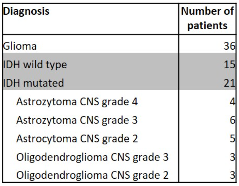
36 adult glioma patients with 7T MRSI scans were included and classified after the WHO CNS5. Details regarding the study cohort are listed in Figure 1.7 Tesla data (Magnetom, Siemens Healthineers) included MP2RAGE, FLAIR and MRSI (acquired in 15 min with a 64 × 64 × 39 matrix with 3.4 mm³ isotropic resolution, a TR of 450 ms and acquisition delay of 1.3 ms) [8,9]. Clinical 3T MRI included FLAIR and pre-/post-contrast T1-weighted sequences (Gadoteridol, 0.1 mmol/kg), used for manual segmentations of contrast-enhancing and non-contrast enhancing tumor parts by a neuroradiologist. IDH mutation status was determined by molecular analysis.
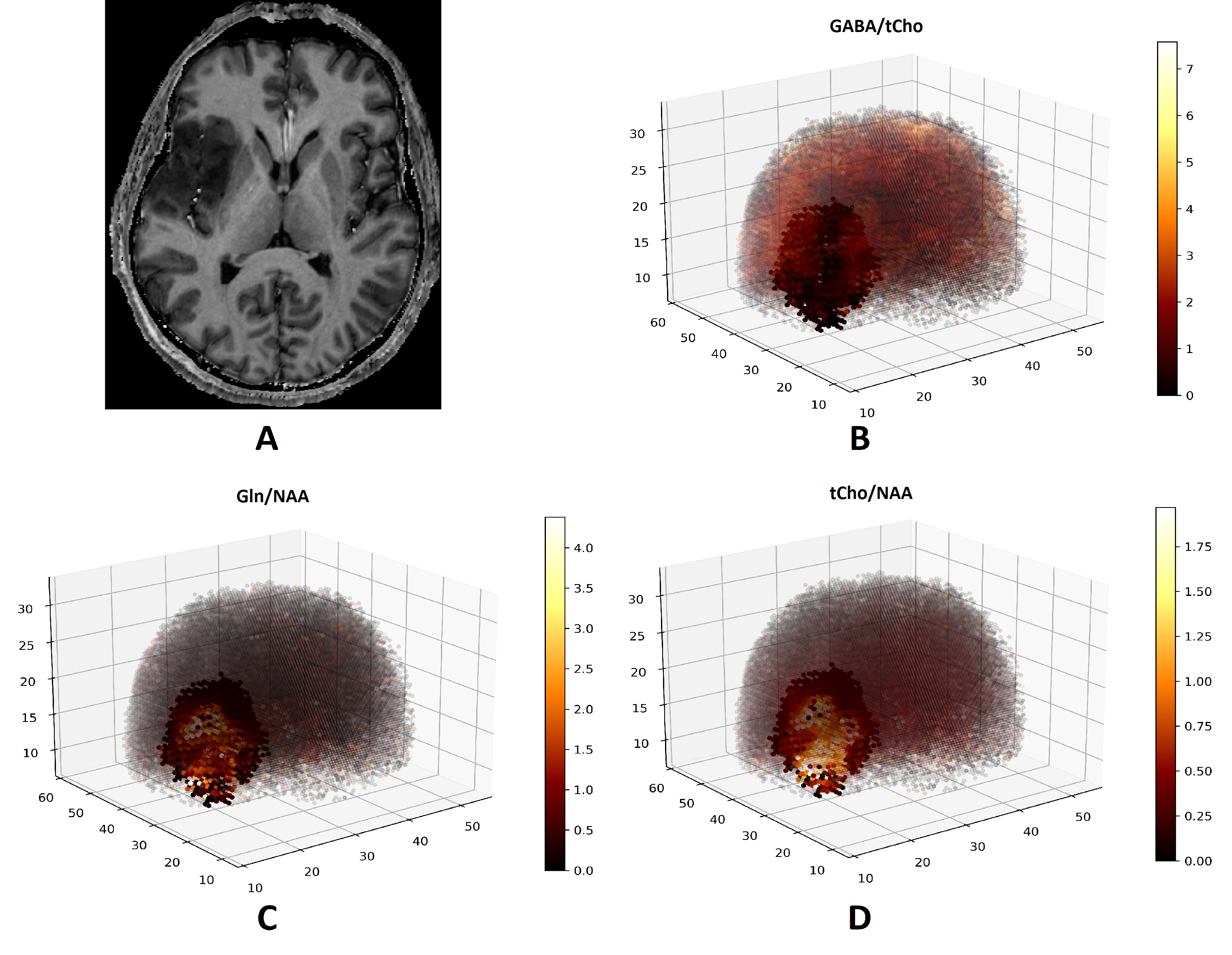
MRSI data were reconstructed using a custom pipeline and quantified with LCModel (basis set with 13 components) [9]. From the results, 50 metabolite ratios (=features) were computed voxel-wise (Figure 2).
The data was divided using a 70:30 stratified train-test split and standardized.
For the prediction of IDH mutation status, voxel feature distribution was analyzed (including mean, standard deviation, quartiles, skewness etc.), resulting in a 550-feature high dimensional feature space per patient. A 5-fold stratified cross validation strategy with embedded feature selection was used to train a Random Forest (RF) classifier.
For voxel-wise CE prediction, a neural network was built consisting of three fully connected layers with L2 regularization and dropout layers to mitigate overfitting and a single output node for classification. A binary focal cross-entropy loss function was employed to handle class imbalance with a 3-fold cross validation strategy used on the 70% training data.
Results
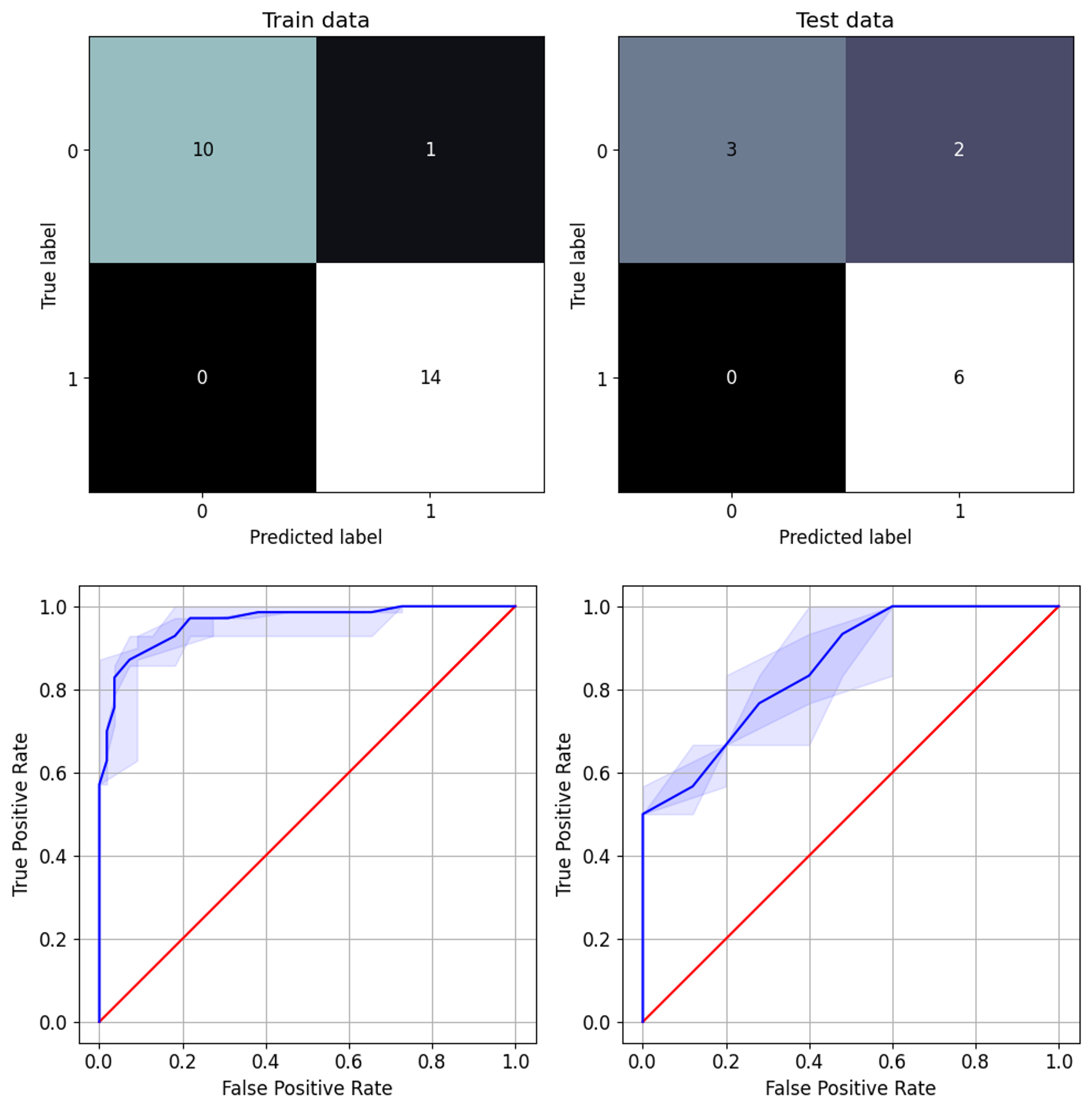
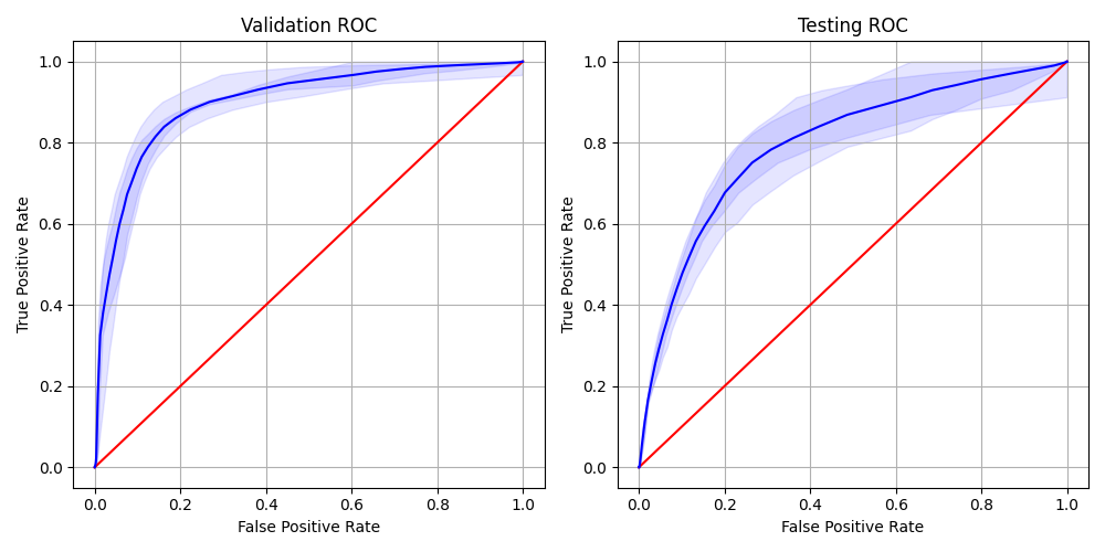
Predicting IDH mutation status: After tuning of hyperparameters, our model achieved high sensitivity (Figure 3), a cross-validation AUC of 0.96 and a test AUC of 0.82.Predicting CE: After tuning the model parameters, the model achieved an average AUC of 0.87 for the training set, 0.82 for the validation set and 0.80 for the testing set. Detailed ROC-curves are shown in Figure 4.
Discussion
In an effort to non-invasively enhance the accuracy of predicting CE and IDH mutation status in diffuse gliomas, we used 7T MRSI in conjunction with machine learning and deep learning techniques thus leveraging cutting-edge technology to contribute to the diagnosis and stratify for treatment strategies in glioma patients. Our models performed well in the training and the testing set (AUC ≥ 0.8) for both, IDH mutation and contrast enhancement prediction.Our IDH classification results are similar to previous work with the same dataset, demonstrating that 7T MRSI is robust with regards to modeling choices [10].
The limitations of this study are the small cohort size and single center study design without an external validation set. Although our test set is derived from the same cohort, we keep it hidden during training to prevent model and developer bias.
Conclusion
Our results show the potential of 7T MRSI to derive useful histopathological and molecular information from biochemical mapping. Application of ML-evaluated 7T MRSI to the research of IDH-inhibitors could offer a way for non-invasive treatment stratification.Acknowledgements
This study was supported by the Austrian Science Fund (FWF) project KLI 1089. The financial support by the Austrian Federal Ministry for Digital and Economic Affairs, the National Foundation for Research, Technology and Development and the Christian Doppler Research Association is gratefully acknowledged.References
1. Hao D, Ai T, Goerner F, Hu X, Runge VM, Tweedle M. MRI contrast agents: basic chemistry and safety. J Magn Reson Imaging. 2012;36(5):1060-1071. doi:10.1002/jmri.23725
2. Forghani R. Adverse Effects of Gadolinium-Based Contrast Agents: Changes in Practice Patterns. Top Magn Reson Imaging. 2016;25(4):163-169. doi:10.1097/RMR.0000000000000095
3. Gulani V, Calamante F, Shellock FG, Kanal E, Reeder SB. Gadolinium deposition in the brain: summary of evidence and recommendations. The Lancet Neurology. 2017;16(7):564-570. doi:10.1016/S1474-4422(17)30158-8
4. Runge VM. Critical Questions Regarding Gadolinium Deposition in the Brain and Body After Injections of the Gadolinium-Based Contrast Agents, Safety, and Clinical Recommendations in Consideration of the EMA’s Pharmacovigilance and Risk Assessment Committee Recommendation for Suspension of the Marketing Authorizations for 4 Linear Agents. Invest Radiol. 2017;52(6):317-323. doi:10.1097/RLI.0000000000000374
5. Louis DN, Perry A, Wesseling P, et al. The 2021 WHO Classification of Tumors of the Central Nervous System: a summary. Neuro Oncol. 2021;23(8):1231-1251. doi:10.1093/neuonc/noab106
6. Brat DJ, Aldape K, Colman H, et al. cIMPACT-NOW update 5: recommended grading criteria and terminologies for IDH-mutant astrocytomas. Acta Neuropathol. 2020;139(3):603-608. doi:10.1007/s00401-020-02127-9
7. Li X, Strasser B, Neuberger U, et al. Deep learning super-resolution magnetic resonance spectroscopic imaging of brain metabolism and mutant isocitrate dehydrogenase glioma. Neuro-Oncology Advances. 2022;4(1):vdac071. doi:10.1093/noajnl/vdac071
8. Hingerl L, Strasser B, Moser P, et al. Clinical High-Resolution 3D-MR Spectroscopic Imaging of the Human Brain at 7 T. Invest Radiol. 2020;55(4):239-248. doi:10.1097/RLI.0000000000000626
9. Hangel G, Cadrien C, Lazen P, et al. High-resolution metabolic imaging of high-grade gliomas using 7T-CRT-FID-MRSI. Neuroimage Clin. 2020;28:102433. doi:10.1016/j.nicl.2020.102433
10. Sharma, S., Cadrien, C., Lazen, P. Glioma Classification With 7T MR Spectroscopic Imaging, Abstract ISMRM 2023, Digital Poster 5197. Published online 2023. https://cds.ismrm.org/protected/23MPresentations/abstracts/5197.html
Figures