3674
A Novel Magnetic Field Gradient Based Thresholding Method to Improve Brain Masking for QSM1Department of Medical Physics and Biomedical Engineering, University College London, London, United Kingdom
Synopsis
Keywords: Electromagnetic Tissue Properties, Quantitative Susceptibility mapping, Echo Planar Imaging, Brain Masking, Artefacts, Electromagnetic Tissue Properties
Motivation: Artefacts in QSM reconstructions can persist even after optimal noise-based masking methods are used, especially in EPI-QSM, due to challenging regions caused by large field gradients rather than noise.
Goal(s): To reduce brain QSM artefacts using a novel, automated brain mask thresholding method.
Approach: Regions with high magnitude of the field gradients (MFG) were removed from the brain mask used for background field removal and/or susceptibility calculation.
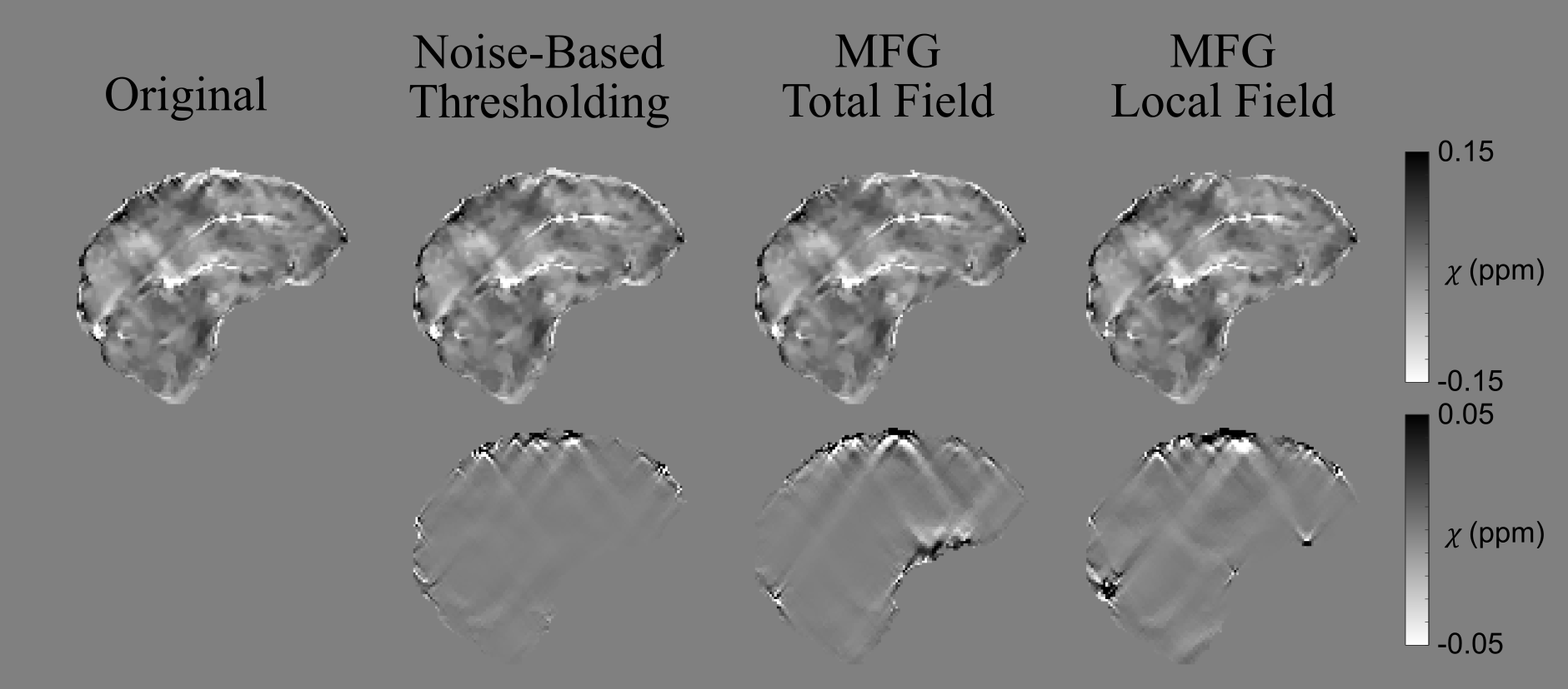
Results: Thresholding the mask based on the MFG of the local field map was superior to thresholding the total field map. MFG-based thresholding reduced artifacts in 2D-EPI and 3D-GRE QSM compared to noise-based thresholding.
Impact: Thresholding the brain mask based on the magnitude of local field gradients improves brain QSM quality by reducing streaking artefacts compared to state-of-the-art noise-based thresholding. This automated MFG-based masking method particularly improves rapid 2D-EPI-QSM as well as conventional 3D-GRE QSM.
Introduction
In QSM, masking is non-trivial and, in regions with large susceptibility ($$$\chi$$$) differences at air-tissue interfaces, large field gradients lead to artefacts, such as streaking, in QSM reconstructions. To improve QSM quality, several masking methods have been introduced including removing voxels with low phase reliability based on noise1,2, signal magnitude3–5 or maximum intensity projections6. Holes in the mask can be filled prior to QSM calculation or assigned susceptibility values from a QSM reconstructed using a non-thresholded mask in a two-pass approach1. However, QSM artefacts can persist due to large field gradients, which can lead to imperfect background field removal and susceptibility calculation. These artefacts are especially prominent in EPI-QSM due to phase accumulation over long echo times (Figure 3, Original QSM). We present a novel, automated method for thresholding the brain mask to improve QSM reconstructions by removing regions with large Magnitude magnetic Field Gradients (MFG)7. This method can be used alone or with other thresholding techniques.Methods
AcquisitionTo test MFG-based masking on EPI-QSM, whole-brain magnitude and phase images were acquired from a healthy volunteer on a Siemens Prisma 3T MR system with a 64-channel head and neck coil using a single-shot 2D-EPI sequence8 with 1.5 mm isotropic resolution; TE=18.2,58.6,98.94,139.3,179.7 ms; TR=25.8 s; TA=41.2 s; FOV=160x160x120; flip angle 90°; partial Fourier 6/8; GRAPPA=2. To investigate if MFG-based thresholding could also improve QSM reconstructions from GRE acquisitions, 3D-GRE whole-brain magnitude and phase images were acquired with 1 mm isotropic resolution; TE1/ΔTE/TE5=12.2/20.9/74.9 ms, chosen for comparability to EPI TEs; TR = 80.0ms; TA = 5min 47s; GRAPPA=4; partial Fourier 6/8 in both PE directions.
MFG Thresholding
The MFG was calculated from7
$$MFG=|ΔB|=\sqrt{\left(\frac{\partial B}{\partial x}\right)^2+\left(\frac{\partial B}{\partial y} \right)^2 + \left(\frac{\partial B}{\partial z} \right)^2}$$
where $$$ΔB = ΔB_{total}$$$ or $$$ΔB_{local}$$$, to investigate whether thresholding MFG calculated from total or local fields (calculated below) was more effective at improving QSM quality.
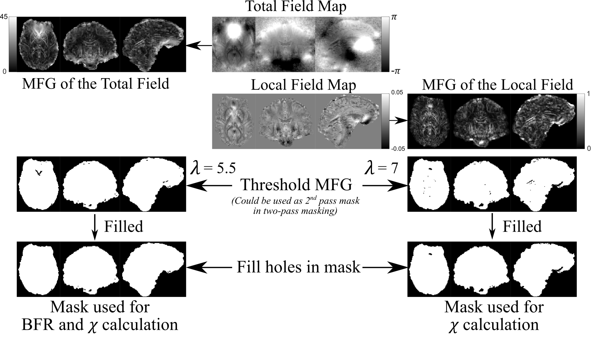
A brain mask, calculated using BET9 on the final echo magnitude image, was thresholded, removing voxels with MFG ≥ mean(MFG)+$$$\lambda\cdot$$$std(MFG), where optimal $$$\lambda$$$ was empirically chosen, using visual inspections of the resulting QSMs, as $$$\lambda=7$$$ and $$$5.5$$$ for the EPI local field map (LFM) and total field map (TFM), respectively, and $$$\lambda=10$$$ and $$$3.5$$$ for the GRE LFM and TFM, respectively. Holes in the mask were then filled (Figure 1) to ensure only brain edges were removed. Note that the unfilled mask could be used for two-pass masking1.
QSM Pipeline
For both EPI and GRE acquisitions, a TFM and a noise map were calculated from a non-linear fit of the complex data over all TEs10. Residual phase wraps were removed using Laplacian unwrapping11 . For comparison, the non-thresholded mask, the hole-filled masks thresholded based on MFG, and at 0.75 times the mean of the inverse noise map1,2 were compared for QSM reconstruction. For 2D-EPI images, background field removal was carried out with 2D+3D V-SHARP12,13 to remove inter-slice phase inconsistencies plus PDF14 to remove residual background fields. For GRE, only PDF was used. QSMs were calculated using non-linear TV15,16 with regularisation parameters $$$1\times 10^{-4}$$$ and $$$2\times10^{-5}$$$ for EPI and GRE, respectively.
Results and Discussion
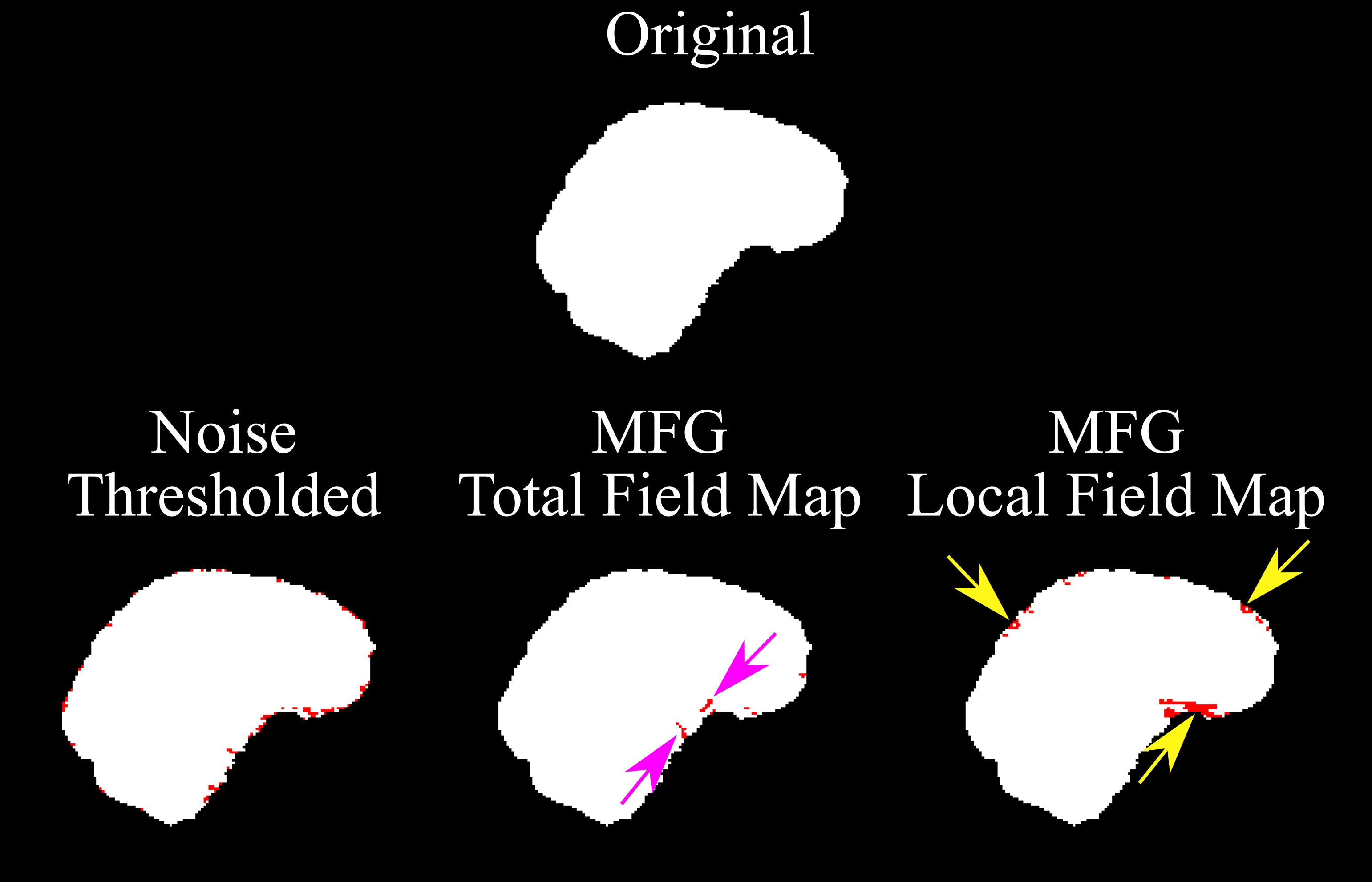
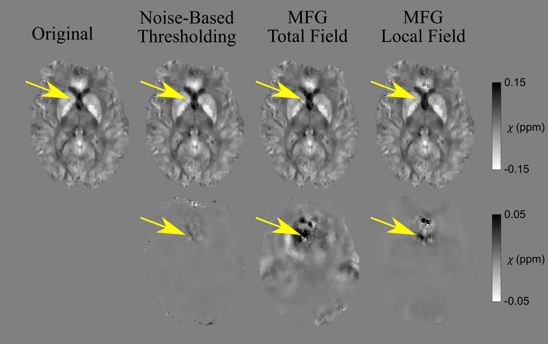
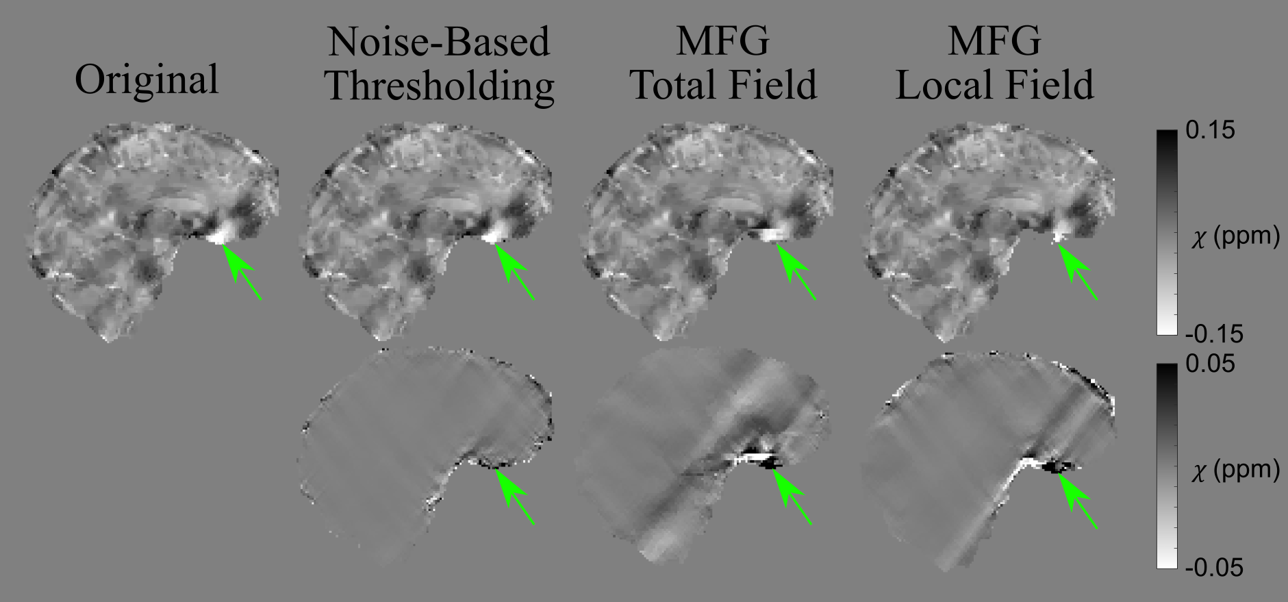
Thresholding the TFM provided a less eroded mask than thresholding the LFM and removed fewer voxels around artefacted areas, especially above the paranasal air sinuses (Figure 2). This is expected given the generally lower spatial frequencies (i.e. smaller MFG) of the total fields compared to the local fields. Thresholding the mask based on the MFG of the LFM led to better artefact removal compared to thresholding the TFM (Figure 4, green arrows), which affected susceptibility values throughout the brain (Figure 3, yellow arrows). This may be because the LFM contains fields localised around the artefacts of interest and, therefore, it was easier to tune for the LFM to solely remove challenging regions without affecting the rest of the brain. For both GRE and EPI, MFG thresholding (on the LFM) reduced streaking more than noise-based thresholding (Figures 4 and 5).Conclusions
Artefacts in QSM reconstructions arising from high susceptibility gradients resulting in large field gradients and phase accumulation were suppressed using masks eroded based on the magnitude of the local field gradients. MFG-based thresholding of the brain mask reduced streaking artefacts more than noise-based thresholding. MFG thresholding the total field map was less effective at removing artefacts and resulted in extensive susceptibility changes throughout the brain that are non-local to the artefact. This novel MFG-based masking method provides an effective way to improve QSM quality, especially for EPI-QSM. It can be combined with two-pass masking methods, alongside noise-based or other thresholding methods to further improve QSM reconstructions.Acknowledgements
Oliver Kiersnowski was supported by EPSRC Doctoral Training Partnership (EP/R513143/1). Karin Shmueli was supported by European Research Council Consolidator Grant DiSCo MRI SFN 770939.References
- Karsa A, Shmueli K. A New, Simple Two-Pass Masking Approach for Streaking Artifact Removal in Any QSM Pipeline (Abstract #2462). In: In Proceedings of ISMRM & SMRT Annual Meeting . ; 2021.
- Karsa A, Punwani S, Shmueli K. An optimized and highly repeatable MRI acquisition and processing pipeline for quantitative susceptibility mapping in the head-and-neck region. Magn Reson Med. 2020;84(6):3206-3222. doi:10.1002/mrm.28377
- Stewart AW, Robinson SD, O’Brien K, et al. QSMxT: Robust masking and artifact reduction for quantitative susceptibility mapping. bioRxiv. Published online 2021:1-19. doi:10.1002/mrm.29048
- Zhao M, Huang L, Zhang Q, Su X, Asundi A, Kemao Q. Quality-Guided Phase Unwrapping Technique: Comparison of Quality Maps and Guiding Strategies.; 2011.
- Bilgic B, Costagli M, Chan KS, et al. Recommended Implementation of Quantitative Susceptibility Mapping for Clinical Research in The Brain: A Consensus of the ISMRM Electro-Magnetic Tissue Properties Study Group. Published online July 5, 2023.
- Boehm C, Diefenbach MN, Makowski MR, Karampinos DC. Improved body quantitative susceptibility mapping by using a variable-layer single-min-cut graph-cut for field-mapping. Magn Reson Med. 2021;85(3):1697-1712. doi:10.1002/mrm.28515
- Cusack R, Russell B, Cox SML, De Panfilis C, Schwarzbauer C, Ansorge R. An evaluation of the use of passive shimming to improve frontal sensitivity in fMRI. Neuroimage. 2005;24(1):82-91. doi:10.1016/j.neuroimage.2004.08.029
- Center for Magnetic Resonance Research Department of Radiology. Multi-Band Accelerated EPI Pulse Sequences. https://www.cmrr.umn.edu/multiband/
- Smith SM. Fast robust automated brain extraction. Hum Brain Mapp. 2002;17(3):143-155. doi:10.1002/hbm.10062
- Liu T, Wisnieff C, Lou M, Chen W, Spincemaille P, Wang Y. Nonlinear formulation of the magnetic field to source relationship for robust quantitative susceptibility mapping. Magn Reson Med. 2013;69(2):467-476. doi:10.1002/mrm.24272
- Schofield MA, Zhu Y. Fast phase unwrapping algorithm for interferometric applications. Opt Lett. Published online 2003. doi:10.1364/ol.28.001194
- Wei H, Zhang Y, Gibbs E, Chen NK, Wang N, Liu C. Joint 2D and 3D phase processing for quantitative susceptibility mapping: application to 2D echo-planar imaging. NMR Biomed. 2017;30(4). doi:10.1002/nbm.3501
- Li W, Wu B, Liu C. Quantitative susceptibility mapping of human brain reflects spatial variation in tissue composition. Neuroimage. 2011;55:1645-1656. doi:10.1016/j.neuroimage.2010.11.088
- Liu T, Khalidov I, de Rochefort L, et al. A novel background field removal method for MRI using projection onto dipole fields (PDF). NMR Biomed. 2011;24(9):1129-1136. doi:10.1002/nbm.1670
- Milovic C, Bilgic B, Zhao B, Acosta-Cabronero J, Tejos C. Fast nonlinear susceptibility inversion with variational regularization. Magn Reson Med. 2018;80(2):814-821. doi:10.1002/mrm.27073
- FANSI Toolbox. https://gitlab.com/cmilovic/FANSI-toolbox
Figures