3673
4D spectral-spatial pulse design for subject-specific fat saturation at 1.5 T1Institute of Radiology, University Hospital Erlangen, Friedrich-Alexander-Universität Erlangen-Nürnberg (FAU), Erlangen, Germany, 2Biomedical Engineering Department, School of Biomedical Engineering and Imaging Sciences, Kings College London, London, United Kingdom, 3Magnetic Resonance, Siemens Healthineers AG, Erlangen, Germany, 4Division of Medical Physics in Radiology, German Cancer Research Center (DKFZ), Heidelberg, Germany
Synopsis
Keywords: Fat & Fat/Water Separation, Spinal Cord, Gradients, Head & Neck /ENT, Parallel Transmission & Multiband, Simulations, System Imperfections: Measurement & Correction
Motivation: Insufficient fat saturation compromises image quality in clinical examinations.
Goal(s): To improve the quality of spectral fat saturation resulting in less residual fat signal in the acquired image.
Approach: Individual 4D spectral-spatial pulses based on subject-specific field maps and a numerically found trajectory are designed within an online workflow. A universal RF solution is also calculated. Performance is compared to Gaussian and SLR pulses on ten cervical spine datasets and one in-vivo measurement.
Results: Simulations show significantly improved fat saturation with individual and universal spectral-spatial pulses, while average water excitation remains low only for individual pulses. The in‑vivo measurement supports the simulation results.
Impact: Customized and universal spectral-spatial fat saturation pulses outperform currently used spectral pre-saturation pulses enabling more definitive interpretation of fat‑suppressed MR images. Potential application to a variety of sequences is straightforward by replacing the pre-saturation pulse with our design.
Introduction
Insufficient fat saturation can deteriorate image quality, e.g., by obscuring pathologies or chemical shift artefacts1,2. There are a variety of fat saturation techniques, each with inherent advantages and disadvantages. Fat-water separation techniques are insensitive to field inhomogeneities but can lead to limitations for different sequences3,4 (e.g., minimum TR, limited contrast, etc.). Spectral pre-saturation methods are dependent on B0/B1 homogeneity, but offer wide versatility5. Advanced spectral-spatial (SPSP) pulse designs compensate for field inhomogeneities while preserving versatile transferability to different sequences6,7. We present an advanced online 4D (3 spatial, 1 spectral) SPSP fat pre-saturation pulse design based on a numerically optimized trajectory and B0/B1 field maps implemented as a “push-button” application. Furthermore, a universal pulse approach is investigated.Methods
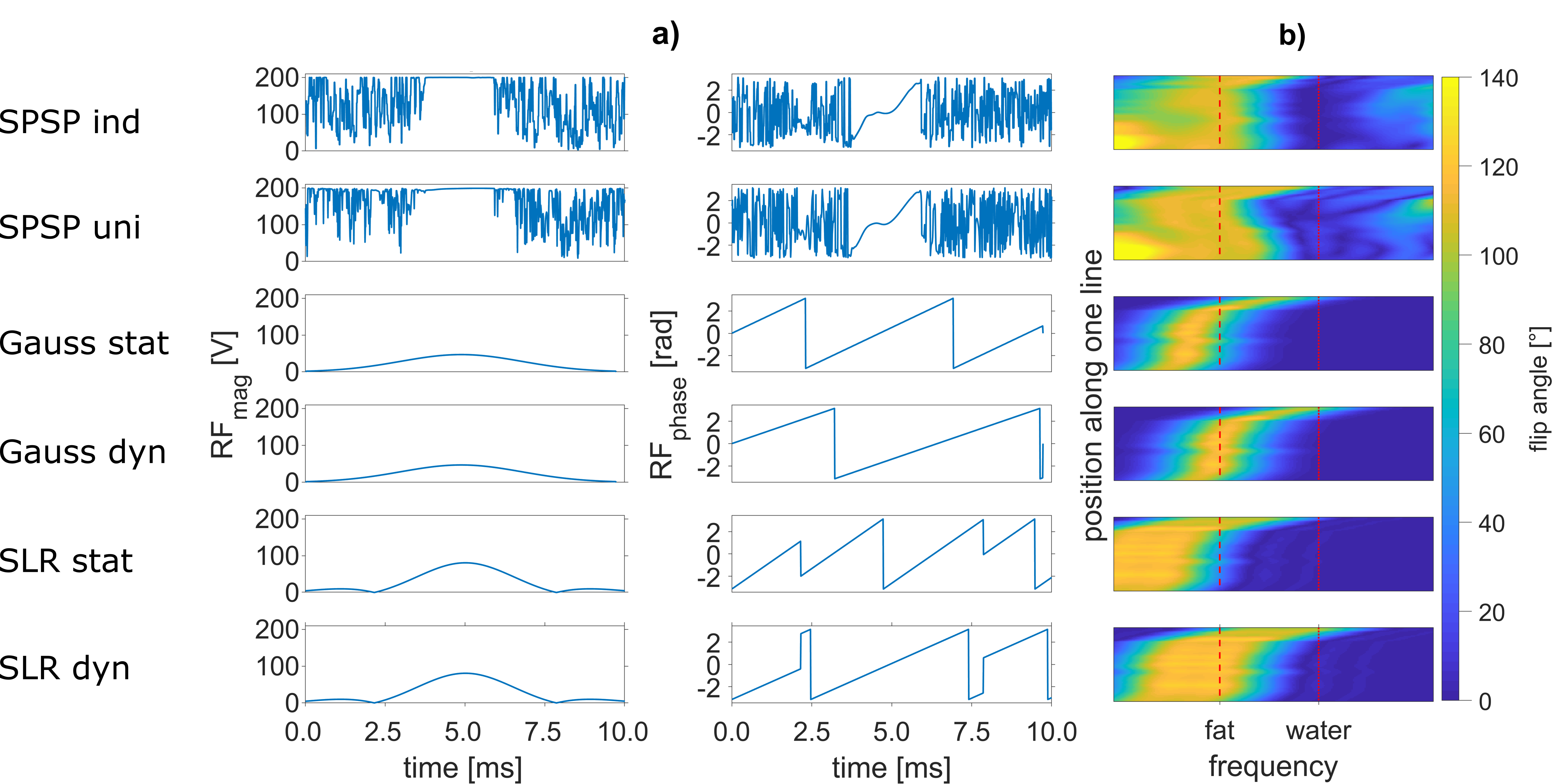
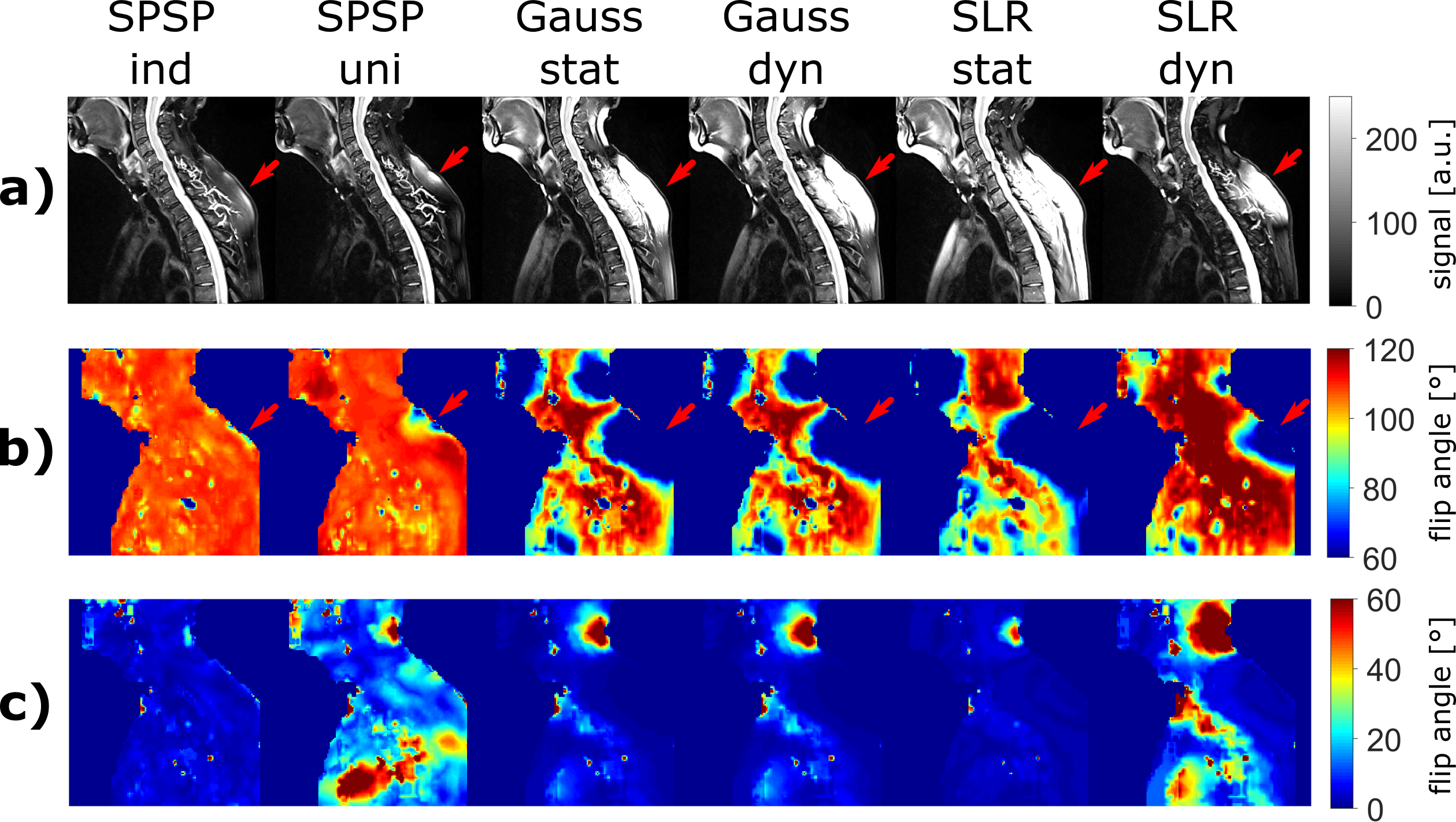
Data is acquired on a 1.5-T MRI system (MAGNETOM Sola, Siemens Healthineers, Erlangen, Germany) in accordance with institutional guidelines. Subjects provided informed consent prior examination. In the proposed method, gradients are applied simultaneously with the RF pulse to manipulate the B0 field during excitation. The gradient and RF shape optimization is based on an interior point solver8, where the target state is 110°/0° for fat (‑3.4 ppm) and water (0.0 ppm), respectively. A universal gradient trajectory and a universal SPSP pulse9 with a duration of 10 ms are trained offline using 15 cervical spine datasets (7 female, median age: 66y, range: 39-78y) including B0 and B1 information and a joint gradient and RF optimization. Furthermore, 10 individual SPSP pulses are optimized, each based on an additional dataset (4 female, median age: 66y, range: 41-81y) and the universal gradient trajectory. All 25 datasets contain field maps with 10 slices and a resolution of (1.76x1.76x3.30) mm3. Gaussian (bandwidth 200 Hz) and SLR10 pulses (bandwidth 300 Hz, https://github.com/mriphysics/Multiband-RF) are employed in two settings. In the static setting, the center frequencies are ‑3.4 ppm (Gauss) and -5.0 ppm (SLR), while in the dynamic setting they are adjusted according to the individual B0 distribution of all slices. To compare performance, complete Bloch simulations are conducted with the same 10 data sets for all pulse types. Fig. 1 shows example pulses with corresponding frequency responses along one line of an exemplary slice. All pulses are implemented in a TSE and tested on one subject (female, 76y).Results
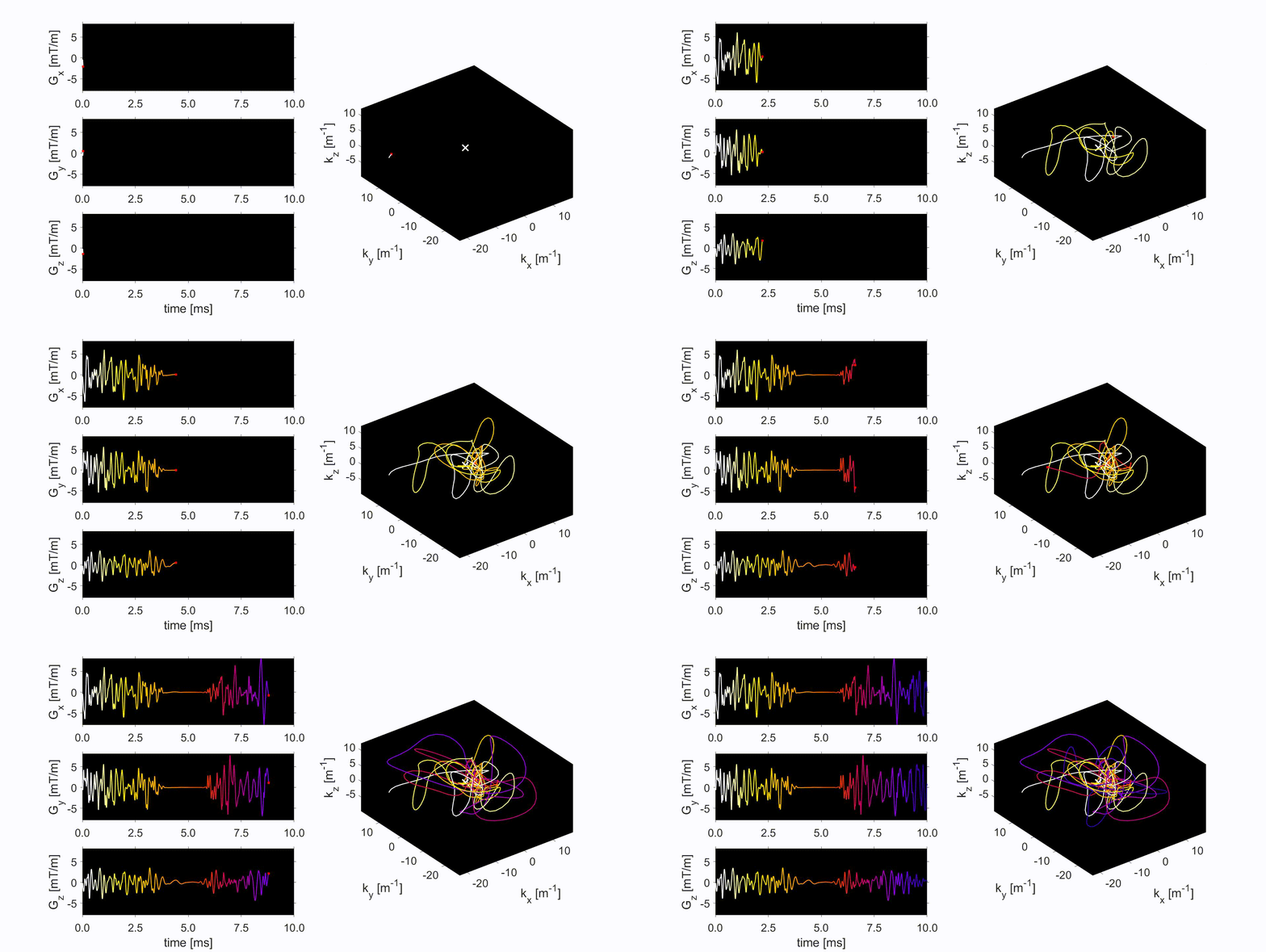
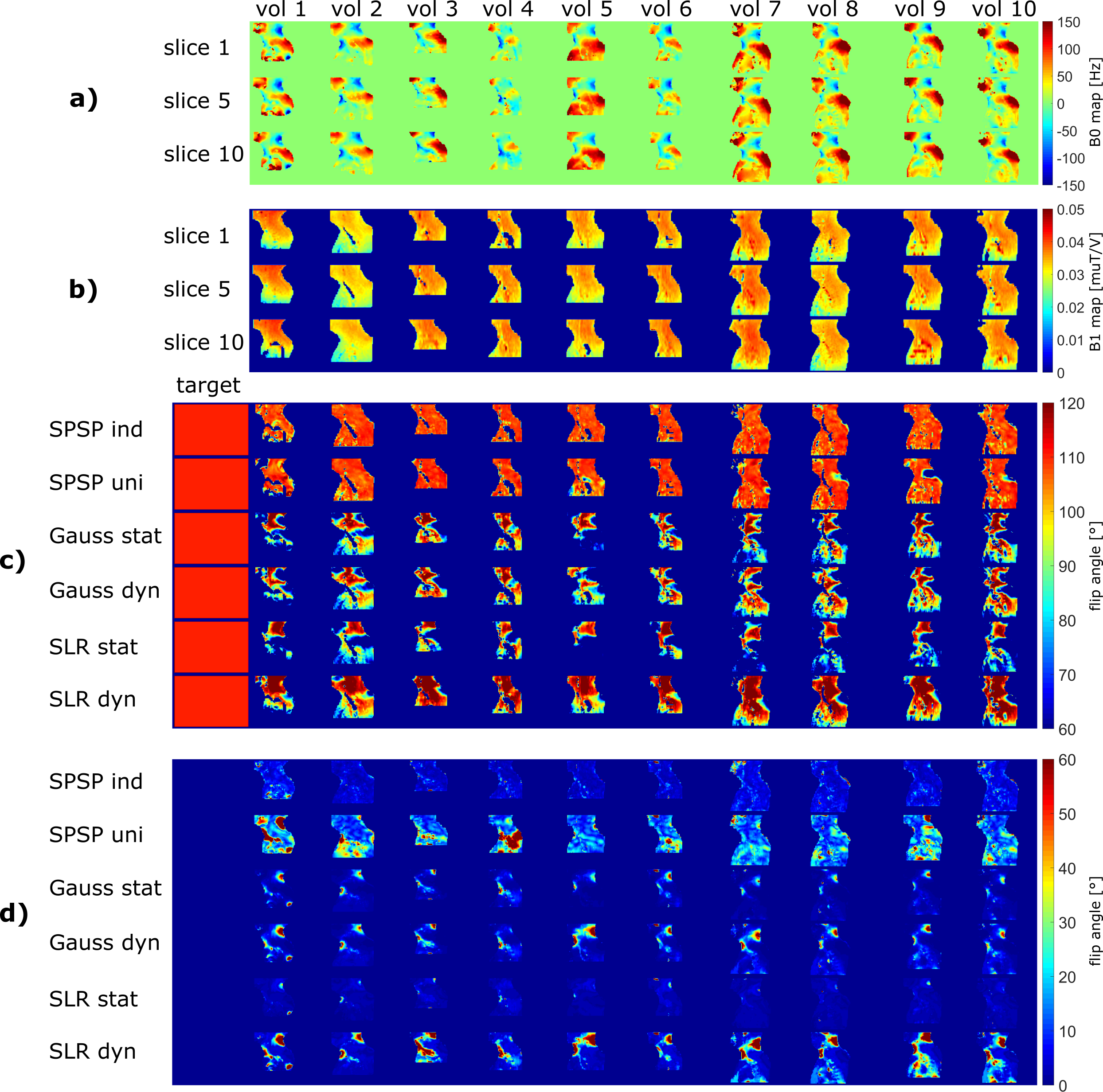
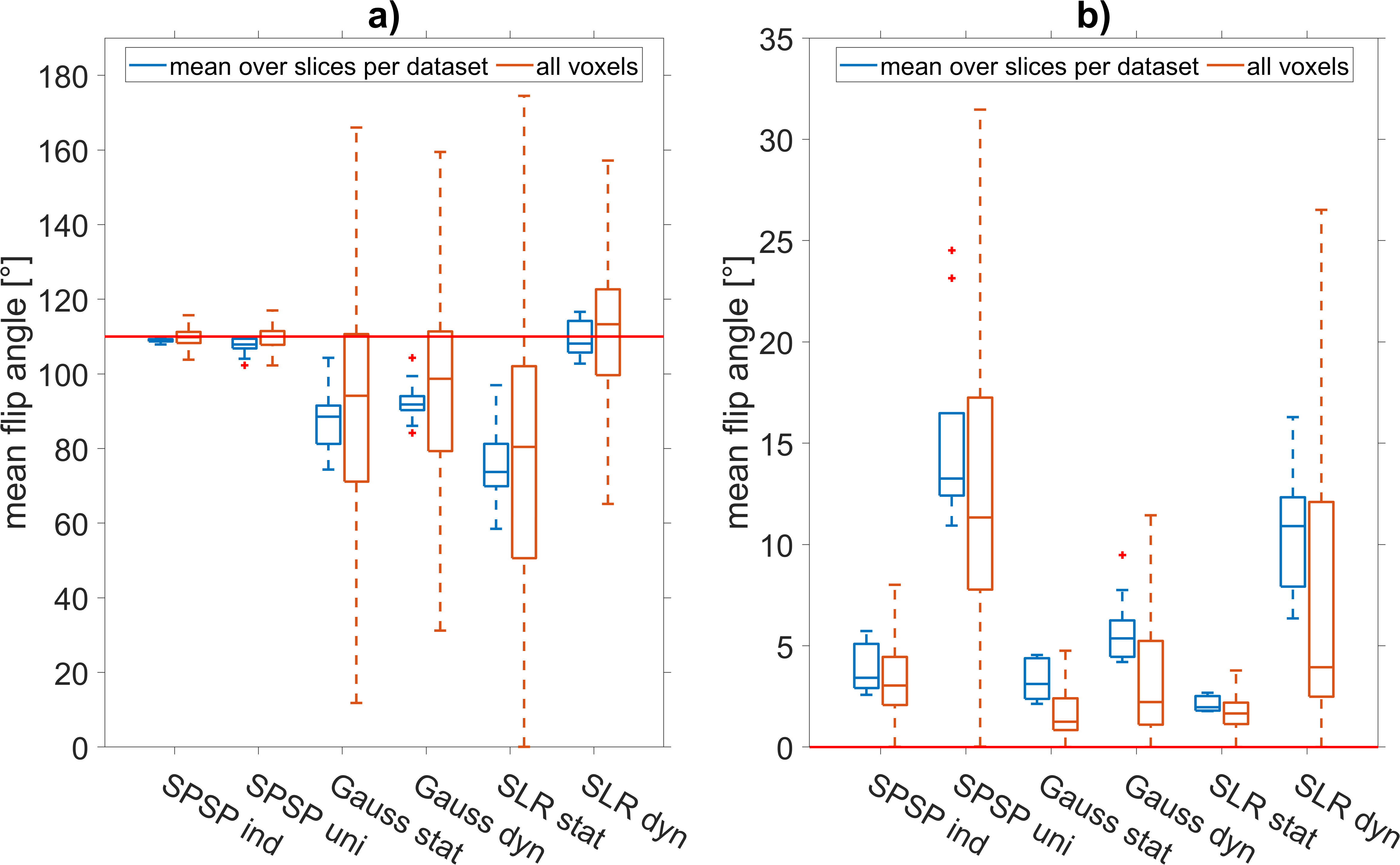
The minimum spatial variations caused by the universal trajectory (Fig. 2) reach 37 mm in-plane and 82 mm in slice direction. The universal pulse and its frequency response are shown in Fig. 1, second row. For all datasets, simulations demonstrate a more homogeneous and stronger fat saturation for SPSP pulses, especially for individually designed pulses (Fig. 3 c). Universal pulse solutions produce good fat saturation across most subjects, with some focal areas of failure, but also generally produce higher (unwanted) water excitation (Fig. 3 d). The average calculation time for individual SPSP pulses on a workstation with Intel Xeon W-2145 @3.70GHz 128 GB RAM is (38.6 ± 8.3) s. Dynamic Gaussian and SLR pulses perform slightly better for fat compared to the static variants but result in stronger excitation of water. Fig. 4 confirms the visual impressions for both an “mean over slices per dataset” and “all voxels” quantitative comparison, with individual SPSP pulses achieving the best fat saturation (a) with a similar level of water excitation as the Gaussian pulse (b). Results of the volunteer measurement are in good agreement with this observation (Fig. 5). This is especially visible in dorsal subcutaneous fat: the universal pulse solution produced clearly worse fat saturation in this region than the individual design but still clearly outperformed the non-SPSP pulses. Online calculation time was 54.0 s.Discussion
Individual and universal SPSP pulses outperform Gaussian and SLR pulses and may therefore serve as an alternative fat saturation method. Further developments in the pulse design such as additional target frequencies could improve robustness (e.g., for motion between B0/B1 acquisition and actual scan). Universal SPSP pulses do not require additional online preparation, but currently show non-negligible water excitation. The volunteer measurement indicates good overall agreement with simulations, with slight deviations for individual SPSP pulses. Further measurements need to be conducted to confirm agreement. The trajectory meets the expectations of low spatial frequencies in the field maps, especially in slice directions. More conservative constraints (e.g. lower slew-rate) might lead to simpler trajectories and thus more robust pulses.Conclusion
This work provides a 4D spectral-spatial pulse design for subject-specific fat saturation implemented directly on the scanner. Simulations show promising results for individual SPSP pulses supported by one in-vivo measurement. Further investigations could result in universal SPSP pulses being a viable alternative.Acknowledgements
No acknowledgement found.References
1. Bley TA, Wieben O, Francois CJ, et al. Fat and water magnetic resonance imaging. J Magn Reson Imaging 2010;31(1):4-18.
2. Delfaut EM, Beltran J, Johnson G, et al. Fat suppression in MR imaging: techniques and pitfalls. Radiographics 1999;19(2):373-382.
3. Cheng C, Zou C, Liang C, et al. Fat-water separation using a region-growing algorithm with self-feeding phasor estimation. Magn Reson Med 2017;77(6):2390-2401.
4. Basty N, Thanaj M, Cule M, et al. Artifact-free fat-water separation in Dixon MRI using deep learning. J Big Data 2023;10(1):4.
5. Haase A, Frahm J, Hanicke W, et al. 1H NMR chemical shift selective (CHESS) imaging. Phys Med Biol 1985;30(4):341-344.
6. Zhao F, Nielsen JF, Noll DC. Four dimensional spectral-spatial fat saturation pulse design. Magn Reson Med 2014;72(6):1637-1647.
7. Levy S, Herrler J, Liebert A, et al. Clinically compatible subject-specific dynamic parallel transmit pulse design for homogeneous fat saturation and water-excitation at 7T: Proof-of-concept for CEST MRI of the brain. Magn Reson Med 2023;89(1):77-94.
8. Majewski K. Simultaneous optimization of radio frequency and gradient waveforms with exact Hessians and slew rate constraints applied to kT-points excitation. J Magn Reson 2021;326:106941.
9. Gras V, Vignaud A, Amadon A, et al. Universal pulses: A new concept for calibration-free parallel transmission. Magn Reson Med 2017;77(2):635-643.
10. Pauly J, Le Roux P, Nishimura D, et al. Parameter relations for the Shinnar-Le Roux selective excitation pulse design algorithm [NMR imaging]. IEEE Trans Med Imaging 1991;10(1):53-65.
Figures